|
Nephelomys Moerex
''Nephelomys moerex'' is a species of rodent in the genus ''Nephelomys'' of family Cricetidae.Weksler et al., 2006, p. 18 The type locality is at Mindo in western Ecuador, where it has been recorded together with three other rodents of the oryzomyine group, '' Sigmodontomys aphrastus'', ''Mindomys hammondi'', and ''Handleyomys alfaroi'', as well as three opossums, ''Chironectes minimus'' and unidentified species of ''Didelphis'' and ''Marmosa''. Mindo is a "tiny agricultural community" located at 0°02'S, 78°48'W and above sea level. It was originally described by Oldfield Thomas as a subspecies of '' Oryzomys albigularis''. It remained synonymized under this species until it was recognized as a separate species when the genus ''Nephelomys'' was established for ''Oryzomys albigularis'' and related species in 2006. Unlike in the type species of the genus, ''N. albigularis'', the lacrimal bone of the skull is connected primarily to the maxillary bone, not equally to the maxillar ... [...More Info...] [...Related Items...] OR: [Wikipedia] [Google] [Baidu] |
Oldfield Thomas
Michael Rogers Oldfield Thomas (21 February 1858 – 16 June 1929) was a British zoologist. Career Thomas worked at the Natural History Museum on mammals, describing about 2,000 new species and subspecies for the first time. He was appointed to the museum secretary's office in 1876, transferring to the zoological department in 1878. In 1891, Thomas married Mary Kane, daughter of Sir Andrew Clark, heiress to a small fortune, which gave him the finances to hire mammal collectors and present their specimens to the museum. He also did field work himself in Western Europe and South America. His wife shared his interest in natural history, and accompanied him on collecting trips. In 1896, when William Henry Flower took control of the department, he hired Richard Lydekker Richard Lydekker (; 25 July 1849 – 16 April 1915) was an English naturalist, geologist and writer of numerous books on natural history. Biography Richard Lydekker was born at Tavistock Square in London. ... [...More Info...] [...Related Items...] OR: [Wikipedia] [Google] [Baidu] |
Synonym (taxonomy)
The Botanical and Zoological Codes of nomenclature treat the concept of synonymy differently. * In botanical nomenclature, a synonym is a scientific name that applies to a taxon that (now) goes by a different scientific name. For example, Linnaeus was the first to give a scientific name (under the currently used system of scientific nomenclature) to the Norway spruce, which he called ''Pinus abies''. This name is no longer in use, so it is now a synonym of the current scientific name, ''Picea abies''. * In zoology, moving a species from one genus to another results in a different binomen, but the name is considered an alternative combination rather than a synonym. The concept of synonymy in zoology is reserved for two names at the same rank that refers to a taxon at that rank - for example, the name ''Papilio prorsa'' Linnaeus, 1758 is a junior synonym of ''Papilio levana'' Linnaeus, 1758, being names for different seasonal forms of the species now referred to as ''Araschnia le ... [...More Info...] [...Related Items...] OR: [Wikipedia] [Google] [Baidu] |
Foramina Of The Skull
This article lists foramina that occur in the human body. __TOC__ Skull The human skull has numerous openings (foramina), through which cranial nerves, arteries, veins, and other structures pass. These foramina vary in size and number, with age. Gray193.png , Base of the skull, upper surface Gray187.png , Base of the skull, inferior surface, attachment of muscles marked in red Spine Within the vertebral column (spine) of vertebrates, including the human spine, each bone has an opening at both its top and bottom to allow nerves, arteries, veins, etc. to pass through. Other * Apical foramen, the opening at the tip of the root of a tooth * Foramen ovale (heart), an opening between the venous and arterial sides of the fetal heart * Foramen transversarium, one of a pair of openings in each cervical vertebra, in which the vertebral artery travels * Greater sciatic foramen, a major foramen of the pelvis * Interventricular foramen, channels connecting ventricles in ... [...More Info...] [...Related Items...] OR: [Wikipedia] [Google] [Baidu] |
Alisphenoid
The greater wing of the sphenoid bone, or alisphenoid, is a bony process of the sphenoid bone; there is one on each side, extending from the side of the body of the sphenoid and curving upward, laterally, and backward. Structure The greater wings of the sphenoid are two strong processes of bone, which arise from the sides of the body, and are curved upward, laterally, and backward; the posterior part of each projects as a triangular process that fits into the angle between the squamous and the petrous part of the temporal bone and presents at its apex a downward-directed process, the spine of sphenoid bone. Cerebral surface The superior or cerebral surface of each greater wing ig. 1forms part of the middle cranial fossa; it is deeply concave, and presents depressions for the convolutions of the temporal lobe of the brain. It has a number of foramina (holes) in it: * The foramen rotundum is a circular aperture at its anterior and medial part; it transmits the maxillary nerve. ... [...More Info...] [...Related Items...] OR: [Wikipedia] [Google] [Baidu] |
Alisphenoid Strut
In some rodents, the alisphenoid strut is an extension of the alisphenoid bone that separates two foramina in the skull, the masticatory–buccinator foramen and the foramen ovale accessorium. The presence or absence of this strut is variable between species, but also within them; some Oryzomyini Oryzomyini is a tribe of rodents in the subfamily Sigmodontinae of the family Cricetidae. It includes about 120 species in about thirty genera,Weksler et al., 2006, table 1 distributed from the eastern United States to the southernmost parts of ... even have a strut on one side of the skull but lack it on the other side.Weksler, 2006, p. 38 References {{reflist Literature cited *Weksler, M. 2006Phylogenetic relationships of oryzomyine rodents (Muroidea: Sigmodontinae): separate and combined analyses of morphological and molecular data Bulletin of the American Museum of Natural History 296:1–149. Rodent anatomy ... [...More Info...] [...Related Items...] OR: [Wikipedia] [Google] [Baidu] |
Nephelomys Nimbosus
''Nephelomys nimbosus'' is a species of rodent in the genus ''Nephelomys'' of family Cricetidae.Weksler et al., 2006, p. 18 Its type locality is at San Antonio on the northeastern slope of the Tungurahua in the Andes of Ecuador, at an altitude of about . The type series included five individuals.Anthony, 1926, p. 4 The fur of the upperparts is brown to blackish, becoming lighter towards the sides. The underparts are grayish, with a white patch at the throat. The long tail virtually lacks hairs and is darker above than below. In the holotype, the total length is , the head and body length is , the combined length of the tail vertebrae is , the hindfoot length (including claws) is , and the length of the skull is . In most species of ''Nephelomys'', the posterolateral palatal pits, perforations of the palate near the third molar, are conspicuous and receded into a depression or fossa, but in ''N. nimbosus'' and '' N. caracolus'', they are much smaller. It was first described, in 1 ... [...More Info...] [...Related Items...] OR: [Wikipedia] [Google] [Baidu] |
Molar (tooth)
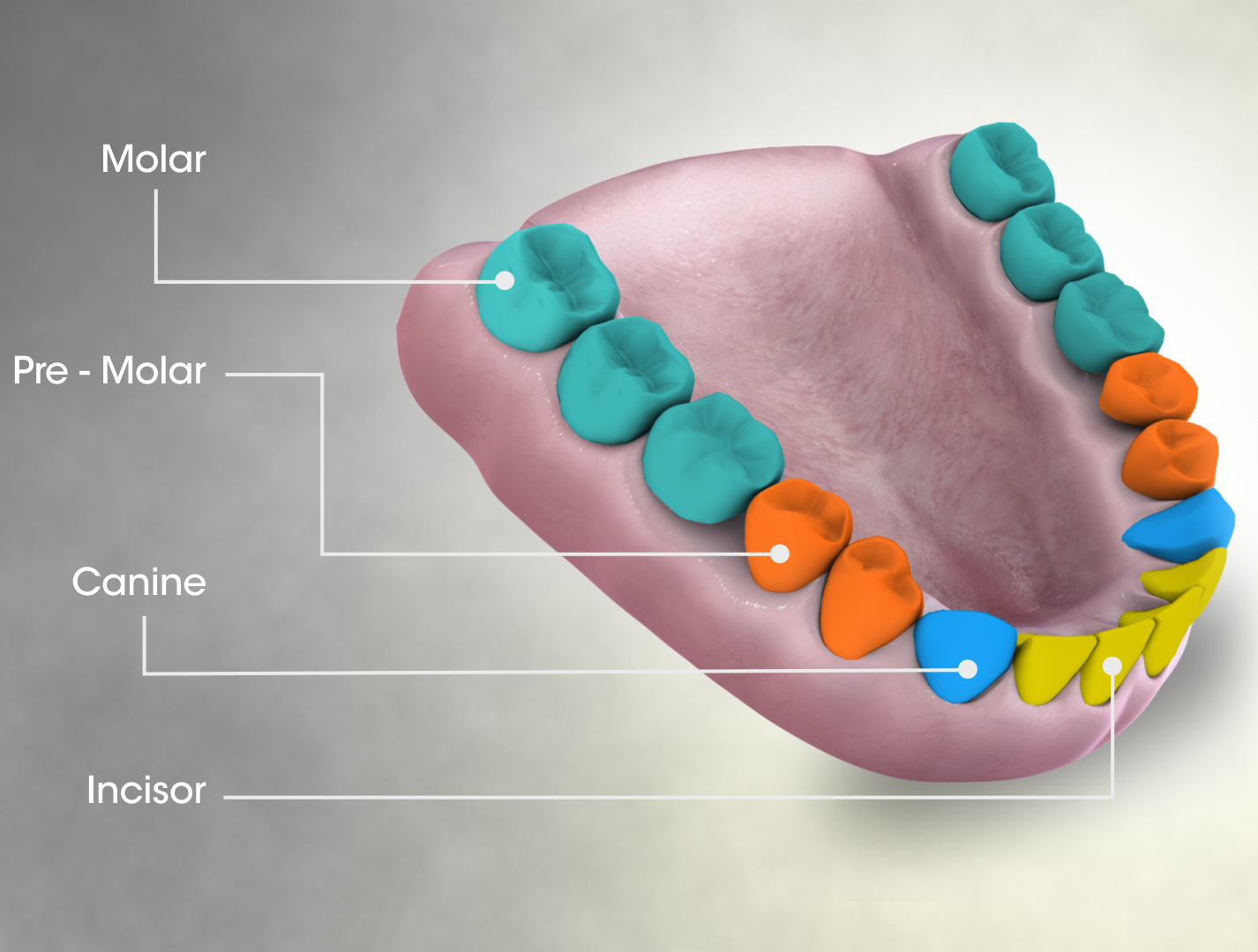
The molars or molar teeth are large, flat teeth at the back of the mouth. They are more developed in mammals. They are used primarily to grind food during chewing. The name ''molar'' derives from Latin, ''molaris dens'', meaning "millstone tooth", from ''mola'', millstone and ''dens'', tooth. Molars show a great deal of diversity in size and shape across mammal groups. The third molar of humans is sometimes vestigial. Human anatomy In humans, the molar teeth have either four or five cusps. Adult humans have 12 molars, in four groups of three at the back of the mouth. The third, rearmost molar in each group is called a wisdom tooth. It is the last tooth to appear, breaking through the front of the gum at about the age of 20, although this varies from individual to individual. Race can also affect the age at which this occurs, with statistical variations between groups. In some cases, it may not even erupt at all. The human mouth contains upper (maxillary) and lower (mandib ... [...More Info...] [...Related Items...] OR: [Wikipedia] [Google] [Baidu] |
Incisor
Incisors (from Latin ''incidere'', "to cut") are the front teeth present in most mammals. They are located in the premaxilla above and on the mandible below. Humans have a total of eight (two on each side, top and bottom). Opossums have 18, whereas armadillos have none. Structure Adult humans normally have eight incisors, two of each type. The types of incisor are: * maxillary central incisor (upper jaw, closest to the center of the lips) * maxillary lateral incisor (upper jaw, beside the maxillary central incisor) * mandibular central incisor (lower jaw, closest to the center of the lips) * mandibular lateral incisor (lower jaw, beside the mandibular central incisor) Children with a full set of deciduous teeth (primary teeth) also have eight incisors, named the same way as in permanent teeth. Young children may have from zero to eight incisors depending on the stage of their tooth eruption and tooth development. Typically, the mandibular central incisors erupt first, followed ... [...More Info...] [...Related Items...] OR: [Wikipedia] [Google] [Baidu] |
Palate
The palate () is the roof of the mouth in humans and other mammals. It separates the oral cavity from the nasal cavity. A similar structure is found in crocodilians, but in most other tetrapods, the oral and nasal cavities are not truly separated. The palate is divided into two parts, the anterior, bony hard palate and the posterior, fleshy soft palate (or velum). Structure Innervation The maxillary nerve branch of the trigeminal nerve supplies sensory innervation to the palate. Development The hard palate forms before birth. Variation If the fusion is incomplete, a cleft palate results. Function When functioning in conjunction with other parts of the mouth, the palate produces certain sounds, particularly velar, palatal, palatalized, postalveolar, alveolopalatal, and uvular consonants. History Etymology The English synonyms palate and palatum, and also the related adjective palatine (as in palatine bone), are all from the Latin ''palatum'' via Old French ''palat ... [...More Info...] [...Related Items...] OR: [Wikipedia] [Google] [Baidu] |
Incisive Foramen
In the human mouth, the incisive foramen (also known as: "''anterior palatine foramen''", or "''nasopalatine foramen''") is the opening of the incisive canals on the hard palate immediately behind the incisor teeth. It gives passage to blood vessels and nerves. The incisive foramen is situated within the incisive fossa of the maxilla. The incisive foramen is used as an anatomical landmark for defining the severity of cleft lip and cleft palate. The incisive foramen exists in a variety of species. Structure The incisive foramen is a funnel-shaped opening of the in the bone of the oral hard palate representing the inferior termination of the incisive canal. An oral prominence - the incisive papilla - overlies the incisive fossa. The incisive foramen is situated immediately behind the incisor teeth, and in between the two premaxillae. Contents The incisive foramen allows for blood vessels and nerves to pass. These include: * the pterygopalatine nerves to the hard palate. * ... [...More Info...] [...Related Items...] OR: [Wikipedia] [Google] [Baidu] |
Frontal Bone
The frontal bone is a bone in the human skull. The bone consists of two portions.''Gray's Anatomy'' (1918) These are the vertically oriented squamous part, and the horizontally oriented orbital part, making up the bony part of the forehead, part of the bony orbital cavity holding the eye, and part of the bony part of the nose respectively. The name comes from the Latin word ''frons'' (meaning " forehead"). Structure of the frontal bone The frontal bone is made up of two main parts. These are the squamous part, and the orbital part. The squamous part marks the vertical, flat, and also the biggest part, and the main region of the forehead. The orbital part is the horizontal and second biggest region of the frontal bone. It enters into the formation of the roofs of the orbital and nasal cavities. Sometimes a third part is included as the nasal part of the frontal bone, and sometimes this is included with the squamous part. The nasal part is between the brow ridges, and ends in ... [...More Info...] [...Related Items...] OR: [Wikipedia] [Google] [Baidu] |

.jpg)