|
Nasal Cavity
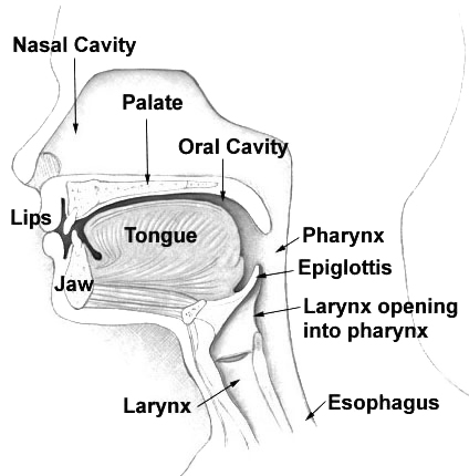
The nasal cavity is a large, air-filled space above and behind the nose in the middle of the face. The nasal septum divides the cavity into two cavities, also known as fossae. Each cavity is the continuation of one of the two nostrils. The nasal cavity is the uppermost part of the respiratory system and provides the nasal passage for inhaled air from the nostrils to the nasopharynx and rest of the respiratory tract. The paranasal sinuses surround and drain into the nasal cavity. Structure The term "nasal cavity" can refer to each of the two cavities of the nose, or to the two sides combined. The lateral wall of each nasal cavity mainly consists of the maxilla. However, there is a deficiency that is compensated for by the perpendicular plate of the palatine bone, the medial pterygoid plate, the labyrinth of ethmoid and the inferior concha. The paranasal sinuses are connected to the nasal cavity through small orifices called ostia. Most of these ostia communicate with the n ... [...More Info...] [...Related Items...] OR: [Wikipedia] [Google] [Baidu] |
Paranasal Sinus
Paranasal sinuses are a group of four paired air-filled spaces that surround the nasal cavity. The maxillary sinuses are located under the eyes; the frontal sinuses are above the eyes; the ethmoidal sinuses are between the eyes and the sphenoidal sinuses are behind the eyes. The sinuses are named for the facial bones in which they are located. Structure Humans possess four pairs of paranasal sinuses, divided into subgroups that are named according to the bones within which the sinuses lie. They are all innervated by branches of the trigeminal nerve (CN V). * The maxillary sinuses, the largest of the paranasal sinuses, are under the eyes, in the maxillary bones (open in the back of the semilunar hiatus of the nose). They are innervated by the maxillary nerve (CN V2). * The frontal sinuses, superior to the eyes, in the frontal bone, which forms the hard part of the forehead. They are innervated by the ophthalmic nerve (CN V1). * The ethmoidal sinuses, which are formed from sever ... [...More Info...] [...Related Items...] OR: [Wikipedia] [Google] [Baidu] |
Vomeronasal Organ
The vomeronasal organ (VNO), or Jacobson's organ, is the paired auxiliary olfactory (smell) sense organ located in the soft tissue of the nasal septum, in the nasal cavity just above the roof of the mouth (the hard palate) in various tetrapods. The name is derived from the fact that it lies adjacent to the unpaired vomer bone (from Latin 'plowshare', for its shape) in the nasal septum. It is present and functional in all snakes and lizards, and in many mammals, including cats, dogs, cattle, pigs, and some primates. Some humans may have physical remnants of a VNO, but it is vestigial and non-functional. The VNO contains the cell bodies of sensory neurons which have receptors that detect specific non-volatile (liquid) organic compounds which are conveyed to them from the environment. These compounds emanate from prey, predators, and the compounds called sex pheromones from potential mates. Activation of the VNO triggers an appropriate behavioral response to the presence of ... [...More Info...] [...Related Items...] OR: [Wikipedia] [Google] [Baidu] |
Olfactory Epithelium
The olfactory epithelium is a specialized epithelial tissue inside the nasal cavity that is involved in smell. In humans, it measures 9 cm2 and lies on the roof of the nasal cavity about 7 cm above and behind the nostrils. The olfactory epithelium is the part of the olfactory system directly responsible for detecting odors. Structure Olfactory epithelium consists of four distinct cell types: * Olfactory sensory neurons * Supporting cells * Basal cells * Brush cells Olfactory sensory neurons The olfactory receptor neurons are sensory neurons of the olfactory epithelium. They are bipolar neurons and their apical poles express odorant receptors on non-motile cilia at the ends of the dendritic knob, which extend out into the airspace to interact with odorants. Odorant receptors bind odorants in the airspace, which are made soluble by the serous secretions from olfactory glands located in the lamina propria of the mucosa.Ross, MH, ''Histology: A Text and Atlas'', 5th Ed ... [...More Info...] [...Related Items...] OR: [Wikipedia] [Google] [Baidu] |
Nasal Concha
In anatomy, a nasal concha (), plural conchae (), also called a nasal turbinate or turbinal, is a long, narrow, curled shelf of bone that protrudes into the breathing passage of the nose in humans and various animals. The conchae are shaped like an elongated seashell, which gave them their name (Latin ''concha'' from Greek ''κόγχη''). A concha is any of the scrolled spongy bones of the nasal passages in vertebrates.''Anatomy of the Human Body'' Gray, Henry (1918) The Nasal Cavity. In humans, the conchae divide the nasal airway into four groove-like air passages, and are responsible for forcing inhaled air to flow in a steady, regular pattern around the largest possible of |
Posterior Nasal Apertures
The choanae (singular choana), posterior nasal apertures or internal nostrils are two openings found at the back of the nasal passage between the nasal cavity and the throat in tetrapods, including humans and other mammals (as well as crocodilians and most skinks). They are considered one of the most important synapomorphies of tetrapodomorphs, that allowed the passage from water to land. In animals with secondary palates, they allow breathing when the mouth is closed. Janvier, Philippe (2004) "Wandering nostrils". ''Nature'', 432 (7013): 23–24. In tetrapods without secondary palates their function relates primarily to olfaction (sense of smell). The choanae are separated in two by the vomer. Boundaries A choana is the opening between the nasal cavity and the nasopharynx. It is therefore not a structure but a space bounded as follows: * anteriorly and inferiorly by the horizontal plate of palatine bone, * superiorly and posteriorly by the sphenoid bone * laterally by th ... [...More Info...] [...Related Items...] OR: [Wikipedia] [Google] [Baidu] |
Nasal Hair
Nasal hair or nose hair, is the hair in the human nose. Adult humans have hair in the nostrils. Nasal hair functions include filtering foreign particles from entering the nasal cavity, and collecting moisture. In support of the first function, the results of a 2011 study indicated that increased nasal hair density decreases the development of asthma in those who have seasonal rhinitis, possibly due to an increased capacity of the hair in the nostrils to filter out pollen and other allergens. Nasal hair is different from the cilia of the ciliated lining of the nasal cavity. These cilia are microtubular-based structures that are found in the respiratory tract, involved in the mucociliary clearance mechanism. Removal A number of devices have been sold to trim nasal hair, including miniature rotary clippers and attachments for electric shavers. The trimmers shorten the hair to such lengths that they do not appear outside of the nasal passage. A pair of tweezers may also be used to ... [...More Info...] [...Related Items...] OR: [Wikipedia] [Google] [Baidu] |
Skin
Skin is the layer of usually soft, flexible outer tissue covering the body of a vertebrate animal, with three main functions: protection, regulation, and sensation. Other cuticle, animal coverings, such as the arthropod exoskeleton, have different cellular differentiation, developmental origin, structure and chemical composition. The adjective cutaneous means "of the skin" (from Latin ''cutis'' 'skin'). In mammals, the skin is an organ (anatomy), organ of the integumentary system made up of multiple layers of ectodermal tissue (biology), tissue and guards the underlying muscles, bones, ligaments, and internal organs. Skin of a different nature exists in amphibians, reptiles, and birds. Skin (including cutaneous and subcutaneous tissues) plays crucial roles in formation, structure, and function of extraskeletal apparatus such as horns of bovids (e.g., cattle) and rhinos, cervids' antlers, giraffids' ossicones, armadillos' osteoderm, and os penis/os clitoris. All mammals have som ... [...More Info...] [...Related Items...] OR: [Wikipedia] [Google] [Baidu] |
Epithelium
Epithelium or epithelial tissue is one of the four basic types of animal tissue, along with connective tissue, muscle tissue and nervous tissue. It is a thin, continuous, protective layer of compactly packed cells with a little intercellular matrix. Epithelial tissues line the outer surfaces of organs and blood vessels throughout the body, as well as the inner surfaces of cavities in many internal organs. An example is the epidermis, the outermost layer of the skin. There are three principal shapes of epithelial cell: squamous (scaly), columnar, and cuboidal. These can be arranged in a singular layer of cells as simple epithelium, either squamous, columnar, or cuboidal, or in layers of two or more cells deep as stratified (layered), or ''compound'', either squamous, columnar or cuboidal. In some tissues, a layer of columnar cells may appear to be stratified due to the placement of the nuclei. This sort of tissue is called pseudostratified. All glands are made up of epithe ... [...More Info...] [...Related Items...] OR: [Wikipedia] [Google] [Baidu] |
Cartilage
Cartilage is a resilient and smooth type of connective tissue. In tetrapods, it covers and protects the ends of long bones at the joints as articular cartilage, and is a structural component of many body parts including the rib cage, the neck and the bronchial tubes, and the intervertebral discs. In other taxa, such as chondrichthyans, but also in cyclostomes, it may constitute a much greater proportion of the skeleton. It is not as hard and rigid as bone, but it is much stiffer and much less flexible than muscle. The matrix of cartilage is made up of glycosaminoglycans, proteoglycans, collagen fibers and, sometimes, elastin. Because of its rigidity, cartilage often serves the purpose of holding tubes open in the body. Examples include the rings of the trachea, such as the cricoid cartilage and carina. Cartilage is composed of specialized cells called chondrocytes that produce a large amount of collagenous extracellular matrix, abundant ground substance that is rich in pro ... [...More Info...] [...Related Items...] OR: [Wikipedia] [Google] [Baidu] |
Nasal Dorsum
The human nose is the most protruding part of the face. It bears the nostrils and is the first organ of the respiratory system. It is also the principal organ in the olfactory system. The shape of the nose is determined by the nasal bones and the nasal cartilages, including the nasal septum which separates the nostrils and divides the nasal cavity into two. On average the nose of a male is larger than that of a female. The nose has an important function in breathing. The nasal mucosa lining the nasal cavity and the paranasal sinuses carries out the necessary conditioning of inhaled air by warming and moistening it. Nasal conchae, shell-like bones in the walls of the cavities, play a major part in this process. Filtering of the air by nasal hair in the nostrils prevents large particles from entering the lungs. Sneezing is a reflex to expel unwanted particles from the nose that irritate the mucosal lining. Sneezing can transmit infections, because aerosols are created in w ... [...More Info...] [...Related Items...] OR: [Wikipedia] [Google] [Baidu] |
Nasal Bone
The nasal bones are two small oblong bones, varying in size and form in different individuals; they are placed side by side at the middle and upper part of the face and by their junction, form the bridge of the upper one third of the nose. Each has two surfaces and four borders. Structure The two nasal bones are joined at the midline internasal suture and make up the bridge of the nose. Surfaces The ''outer surface'' is concavo-convex from above downward, convex from side to side; it is covered by the procerus and nasalis muscles, and perforated about its center by a foramen, for the transmission of a small vein. The ''inner surface'' is concave from side to side, and is traversed from above downward, by a groove for the passage of a branch of the nasociliary nerve. Articulations The nasal articulates with four bones: two of the cranium, the frontal and ethmoid, and two of the face, the opposite nasal and the maxilla. Other animals In primitive bony fish and tetrapod ... [...More Info...] [...Related Items...] OR: [Wikipedia] [Google] [Baidu] |


