|
Nanotomography
Nanotomography, much like its related modalities tomography and microtomography, uses x-rays to create cross-sections from a 3D-object that later can be used to recreate a virtual model without destroying the original model, applying Nondestructive testing. The term nano is used to indicate that the pixel sizes of the cross-sections are in the nanometer range Nano-CT beamlines have been built at 3rd generation synchrotron radiation facilities, including the Advanced Photon Source of Argonne National Laboratory, SPring-8, and ESRF from early 2000s. They have been applied to wide variety of three-dimensional visualization studies, such as those of comet samples returned by the Startdust mission, mechanical degradation in lithium-ion batteries, and neuron deformation in schizophrenic brains. Although a lot of research is done to create nano-CT scanners, currently there are only a few available commercially. The SkyScan-201has a range of about 150 to 250 nanometers per pixel with a ... [...More Info...] [...Related Items...] OR: [Wikipedia] [Google] [Baidu] |
Tomography
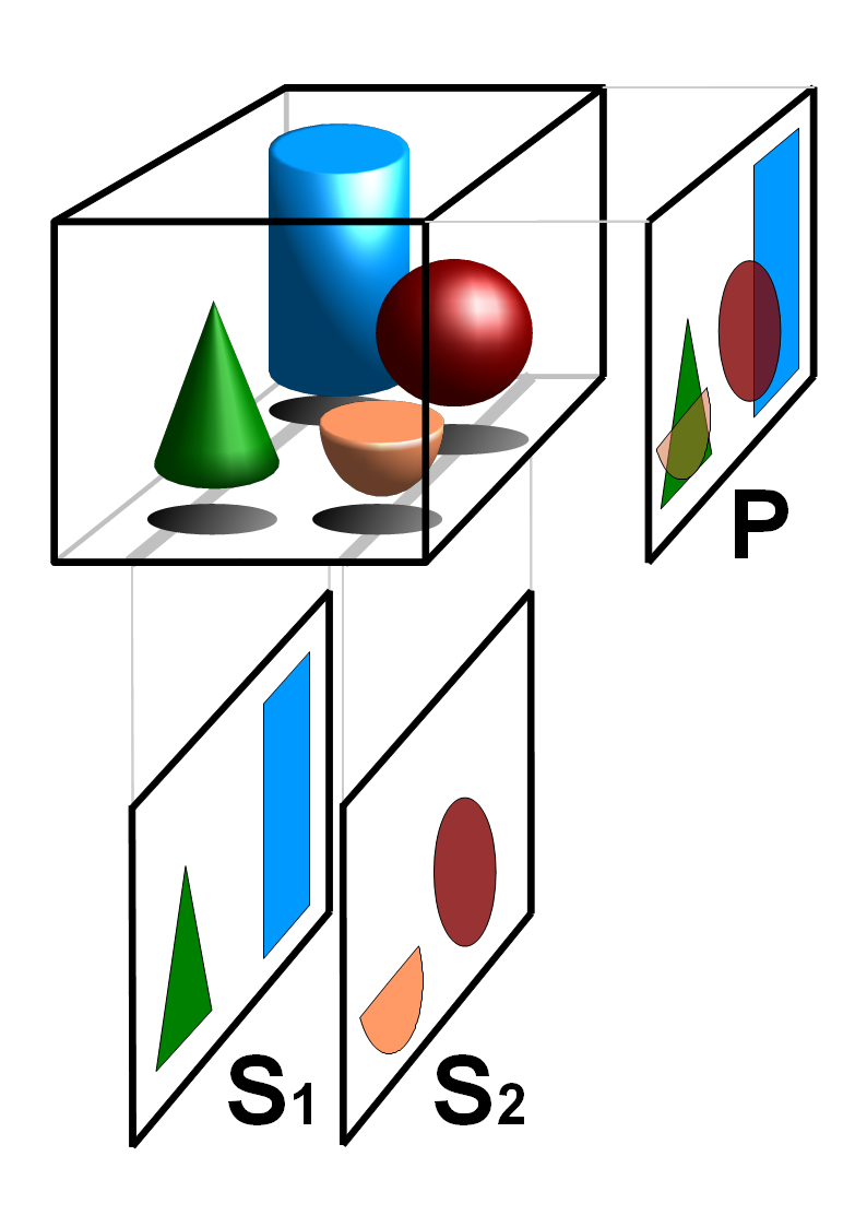
Tomography is imaging by sections or sectioning that uses any kind of penetrating wave. The method is used in radiology, archaeology, biology, atmospheric science, geophysics, oceanography, plasma physics, materials science, astrophysics, quantum information, and other areas of science. The word ''tomography'' is derived from Ancient Greek τόμος ''tomos'', "slice, section" and γράφω ''graphō'', "to write" or, in this context as well, "to describe." A device used in tomography is called a tomograph, while the image produced is a tomogram. In many cases, the production of these images is based on the mathematical procedure tomographic reconstruction, such as X-ray computed tomography technically being produced from multiple projectional radiographs. Many different reconstruction algorithms exist. Most algorithms fall into one of two categories: filtered back projection (FBP) and iterative reconstruction (IR). These procedures give inexact results: they represent a compr ... [...More Info...] [...Related Items...] OR: [Wikipedia] [Google] [Baidu] |
Microtomography
X-ray microtomography, like tomography and X-ray computed tomography, uses X-rays to create cross-sections of a physical object that can be used to recreate a virtual model (3D model) without destroying the original object. The prefix ''micro-'' (symbol: µ) is used to indicate that the pixel sizes of the cross-sections are in the micrometre range. These pixel sizes have also resulted in the terms high-resolution X-ray tomography, micro–computed tomography (micro-CT or µCT), and similar terms. Sometimes the terms high-resolution CT (HRCT) and micro-CT are differentiated, but in other cases the term high-resolution micro-CT is used. Virtually all tomography today is computed tomography. Micro-CT has applications both in medical imaging and in industrial computed tomography. In general, there are two types of scanner setups. In one setup, the X-ray source and detector are typically stationary during the scan while the sample/animal rotates. The second setup, much more like a cli ... [...More Info...] [...Related Items...] OR: [Wikipedia] [Google] [Baidu] |
X-ray
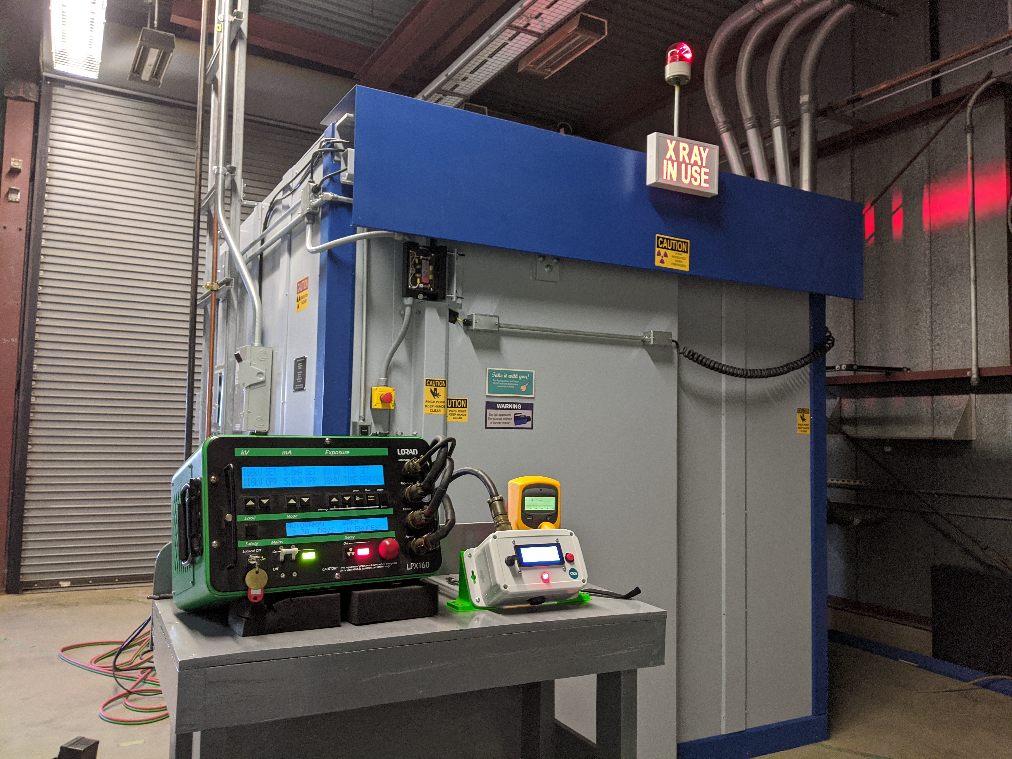
An X-ray, or, much less commonly, X-radiation, is a penetrating form of high-energy electromagnetic radiation. Most X-rays have a wavelength ranging from 10 picometers to 10 nanometers, corresponding to frequencies in the range 30 petahertz to 30 exahertz ( to ) and energies in the range 145 eV to 124 keV. X-ray wavelengths are shorter than those of UV rays and typically longer than those of gamma rays. In many languages, X-radiation is referred to as Röntgen radiation, after the German scientist Wilhelm Conrad Röntgen, who discovered it on November 8, 1895. He named it ''X-radiation'' to signify an unknown type of radiation.Novelline, Robert (1997). ''Squire's Fundamentals of Radiology''. Harvard University Press. 5th edition. . Spellings of ''X-ray(s)'' in English include the variants ''x-ray(s)'', ''xray(s)'', and ''X ray(s)''. The most familiar use of X-rays is checking for fractures (broken bones), but X-rays are also used in other ways. ... [...More Info...] [...Related Items...] OR: [Wikipedia] [Google] [Baidu] |
Nondestructive Testing
Nondestructive testing (NDT) is any of a wide group of analysis techniques used in science and technology industry to evaluate the properties of a material, component or system without causing damage. The terms nondestructive examination (NDE), nondestructive inspection (NDI), and nondestructive evaluation (NDE) are also commonly used to describe this technology. Because NDT does not permanently alter the article being inspected, it is a highly valuable technique that can save both money and time in product evaluation, troubleshooting, and research. The six most frequently used NDT methods are eddy-current, magnetic-particle, liquid penetrant, radiographic, ultrasonic, and visual testing. NDT is commonly used in forensic engineering, mechanical engineering, petroleum engineering, electrical engineering, civil engineering, systems engineering, aeronautical engineering, medicine, and art. Innovations in the field of nondestructive testing have had a profound impact on medical im ... [...More Info...] [...Related Items...] OR: [Wikipedia] [Google] [Baidu] |
Field Of View
The field of view (FoV) is the extent of the observable world that is seen at any given moment. In the case of optical instruments or sensors it is a solid angle through which a detector is sensitive to electromagnetic radiation. Humans and animals In the context of human and primate vision, the term "field of view" is typically only used in the sense of a restriction to what is visible by external apparatus, like when wearing spectacles or virtual reality goggles. Note that eye movements are allowed in the definition but do not change the field of view when understood this way. If the analogy of the eye's retina working as a sensor is drawn upon, the corresponding concept in human (and much of animal vision) is the visual field. It is defined as "the number of degrees of visual angle during stable fixation of the eyes".Strasburger, Hans; Pöppel, Ernst (2002). Visual Field. In G. Adelman & B.H. Smith (Eds): ''Encyclopedia of Neuroscience''; 3rd edition, on CD-ROM. El ... [...More Info...] [...Related Items...] OR: [Wikipedia] [Google] [Baidu] |
Medical Imaging
Medical imaging is the technique and process of imaging the interior of a body for clinical analysis and medical intervention, as well as visual representation of the function of some organs or tissues (physiology). Medical imaging seeks to reveal internal structures hidden by the skin and bones, as well as to diagnose and treat disease. Medical imaging also establishes a database of normal anatomy and physiology to make it possible to identify abnormalities. Although imaging of removed organs and tissues can be performed for medical reasons, such procedures are usually considered part of pathology instead of medical imaging. Measurement and recording techniques that are not primarily designed to produce images, such as electroencephalography (EEG), magnetoencephalography (MEG), electrocardiography (ECG), and others, represent other technologies that produce data susceptible to representation as a parameter graph versus time or maps that contain data about the measurement loca ... [...More Info...] [...Related Items...] OR: [Wikipedia] [Google] [Baidu] |


