|
Nanosims
NanoSIMS (nanoscale secondary ion mass spectrometry) is an analytical instrument manufactured by CAMECA which operates on the principle of secondary ion mass spectrometry. The NanoSIMS is used to acquire nanoscale resolution measurements of the elemental and isotopic composition of a sample. The NanoSIMS is able to create nanoscale maps of elemental or isotopic distribution, parallel acquisition of up to seven masses, isotopic identification, high mass resolution, subparts-per-million sensitivity with spatial resolution down to 50 nm. The original design of the NanoSIMS instrument was conceived by Georges Slodzian at the University of Paris Sud in France. There are currently around 50 NanoSIMS instruments worldwide.Li, K., Liu, J., Grovenor, C. R. M., & Moore, K. L. (2020). NanoSIMS Imaging and Analysis in Materials Science. Annual Review of Analytical Chemistry, 13, 273-292. https://doi.org/10.1146/annurev-anchem-092019-032524 How it works The NanoSIMS uses an ion sour ... [...More Info...] [...Related Items...] OR: [Wikipedia] [Google] [Baidu] |
Secondary Ion Mass Spectrometry
Secondary-ion mass spectrometry (SIMS) is a technique used to analyze the composition of solid surfaces and thin films by sputtering the surface of the specimen with a focused primary ion beam and collecting and analyzing ejected secondary ions. The mass/charge ratios of these secondary ions are measured with a mass spectrometer to determine the elemental, isotopic, or molecular composition of the surface to a depth of 1 to 2 nm. Due to the large variation in ionization probabilities among elements sputtered from different materials, comparison against well-calibrated standards is necessary to achieve accurate quantitative results. SIMS is the most sensitive surface analysis technique, with elemental detection limits ranging from parts per million to parts per billion. History In 1910 British physicist J. J. Thomson observed a release of positive ions and neutral atoms from a solid surface induced by ion bombardment. Improved vacuum pump technology in the 1940s enabled the fi ... [...More Info...] [...Related Items...] OR: [Wikipedia] [Google] [Baidu] |
Hydrogen Embrittlement
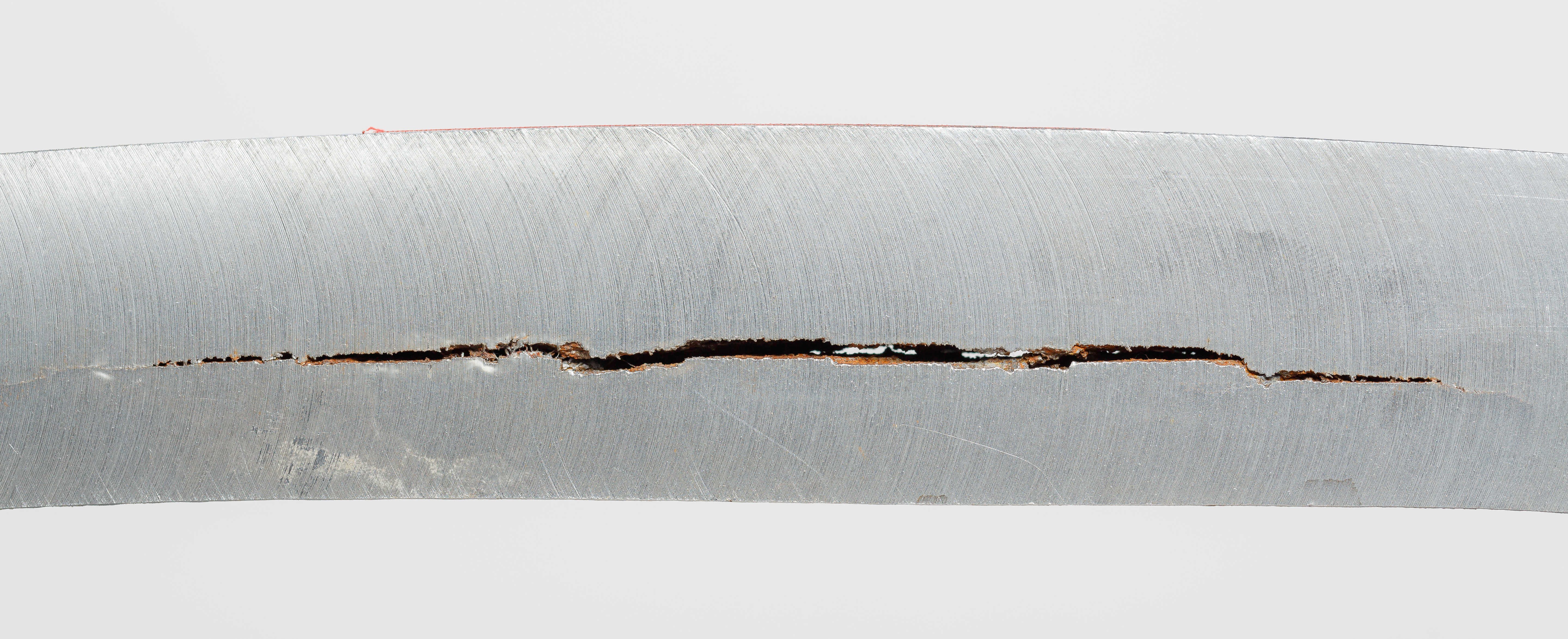
Hydrogen embrittlement (HE), also known as hydrogen-assisted cracking or hydrogen-induced cracking (HIC), is a reduction in the ductility of a metal due to absorbed hydrogen. Hydrogen atoms are small and can permeate solid metals. Once absorbed, hydrogen lowers the stress required for cracks in the metal to initiate and propagate, resulting in embrittlement. Hydrogen embrittlement occurs most notably in steels, as well as in iron, nickel, titanium, cobalt, and their alloys. Copper, aluminium, and stainless steels are less susceptible to hydrogen embrittlement. The essential facts about the nature of hydrogen embrittlement have been known since the 19th century. Hydrogen embrittlement is maximised at around room temperature in steels, and most metals are relatively immune to hydrogen embrittlement at temperatures above 150 °C. Hydrogen embrittlement requires the presence of both atomic ("diffusible") hydrogen and a mechanical stress to induce crack growth, although that str ... [...More Info...] [...Related Items...] OR: [Wikipedia] [Google] [Baidu] |
Intracellular
This glossary of biology terms is a list of definitions of fundamental terms and concepts used in biology, the study of life and of living organisms. It is intended as introductory material for novices; for more specific and technical definitions from sub-disciplines and related fields, see Glossary of genetics, Glossary of evolutionary biology, Glossary of ecology, and Glossary of scientific naming, or any of the organism-specific glossaries in :Glossaries of biology. A B C D E ... [...More Info...] [...Related Items...] OR: [Wikipedia] [Google] [Baidu] |
Transmission Electron Microscopy
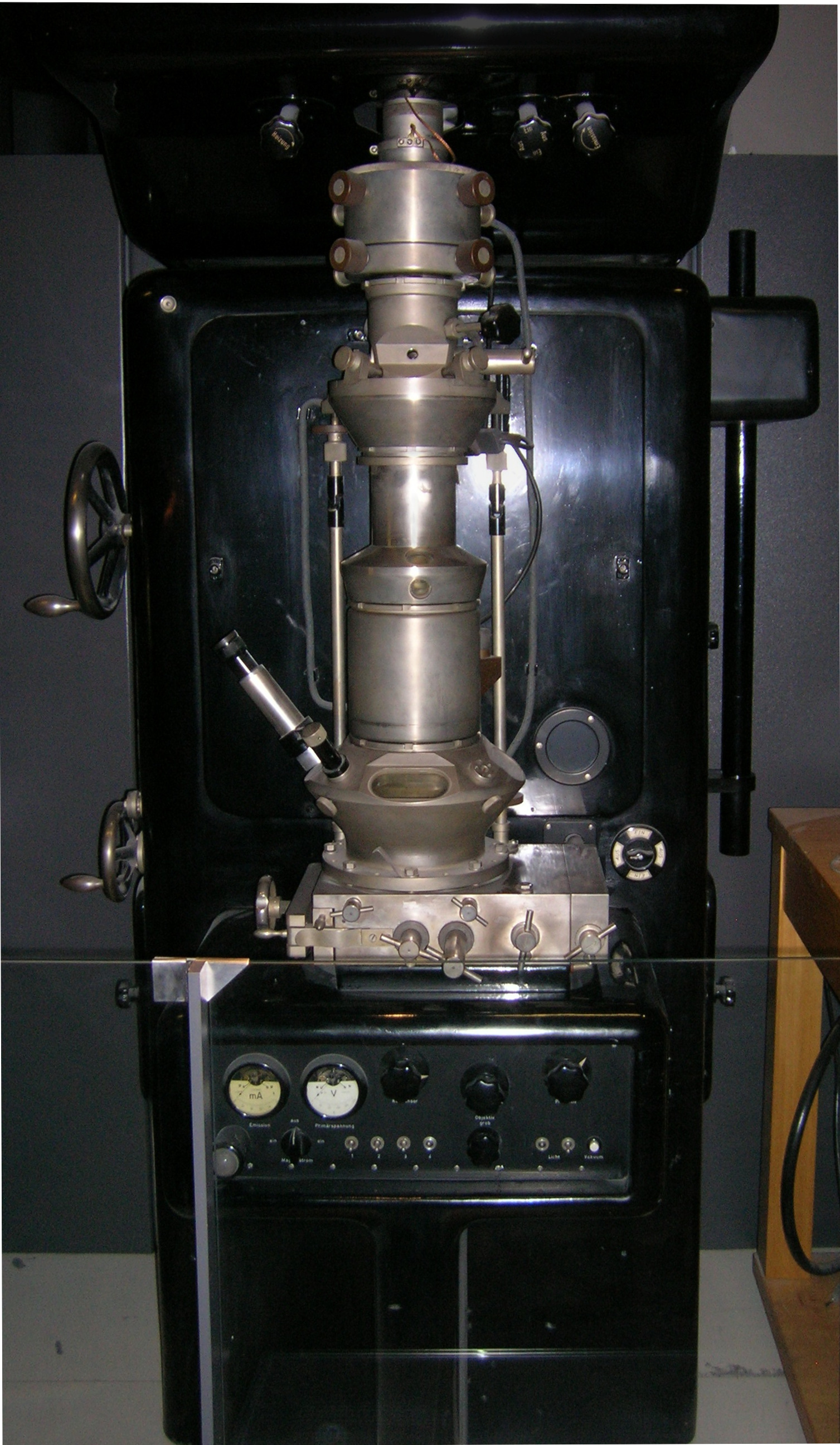
Transmission electron microscopy (TEM) is a microscopy technique in which a beam of electrons is transmitted through a specimen to form an image. The specimen is most often an ultrathin section less than 100 nm thick or a suspension on a grid. An image is formed from the interaction of the electrons with the sample as the beam is transmitted through the specimen. The image is then magnified and focused onto an imaging device, such as a fluorescent screen, a layer of photographic film, or a sensor such as a scintillator attached to a charge-coupled device. Transmission electron microscopes are capable of imaging at a significantly higher resolution than light microscopes, owing to the smaller de Broglie wavelength of electrons. This enables the instrument to capture fine detail—even as small as a single column of atoms, which is thousands of times smaller than a resolvable object seen in a light microscope. Transmission electron microscopy is a major analytical method i ... [...More Info...] [...Related Items...] OR: [Wikipedia] [Google] [Baidu] |
Focused Ion Beam
Focused ion beam, also known as FIB, is a technique used particularly in the semiconductor industry, materials science and increasingly in the biological field for site-specific analysis, deposition, and ablation of materials. A FIB setup is a scientific instrument that resembles a scanning electron microscope (SEM). However, while the SEM uses a focused beam of electrons to image the sample in the chamber, a FIB setup uses a focused beam of ions instead. FIB can also be incorporated in a system with both electron and ion beam columns, allowing the same feature to be investigated using either of the beams. FIB should not be confused with using a beam of focused ions for direct write lithography (such as in proton beam writing). These are generally quite different systems where the material is modified by other mechanisms. Ion beam source Most widespread instruments are using liquid metal ion sources (LMIS), especially gallium ion sources. Ion sources based on elemental gold an ... [...More Info...] [...Related Items...] OR: [Wikipedia] [Google] [Baidu] |
Microtome
A microtome (from the Greek ''mikros'', meaning "small", and ''temnein'', meaning "to cut") is a cutting tool used to produce extremely thin slices of material known as ''sections''. Important in science, microtomes are used in microscopy, allowing for the preparation of samples for observation under transmitted light or electron radiation. Microtomes use steel, glass or diamond blades depending upon the specimen being sliced and the desired thickness of the sections being cut. Steel blades are used to prepare histological sections of animal or plant tissues for light microscopy. Glass knives are used to slice sections for light microscopy and to slice very thin sections for electron microscopy. Industrial grade diamond knives are used to slice hard materials such as bone, teeth and tough plant matter for both light microscopy and for electron microscopy. Gem-quality diamond knives are also used for slicing thin sections for electron microscopy. Microtomy is a method for the p ... [...More Info...] [...Related Items...] OR: [Wikipedia] [Google] [Baidu] |
Cosmochemistry
Cosmochemistry (from Greek κόσμος ''kósmos'', "universe" and χημεία ''khemeía'') or chemical cosmology is the study of the chemical composition of matter in the universe and the processes that led to those compositions. This is done primarily through the study of the chemical composition of meteorites and other physical samples. Given that the asteroid parent bodies of meteorites were some of the first solid material to condense from the early solar nebula, cosmochemists are generally, but not exclusively, concerned with the objects contained within the Solar System. History In 1938, Swiss mineralogist Victor Goldschmidt and his colleagues compiled a list of what they called "cosmic abundances" based on their analysis of several terrestrial and meteorite samples. Goldschmidt justified the inclusion of meteorite composition data into his table by claiming that terrestrial rocks were subjected to a significant amount of chemical change due to the inherent processes of ... [...More Info...] [...Related Items...] OR: [Wikipedia] [Google] [Baidu] |
Nano Structure
A nanostructure is a structure of intermediate size between microscopic and molecular structures. Nanostructural detail is microstructure at nanoscale. In describing nanostructures, it is necessary to differentiate between the number of dimensions in the volume of an object which are on the nanoscale. Nanotextured surfaces have ''one dimension'' on the nanoscale, i.e., only the thickness of the surface of an object is between 0.1 and 100 nm. Nanotubes have ''two dimensions'' on the nanoscale, i.e., the diameter of the tube is between 0.1 and 100 nm; its length can be far more. Finally, spherical nanoparticles have ''three dimensions'' on the nanoscale, i.e., the particle is between 0.1 and 100 nm in each spatial dimension. The terms nanoparticles and ultrafine particles (UFP) are often used synonymously although UFP can reach into the micrometre range. The term ''nanostructure'' is often used when referring to magnetic technology. Nanoscale structure in biology is ... [...More Info...] [...Related Items...] OR: [Wikipedia] [Google] [Baidu] |
Polishing
Polishing is the process of creating a smooth and shiny surface by rubbing it or by applying a chemical treatment, leaving a clean surface with a significant specular reflection (still limited by the index of refraction of the material according to the Fresnel equations). In some materials (such as metals, glasses, black or transparent stones), polishing is also able to reduce diffuse reflection to minimal values. When an unpolished surface is magnified thousands of times, it usually looks like a succession of mountains and valleys. By repeated abrasion, those "mountains" are worn down until they are flat or just small "hills." The process of polishing with abrasives starts with a coarse grain size and gradually proceeds to the finer ones to efficiently flatten the surface imperfections and to obtain optimal results. Mechanical properties The strength of polished products can be higher than their unpolished counterparts owing to the removal of stress concentrations present ... [...More Info...] [...Related Items...] OR: [Wikipedia] [Google] [Baidu] |
Cryopreservation
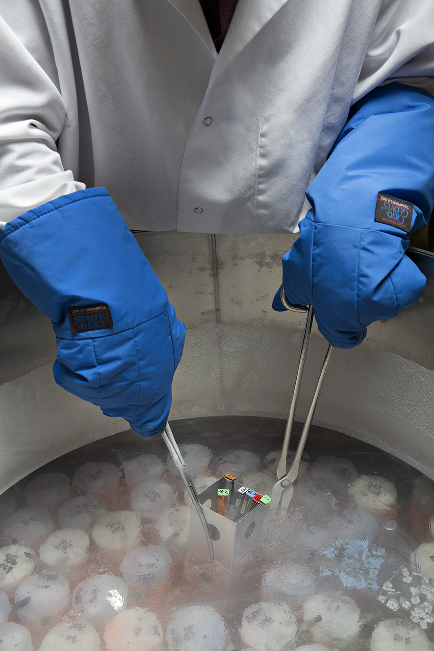
Cryo-preservation or cryo-conservation is a process where Organism, organisms, organelles, cell (biology), cells, Biological tissue, tissues, extracellular matrix, Organ (anatomy), organs, or any other biological constructs susceptible to damage caused by unregulated chemical kinetics are preserved by cooling to very low temperatures (typically using solid carbon dioxide or using liquid nitrogen). At low enough temperatures, any Enzyme, enzymatic or chemical activity which might cause damage to the biological material in question is effectively stopped. Cryopreservation methods seek to reach low temperatures without causing additional damage caused by the formation of ice crystals during freezing. Traditional cryopreservation has relied on coating the material to be frozen with a class of molecules termed cryoprotectants. New methods are being investigated due to the inherent toxicity of many cryoprotectants. Cryoconservation of animal genetic resources is done with the intentio ... [...More Info...] [...Related Items...] OR: [Wikipedia] [Google] [Baidu] |
Fixation (histology)
In the fields of histology, pathology, and cell biology, fixation is the preservation of biological tissues from decay due to autolysis or putrefaction. It terminates any ongoing biochemical reactions and may also increase the treated tissues' mechanical strength or stability. Tissue fixation is a critical step in the preparation of histological sections, its broad objective being to preserve cells and tissue components and to do this in such a way as to allow for the preparation of thin, stained sections. This allows the investigation of the tissues' structure, which is determined by the shapes and sizes of such macromolecules (in and around cells) as proteins and nucleic acids. Purposes In performing their protective role, fixatives denature proteins by coagulation, by forming additive compounds, or by a combination of coagulation and additive processes. A compound that adds chemically to macromolecules stabilizes structure most effectively if it is able to combine with part ... [...More Info...] [...Related Items...] OR: [Wikipedia] [Google] [Baidu] |


