|
NCR3
Natural cytotoxicity triggering receptor 3 is a protein that in humans is encoded by the ''NCR3'' gene. NCR3 has also been designated as CD337 (cluster of differentiation 337) and as NKp30. NCR3 belongs to the family of NCR membrane receptors together with NCR1 (NKp46) and NCR2 (NKp44). Identification NKp30 receptor was first identified in 1999. According to Western blot analysis specific monoclonal antibodies reacted with 30kDa molecule, therefore was the protein named NKp30. Structure Gene for NKp30 is located in the MHC class III region of the human MHC locus and encodes 190 amino acid long type I transmembrane receptor which belongs to immunoglobulin super family (IgSF). NKp30 has a mass of 30 kDa and includes one Ig-like extracellular domain which is 138 amino acids long, a 19 amino acid transmembrane (TM) domain and a 33 amino acid cytoplasmic tail. The Ig-like domain consists of 2 antiparallel beta-sheets linked by a disulphide bond. The extracellular domain conta ... [...More Info...] [...Related Items...] OR: [Wikipedia] [Google] [Baidu] |
MHC Class III
MHC class III is a group of proteins belonging the class of major histocompatibility complex (MHC). Unlike other MHC types such as MHC class I and MHC class II, of which their structure and functions in immune response are well defined, MHC class III are poorly defined structurally and functionally. They are not involved in antigen binding (the process called antigen presentation, a classic function of MHC proteins). Only few of them are actually involved in immunity while many are signalling molecules in other cell communications. They are mainly known from their genes because their gene cluster is present between those of class I and class II. The gene cluster was discovered when genes (specifically those of complement components C2, C4, and factor B) were found in between class I and class II genes on the short (p) arm of human chromosome 6. It was later found that it contains many genes for different signalling molecules such as tumour necrosis factors (TNFs) and heat sho ... [...More Info...] [...Related Items...] OR: [Wikipedia] [Google] [Baidu] |
Protein
Proteins are large biomolecules and macromolecules that comprise one or more long chains of amino acid residues. Proteins perform a vast array of functions within organisms, including catalysing metabolic reactions, DNA replication, responding to stimuli, providing structure to cells and organisms, and transporting molecules from one location to another. Proteins differ from one another primarily in their sequence of amino acids, which is dictated by the nucleotide sequence of their genes, and which usually results in protein folding into a specific 3D structure that determines its activity. A linear chain of amino acid residues is called a polypeptide. A protein contains at least one long polypeptide. Short polypeptides, containing less than 20–30 residues, are rarely considered to be proteins and are commonly called peptides. The individual amino acid residues are bonded together by peptide bonds and adjacent amino acid residues. The sequence of amino acid residue ... [...More Info...] [...Related Items...] OR: [Wikipedia] [Google] [Baidu] |
CD3ζ
T-cell surface glycoprotein CD3 zeta chain also known as T-cell receptor T3 zeta chain or CD247 (Cluster of Differentiation 247) is a protein that in humans is encoded by the ''CD247'' gene. Some older literature mention a similar protein called "CD3 eta" in mice. It is now understood to be an isoform differing in the last exon. Genomics The gene is located on the long arm of chromosome 1 at location 1q22-q25 on the Crick (negative) strand. The encoded protein is 164 amino acids long with a predicted weight of 18.696 kilo Daltons. Function T-cell receptor zeta (ζ), together with T-cell receptor alpha/beta and gamma/delta heterodimers and CD3-gamma, -delta, and -epsilon, forms the T-cell receptor-CD3 complex. The zeta chain plays an important role in coupling antigen recognition to several intracellular signal-transduction pathways. Low expression of the antigen results in impaired immune response. Two alternatively spliced transcript variants encoding distinct isoforms h ... [...More Info...] [...Related Items...] OR: [Wikipedia] [Google] [Baidu] |
Cytotoxicity
Cytotoxicity is the quality of being toxic to cells. Examples of toxic agents are an immune cell or some types of venom, e.g. from the puff adder (''Bitis arietans'') or brown recluse spider (''Loxosceles reclusa''). Cell physiology Treating cells with the cytotoxic compound can result in a variety of cell fates. The cells may undergo necrosis, in which they lose membrane integrity and die rapidly as a result of cell lysis. The cells can stop actively growing and dividing (a decrease in cell viability), or the cells can activate a genetic program of controlled cell death (apoptosis). Cells undergoing necrosis typically exhibit rapid swelling, lose membrane integrity, shut down metabolism, and release their contents into the environment. Cells that undergo rapid necrosis in vitro do not have sufficient time or energy to activate apoptotic machinery and will not express apoptotic markers. Apoptosis is characterized by well defined cytological and molecular events including a change i ... [...More Info...] [...Related Items...] OR: [Wikipedia] [Google] [Baidu] |
Immunosurveillance
The immune system is a network of biological processes that protects an organism from diseases. It detects and responds to a wide variety of pathogens, from viruses to parasitic worms, as well as cancer cells and objects such as wood splinters, distinguishing them from the organism's own healthy tissue. Many species have two major subsystems of the immune system. The innate immune system provides a preconfigured response to broad groups of situations and stimuli. The adaptive immune system provides a tailored response to each stimulus by learning to recognize molecules it has previously encountered. Both use molecules and cells to perform their functions. Nearly all organisms have some kind of immune system. Bacteria have a rudimentary immune system in the form of enzymes that protect against virus infections. Other basic immune mechanisms evolved in ancient plants and animals and remain in their modern descendants. These mechanisms include phagocytosis, antimicrobial peptides ... [...More Info...] [...Related Items...] OR: [Wikipedia] [Google] [Baidu] |
Endometrium
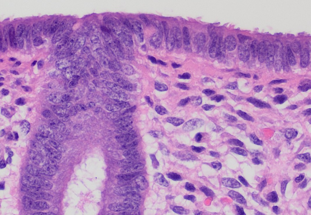
The endometrium is the inner epithelial layer, along with its mucous membrane, of the mammalian uterus. It has a basal layer and a functional layer: the basal layer contains stem cells which regenerate the functional layer. The functional layer thickens and then is shed during menstruation in humans and some other mammals, including apes, Old World monkeys, some species of bat, the elephant shrew and the Cairo spiny mouse. In most other mammals, the endometrium is reabsorbed in the estrous cycle. During pregnancy, the glands and blood vessels in the endometrium further increase in size and number. Vascular spaces fuse and become interconnected, forming the placenta, which supplies oxygen and nutrition to the embryo and fetus.Blue Histology - Female Reproductive System . School ... [...More Info...] [...Related Items...] OR: [Wikipedia] [Google] [Baidu] |
Interleukin 2
Interleukin-2 (IL-2) is an interleukin, a type of cytokine signaling molecule in the immune system. It is a 15.5–16 kDa protein that regulates the activities of white blood cells (leukocytes, often lymphocytes) that are responsible for immunity. IL-2 is part of the body's natural response to microbial infection, and in discriminating between foreign ("non-self") and "self". IL-2 mediates its effects by binding to IL-2 receptors, which are expressed by lymphocytes. The major sources of IL-2 are activated CD4+ T cells and activated CD8+ T cells. IL-2 receptor IL-2 is a member of a cytokine family, each member of which has a four alpha helix bundle; the family also includes IL-4, IL-7, IL-9, IL-15 and IL-21. IL-2 signals through the IL-2 receptor, a complex consisting of three chains, termed alpha (CD25), beta (CD122) and gamma ( CD132). The gamma chain is shared by all family members. The IL-2 receptor (IL-2R) α subunit binds IL-2 with low affinity (Kd~ 10� ... [...More Info...] [...Related Items...] OR: [Wikipedia] [Google] [Baidu] |
Interleukin 15
Interleukin-15 (IL-15) is a cytokine with structural similarity to Interleukin-2 (IL-2). Like IL-2, IL-15 binds to and signals through a complex composed of IL-2/IL-15 receptor beta chain (CD122) and the common gamma chain (gamma-C, CD132). IL-15 is secreted by mononuclear phagocytes (and some other cells) following infection by virus(es). This cytokine induces the proliferation of natural killer cells, i.e. cells of the innate immune system whose principal role is to kill virally infected cells. Expression IL-15 was discovered in 1994 by two different laboratories, and characterized as T cell growth factor. Together with Interleukin-2 (IL-2), Interleukin-4 ( IL-4), Interleukin-7 ( IL-7), Interleukin-9 ( IL-9), granulocyte colony-stimulating factor (G-CSF), and granulocyte-macrophage colony-stimulating factor (GM-CSF), IL-15 belongs to the four α-helix bundle family of cytokines. IL-15 is constitutively expressed by a large number of cell types and tissues, including mo ... [...More Info...] [...Related Items...] OR: [Wikipedia] [Google] [Baidu] |
ILC2
ILC2 cells, or type 2 innate lymphoid cells are a type of innate lymphoid cell. Not to be confused with the ILC. They are derived from common lymphoid progenitor and belong to the lymphoid lineage. These cells lack antigen specific B or T cell receptor because of the lack of recombination activating gene. ILC2s produce type 2 cytokines (e.g. IL-4, IL-5, IL-9, IL-13) and are involved in responses to helminths, allergens, some viruses, such as influenza virus and cancer. The cell type was first described in 2001 as non-B/non-T cells, which produced IL-5 and IL-13 in response to IL-25 and expressed MHC class II and CD11c. In 2006, a similar cell population was identified in a case of helminthic infection. The name "ILC2" was not proposed until 2013. They were previously identified in literature as natural helper cells, nuocytes, or innate helper 2 cells. It is believed that ILC2s are rather old cell type with ancestor populations emerging in lamprey and bony fish. Parasitic ... [...More Info...] [...Related Items...] OR: [Wikipedia] [Google] [Baidu] |
T-cell Receptor
The T-cell receptor (TCR) is a protein complex found on the surface of T cells, or T lymphocytes, that is responsible for recognizing fragments of antigen as peptides bound to major histocompatibility complex (MHC) molecules. The binding between TCR and antigen peptides is of relatively low affinity and is degenerate: that is, many TCRs recognize the same antigen peptide and many antigen peptides are recognized by the same TCR. The TCR is composed of two different protein chains (that is, it is a heterodimer). In humans, in 95% of T cells the TCR consists of an alpha (α) chain and a beta (β) chain (encoded by '' TRA'' and ''TRB'', respectively), whereas in 5% of T cells the TCR consists of gamma and delta (γ/δ) chains (encoded by '' TRG'' and '' TRD'', respectively). This ratio changes during ontogeny and in diseased states (such as leukemia). It also differs between species. Orthologues of the 4 loci have been mapped in various species. Each locus can produce a vari ... [...More Info...] [...Related Items...] OR: [Wikipedia] [Google] [Baidu] |
Gamma Delta T Cell
Gamma delta T cells (γδ T cells) are T cells that have a γδ T-cell receptor (TCR) on their surface. Most T cells are αβ (alpha beta) T cells with TCR composed of two glycoprotein chains called α (alpha) and β (beta) TCR chains. In contrast, γδ T cells have a TCR that is made up of one γ (gamma) chain and one δ (delta) chain. This group of T cells is usually less common than αβ T cells, but are at their highest abundance in the gut mucosa, within a population of lymphocytes known as intraepithelial lymphocytes (IELs). The antigenic molecules that activate gamma delta T cells are still largely unknown. However, γδ T cells are peculiar in that they do not seem to require antigen processing and major-histocompatibility-complex (MHC) presentation of peptide epitopes, although some recognize MHC class Ib molecules. γδ T cells are believed to have a prominent role in recognition of lipid antigens. They are of an invariant nature and may be triggered by alarm signals, ... [...More Info...] [...Related Items...] OR: [Wikipedia] [Google] [Baidu] |
Cytotoxic T Cell
A cytotoxic T cell (also known as TC, cytotoxic T lymphocyte, CTL, T-killer cell, cytolytic T cell, CD8+ T-cell or killer T cell) is a T lymphocyte (a type of white blood cell) that kills cancer cells, cells that are infected by intracellular pathogens (such as viruses or bacteria), or cells that are damaged in other ways. Most cytotoxic T cells express T-cell receptors (TCRs) that can recognize a specific antigen. An antigen is a molecule capable of stimulating an immune response and is often produced by cancer cells, viruses, bacteria or intracellular signals. Antigens inside a cell are bound to class I MHC molecules, and brought to the surface of the cell by the class I MHC molecule, where they can be recognized by the T cell. If the TCR is specific for that antigen, it binds to the complex of the class I MHC molecule and the antigen, and the T cell destroys the cell. In order for the TCR to bind to the class I MHC molecule, the former must be accompanied by a glycoprotein ... [...More Info...] [...Related Items...] OR: [Wikipedia] [Google] [Baidu] |


