|
Mycorrhaphium Adustum
''Mycorrhaphium'' is a genus of fungi in the family Steccherinaceae. The genus was circumscribed by Dutch mycologist Rudolph Arnold Maas Geesteranus in 1962. The type species is ''Mycorrhaphium adustum'' (formerly referred to '' Hydnum''). Fruit bodies of species in the genus have caps, stipes, and a hydnoid (tooth-like) hymenophore. There is a dimitic hyphal system, where the skeletal hyphae are found only in the tissue of the "teeth", and a lack of cystidia. The spores are smooth, hyaline (translucent), and inamyloid. Walter Jülich created the family Mycorrhaphiaceae to contain the type genus ''Mycorrhaphium''. This family is now placed in synonymy with Steccherinaceae. Species *'' M. adustulum'' (Banker) Ryvarden (1989) – Europe, North America *'' M. adustum'' (Schwein.) Maas Geest. (1962) *''M. africanum'' Mossebo & Ryvarden (2003)– Africa (Cameroon) *'' M. citrinum'' Ryvarden (1989) – Africa (Zambia Zambia (), officially the Republic of Zambia, is a lan ... [...More Info...] [...Related Items...] OR: [Wikipedia] [Google] [Baidu] |
Fungi
A fungus ( : fungi or funguses) is any member of the group of eukaryotic organisms that includes microorganisms such as yeasts and molds, as well as the more familiar mushrooms. These organisms are classified as a kingdom, separately from the other eukaryotic kingdoms, which by one traditional classification include Plantae, Animalia, Protozoa, and Chromista. A characteristic that places fungi in a different kingdom from plants, bacteria, and some protists is chitin in their cell walls. Fungi, like animals, are heterotrophs; they acquire their food by absorbing dissolved molecules, typically by secreting digestive enzymes into their environment. Fungi do not photosynthesize. Growth is their means of mobility, except for spores (a few of which are flagellated), which may travel through the air or water. Fungi are the principal decomposers in ecological systems. These and other differences place fungi in a single group of related organisms, named the ''Eumycota'' (''t ... [...More Info...] [...Related Items...] OR: [Wikipedia] [Google] [Baidu] |
Rudolph Arnold Maas Geesteranus
Rudolf Arnold Maas Geesteranus (20 January 1911 in The Hague – May 18, 2003 in Oegstgeest), was a Dutch mycologist Mycology is the branch of biology concerned with the study of fungus, fungi, including their genetics, genetic and biochemistry, biochemical properties, their Taxonomy (biology), taxonomy and ethnomycology, their use to humans, including as a so .... See also * :Taxa named by Rudolf Arnold Maas Geesteranus References {{DEFAULTSORT:Maas Geesteranus, Rudolph Arnold 1911 births 2003 deaths Dutch mycologists Scientists from The Hague Leiden University alumni Academic staff of Leiden University ... [...More Info...] [...Related Items...] OR: [Wikipedia] [Google] [Baidu] |
Synonym (biology)
The Botanical and Zoological Codes of nomenclature treat the concept of synonymy differently. * In botanical nomenclature, a synonym is a scientific name that applies to a taxon that (now) goes by a different scientific name. For example, Linnaeus was the first to give a scientific name (under the currently used system of scientific nomenclature) to the Norway spruce, which he called ''Pinus abies''. This name is no longer in use, so it is now a synonym of the current scientific name, ''Picea abies''. * In zoology, moving a species from one genus to another results in a different binomen, but the name is considered an alternative combination rather than a synonym. The concept of synonymy in zoology is reserved for two names at the same rank that refers to a taxon at that rank - for example, the name ''Papilio prorsa'' Linnaeus, 1758 is a junior synonym of ''Papilio levana'' Linnaeus, 1758, being names for different seasonal forms of the species now referred to as ''Araschnia leva ... [...More Info...] [...Related Items...] OR: [Wikipedia] [Google] [Baidu] |
Inamyloid
In mycology a tissue or feature is said to be amyloid if it has a positive amyloid reaction when subjected to a crude chemical test using iodine as an ingredient of either Melzer's reagent or Lugol's solution, producing a blue to blue-black staining. The term "amyloid" is derived from the Latin ''amyloideus'' ("starch-like"). It refers to the fact that starch gives a similar reaction, also called an amyloid reaction. The test can be on microscopic features, such as spore walls or hyphal walls, or the apical apparatus or entire ascus wall of an ascus, or be a macroscopic reaction on tissue where a drop of the reagent is applied. Negative reactions, called inamyloid or nonamyloid, are for structures that remain pale yellow-brown or clear. A reaction producing a deep reddish to reddish-brown staining is either termed a dextrinoid reaction (pseudoamyloid is a synonym) or a hemiamyloid reaction. Melzer's reagent reactions Hemiamyloidity Hemiamyloidity in mycology refers to a special ... [...More Info...] [...Related Items...] OR: [Wikipedia] [Google] [Baidu] |
Hyaline
A hyaline substance is one with a glassy appearance. The word is derived from el, ὑάλινος, translit=hyálinos, lit=transparent, and el, ὕαλος, translit=hýalos, lit=crystal, glass, label=none. Histopathology Hyaline cartilage is named after its glassy appearance on fresh gross pathology. On light microscopy of H&E stained slides, the extracellular matrix of hyaline cartilage looks homogeneously pink, and the term "hyaline" is used to describe similarly homogeneously pink material besides the cartilage. Hyaline material is usually acellular and proteinaceous. For example, arterial hyaline is seen in aging, high blood pressure, diabetes mellitus and in association with some drugs (e.g. calcineurin inhibitors). It is bright pink with PAS staining. Ichthyology and entomology In ichthyology and entomology, ''hyaline'' denotes a colorless, transparent substance, such as unpigmented fins of fishes or clear insect wings. Resh, Vincent H. and R. T. Cardé, Eds. Encyclo ... [...More Info...] [...Related Items...] OR: [Wikipedia] [Google] [Baidu] |
Basidiospore
A basidiospore is a reproductive spore produced by Basidiomycete fungi, a grouping that includes mushrooms, shelf fungi, rusts, and smuts. Basidiospores typically each contain one haploid nucleus that is the product of meiosis, and they are produced by specialized fungal cells called basidia. Typically, four basidiospores develop on appendages from each basidium, of which two are of one strain and the other two of its opposite strain. In gills under a cap of one common species, there exist millions of basidia. Some gilled mushrooms in the order Agaricales have the ability to release billions of spores. The puffball fungus ''Calvatia gigantea'' has been calculated to produce about five trillion basidiospores. Most basidiospores are forcibly discharged, and are thus considered ballistospores. These spores serve as the main air dispersal units for the fungi. The spores are released during periods of high humidity and generally have a night-time or pre-dawn peak concentration in the ... [...More Info...] [...Related Items...] OR: [Wikipedia] [Google] [Baidu] |
Cystidia
A cystidium (plural cystidia) is a relatively large cell found on the sporocarp of a basidiomycete (for example, on the surface of a mushroom gill), often between clusters of basidia. Since cystidia have highly varied and distinct shapes that are often unique to a particular species or genus, they are a useful micromorphological characteristic in the identification of basidiomycetes. In general, the adaptive significance of cystidia is not well understood. Classification of cystidia By position Cystidia may occur on the edge of a lamella (or analogous hymenophoral structure) (cheilocystidia), on the face of a lamella (pleurocystidia), on the surface of the cap (dermatocystidia or pileocystidia), on the margin of the cap (circumcystidia) or on the stipe (caulocystidia). Especially the pleurocystidia and cheilocystidia are important for identification within many genera. Sometimes the cheilocystidia give the gill edge a distinct colour which is visible to the naked eye or wit ... [...More Info...] [...Related Items...] OR: [Wikipedia] [Google] [Baidu] |
Hypha
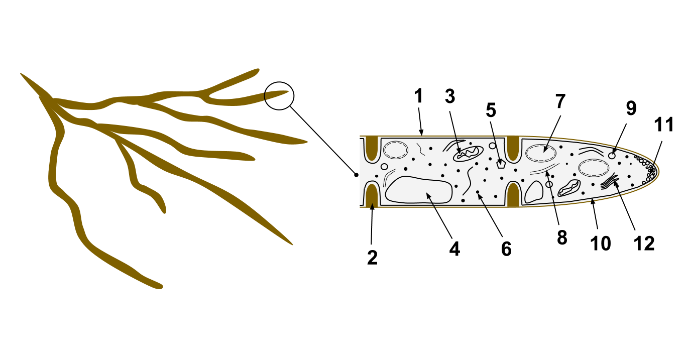
A hypha (; ) is a long, branching, filamentous structure of a fungus, oomycete, or actinobacterium. In most fungi, hyphae are the main mode of vegetative growth, and are collectively called a mycelium. Structure A hypha consists of one or more cells surrounded by a tubular cell wall. In most fungi, hyphae are divided into cells by internal cross-walls called "septa" (singular septum). Septa are usually perforated by pores large enough for ribosomes, mitochondria, and sometimes nuclei to flow between cells. The major structural polymer in fungal cell walls is typically chitin, in contrast to plants and oomycetes that have cellulosic cell walls. Some fungi have aseptate hyphae, meaning their hyphae are not partitioned by septa. Hyphae have an average diameter of 4–6 µm. Growth Hyphae grow at their tips. During tip growth, cell walls are extended by the external assembly and polymerization of cell wall components, and the internal production of new cell membrane. The S ... [...More Info...] [...Related Items...] OR: [Wikipedia] [Google] [Baidu] |
Hymenophore
A hymenophore refers to the hymenium-bearing structure of a fungal fruiting body. Hymenophores can be smooth surfaces, lamellae, folds, tubes, or teeth. The term was coined by Robert Hooke Robert Hooke FRS (; 18 July 16353 March 1703) was an English polymath active as a scientist, natural philosopher and architect, who is credited to be one of two scientists to discover microorganisms in 1665 using a compound microscope that ... in 1665. References {{Mycology-stub Mycology Fungal morphology and anatomy ... [...More Info...] [...Related Items...] OR: [Wikipedia] [Google] [Baidu] |
Hydnoid Fungi
The hydnoid fungi are a group of fungi in the Basidiomycota with basidiocarps (fruit bodies) producing spores on pendant, tooth-like or spine-like projections. They are colloquially called tooth fungi. Originally such fungi were referred to the genus ''Hydnum'' ("hydnoid" means ''Hydnum''-like), but it is now known that not all hydnoid species are closely related. History ''Hydnum'' was one of the original genera created by Linnaeus in his ''Species Plantarum'' of 1753. It contained all species of fungi with fruit bodies bearing pendant, tooth-like projections. Subsequent authors described around 900 species in the genus. With increasing use of the microscope, it became clear that not all tooth fungi were closely related and most ''Hydnum'' species were gradually moved to other genera. The Dutch mycologist Rudolph Arnold Maas Geesteranus paid particular attention to the group, producing a series of papers reviewing the taxonomy of hydnoid fungi. The original genus ''Hydnum'' is ... [...More Info...] [...Related Items...] OR: [Wikipedia] [Google] [Baidu] |
Stipe (mycology)
In mycology, a stipe () is the stem or stalk-like feature supporting the cap of a mushroom. Like all tissues of the mushroom other than the hymenium, the stipe is composed of sterile hyphal tissue. In many instances, however, the fertile hymenium extends down the stipe some distance. Fungi that have stipes are said to be stipitate. The evolutionary benefit of a stipe is generally considered to be in mediating spore dispersal. An elevated mushroom will more easily release its spores into wind currents or onto passing animals. Nevertheless, many mushrooms do not have stipes, including cup fungi, puffballs, earthstars, some polypores, jelly fungi, ergots, and smuts. It is often the case that features of the stipe are required to make a positive identification of a mushroom. Such distinguishing characters include: # the texture of the stipe (fibrous, brittle, chalky, leathery, firm, etc.) # whether it has remains of a partial veil (such as an annulus or cortina) or universal ve ... [...More Info...] [...Related Items...] OR: [Wikipedia] [Google] [Baidu] |
Pileus (mycology)
The pileus is the technical name for the cap, or cap-like part, of a basidiocarp or ascocarp (fungal fruiting body) that supports a spore-bearing surface, the hymenium.Moore-Landecker, E: "Fundamentals of the Fungi", page 560. Prentice Hall, 1972. The hymenium (hymenophore) may consist of lamellae, tubes, or teeth, on the underside of the pileus. A pileus is characteristic of agarics, boletes, some polypores, tooth fungi, and some ascomycetes. Classification Pilei can be formed in various shapes, and the shapes can change over the course of the developmental cycle of a fungus. The most familiar pileus shape is hemispherical or ''convex.'' Convex pilei often continue to expand as they mature until they become flat. Many well-known species have a convex pileus, including the button mushroom, various ''Amanita'' species and boletes. Some, such as the parasol mushroom, have distinct bosses or umbos and are described as ''umbonate''. An umbo is a knobby protrusion at the center of th ... [...More Info...] [...Related Items...] OR: [Wikipedia] [Google] [Baidu] |


