|
Mycena Fonticola
''Mycena fonticola'' is a species of fungus in the family Mycenaceae. First reported in 2007, it is known only from central Honshu, in Japan, where it grows on dead leaves and twigs in low-elevation forests dominated by oak trees. The fruit body of the fungus has a smooth, violet-brown cap up to in diameter, and a slender stem up to long. Distinguishing microscopic characteristics of the mushroom include the relatively large, distinctly amyloid spores (turning blue to black when stained with Melzer's reagent), the smooth, spindle-shaped cheilocystidia ( cystidia on the gill edge), the absence of pleurocystidia (cystidia on the gill face), the diverticulate hyphae of the cap cuticle, and the absence of clamp connections. Taxonomy, naming, and classification The fungus was first collected by Japanese mycologist Haruki Takahashi in 1999, and described as a new species along with seven other Japanese Mycenas in a 2007 publication. The mushroom's Japanese name is ''Izumino-as ... [...More Info...] [...Related Items...] OR: [Wikipedia] [Google] [Baidu] |
Kanagawa Prefecture
is a prefecture of Japan located in the Kantō region of Honshu. Kanagawa Prefecture is the second-most populous prefecture of Japan at 9,221,129 (1 April 2022) and third-densest at . Its geographic area of makes it fifth-smallest. Kanagawa Prefecture borders Tokyo to the north, Yamanashi Prefecture to the northwest and Shizuoka Prefecture to the west. Yokohama is the capital and largest city of Kanagawa Prefecture and the second-largest city in Japan, with other major cities including Kawasaki, Sagamihara, and Fujisawa. Kanagawa Prefecture is located on Japan's eastern Pacific coast on Tokyo Bay and Sagami Bay, separated by the Miura Peninsula, across from Chiba Prefecture on the Bōsō Peninsula. Kanagawa Prefecture is part of the Greater Tokyo Area, the most populous metropolitan area in the world, with Yokohama and many of its cities being major commercial hubs and southern suburbs of Tokyo. Kanagawa Prefecture was the political and economic center of Japan du ... [...More Info...] [...Related Items...] OR: [Wikipedia] [Google] [Baidu] |
Section (biology)
In biology a section ( la, sectio) is a taxonomic rank that is applied differently in botany and zoology. In botany Within flora (plants), 'section' refers to a ''botanical'' rank below the genus, but above the species: * Domain > Kingdom > Division > Class > Order > Family > Tribe > Genus > Subgenus > Section > Subsection > Species In zoology Within fauna (animals), 'section' refers to a ''zoological'' rank below the order, but above the family: * Domain > Kingdom > Phylum > Class > Order > Section > Family > Tribe > Genus > Species In bacteriology The International Code of Nomenclature for Bacteria The International Code of Nomenclature of Prokaryotes (ICNP) formerly the International Code of Nomenclature of Bacteria (ICNB) or Bacteriological Code (BC) governs the scientific names for Bacteria and Archaea.P. H. A. Sneath, 2003. A short histor ... states that the Section rank is an informal one, between the subgenus and species (as in botany). References Botanical no ... [...More Info...] [...Related Items...] OR: [Wikipedia] [Google] [Baidu] |
Hymenium
The hymenium is the tissue layer on the hymenophore of a fungal fruiting body where the cells develop into basidia or asci, which produce spores. In some species all of the cells of the hymenium develop into basidia or asci, while in others some cells develop into sterile cells called cystidia (basidiomycetes) or paraphyses (ascomycetes). Cystidia are often important for microscopic identification. The subhymenium consists of the supportive hyphae from which the cells of the hymenium grow, beneath which is the hymenophoral trama, the hyphae that make up the mass of the hymenophore. The position of the hymenium is traditionally the first characteristic used in the classification and identification of mushrooms. Below are some examples of the diverse types which exist among the macroscopic Basidiomycota and Ascomycota. * In agarics, the hymenium is on the vertical faces of the gills. * In boletes and polypores, it is in a spongy mass of downward-pointing tubes. * In puffballs, ... [...More Info...] [...Related Items...] OR: [Wikipedia] [Google] [Baidu] |
Hymenophore
A hymenophore refers to the hymenium-bearing structure of a fungal fruiting body. Hymenophores can be smooth surfaces, lamellae, folds, tubes, or teeth. The term was coined by Robert Hooke Robert Hooke FRS (; 18 July 16353 March 1703) was an English polymath active as a scientist, natural philosopher and architect, who is credited to be one of two scientists to discover microorganisms in 1665 using a compound microscope that ... in 1665. References {{Mycology-stub Mycology Fungal morphology and anatomy ... [...More Info...] [...Related Items...] OR: [Wikipedia] [Google] [Baidu] |
Basidia
A basidium () is a microscopic sporangium (a spore-producing structure) found on the hymenophore of fruiting bodies of basidiomycete fungi which are also called tertiary mycelium, developed from secondary mycelium. Tertiary mycelium is highly-coiled secondary myceliuma dikaryon. The presence of basidia is one of the main characteristic features of the Basidiomycota. A basidium usually bears four sexual spores called basidiospores; occasionally the number may be two or even eight. In a typical basidium, each basidiospore is borne at the tip of a narrow prong or horn called a sterigma (), and is forcibly discharged upon maturity. The word ''basidium'' literally means "little pedestal", from the way in which the basidium supports the spores. However, some biologists suggest that the structure more closely resembles a club. An immature basidium is known as a basidiole. Structure Most basidiomycota have single celled basidia (holobasidia), but in some groups basidia can be multice ... [...More Info...] [...Related Items...] OR: [Wikipedia] [Google] [Baidu] |
Micrometre
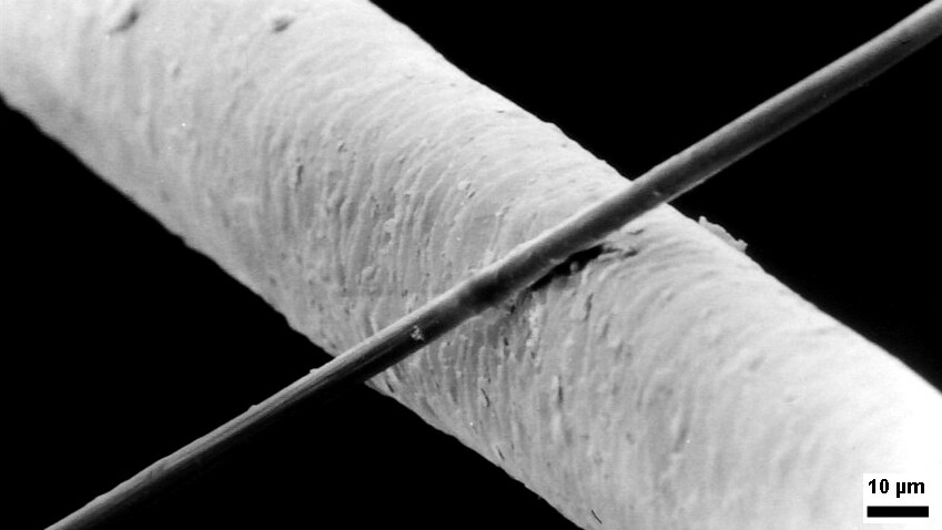
The micrometre ( international spelling as used by the International Bureau of Weights and Measures; SI symbol: μm) or micrometer (American spelling), also commonly known as a micron, is a unit of length in the International System of Units (SI) equalling (SI standard prefix "micro-" = ); that is, one millionth of a metre (or one thousandth of a millimetre, , or about ). The nearest smaller common SI unit is the nanometre, equivalent to one thousandth of a micrometre, one millionth of a millimetre or one billionth of a metre (). The micrometre is a common unit of measurement for wavelengths of infrared radiation as well as sizes of biological cells and bacteria, and for grading wool by the diameter of the fibres. The width of a single human hair ranges from approximately 20 to . The longest human chromosome, chromosome 1, is approximately in length. Examples Between 1 μm and 10 μm: * 1–10 μm – length of a typical bacterium * 3–8 μm – width of ... [...More Info...] [...Related Items...] OR: [Wikipedia] [Google] [Baidu] |
Amyloid (mycology)
In mycology a tissue or feature is said to be amyloid if it has a positive amyloid reaction when subjected to a crude chemical test using iodine as an ingredient of either Melzer's reagent or Lugol's solution, producing a blue to blue-black staining. The term "amyloid" is derived from the Latin ''amyloideus'' ("starch-like"). It refers to the fact that starch gives a similar reaction, also called an amyloid reaction. The test can be on microscopic features, such as spore walls or hyphal walls, or the apical apparatus or entire ascus wall of an ascus, or be a macroscopic reaction on tissue where a drop of the reagent is applied. Negative reactions, called inamyloid or nonamyloid, are for structures that remain pale yellow-brown or clear. A reaction producing a deep reddish to reddish-brown staining is either termed a dextrinoid reaction (pseudoamyloid is a synonym) or a hemiamyloid reaction. Melzer's reagent reactions Hemiamyloidity Hemiamyloidity in mycology refers to a special ... [...More Info...] [...Related Items...] OR: [Wikipedia] [Google] [Baidu] |
Ellipsoid
An ellipsoid is a surface that may be obtained from a sphere by deforming it by means of directional scalings, or more generally, of an affine transformation. An ellipsoid is a quadric surface; that is, a surface that may be defined as the zero set of a polynomial of degree two in three variables. Among quadric surfaces, an ellipsoid is characterized by either of the two following properties. Every planar cross section is either an ellipse, or is empty, or is reduced to a single point (this explains the name, meaning "ellipse-like"). It is bounded, which means that it may be enclosed in a sufficiently large sphere. An ellipsoid has three pairwise perpendicular axes of symmetry which intersect at a center of symmetry, called the center of the ellipsoid. The line segments that are delimited on the axes of symmetry by the ellipsoid are called the ''principal axes'', or simply axes of the ellipsoid. If the three axes have different lengths, the figure is a triaxial ellipsoid (r ... [...More Info...] [...Related Items...] OR: [Wikipedia] [Google] [Baidu] |
Spore
In biology, a spore is a unit of sexual or asexual reproduction that may be adapted for dispersal and for survival, often for extended periods of time, in unfavourable conditions. Spores form part of the life cycles of many plants, algae, fungi and protozoa. Bacterial spores are not part of a sexual cycle, but are resistant structures used for survival under unfavourable conditions. Myxozoan spores release amoeboid infectious germs ("amoebulae") into their hosts for parasitic infection, but also reproduce within the hosts through the pairing of two nuclei within the plasmodium, which develops from the amoebula. In plants, spores are usually haploid and unicellular and are produced by meiosis in the sporangium of a diploid sporophyte. Under favourable conditions the spore can develop into a new organism using mitotic division, producing a multicellular gametophyte, which eventually goes on to produce gametes. Two gametes fuse to form a zygote which develops into a new s ... [...More Info...] [...Related Items...] OR: [Wikipedia] [Google] [Baidu] |
Lamellae (mycology)
In mycology, a lamella, or gill, is a papery hymenophore rib under the cap of some mushroom species, most often agarics. The gills are used by the mushrooms as a means of spore dispersal, and are important for species identification. The attachment of the gills to the stem is classified based on the shape of the gills when viewed from the side, while color, crowding and the shape of individual gills can also be important features. Additionally, gills can have distinctive microscopic or macroscopic features. For instance, ''Lactarius'' species typically seep latex from their gills. It was originally believed that all gilled fungi were Agaricales, but as fungi were studied in more detail, some gilled species were demonstrated not to be. It is now clear that this is a case of convergent evolution (i.e. gill-like structures evolved separately) rather than being an anatomic feature that evolved only once. The apparent reason that various basidiomycetes have evolved gills is that i ... [...More Info...] [...Related Items...] OR: [Wikipedia] [Google] [Baidu] |
Pruinose
Pruinescence , or pruinosity, is a "frosted" or dusty-looking coating on top of a surface. It may also be called a pruina (plural: ''pruinae''), from the Latin word for hoarfrost. The adjectival form is pruinose . Entomology In insects, a "bloom" caused by wax particles on top of an insect's cuticle covers up the underlying coloration, giving a dusty or frosted appearance. The pruinescence is commonly white to pale blue in color but can be gray, pink, purple, or red; these colors may be produced by Tyndall scattering of light. When pale in color, pruinescence often strongly reflects ultraviolet. Pruinescence is found in many species of Odonata, particularly damselflies of the families Lestidae and Coenagrionidae, where it occurs on the wings and body. Among true dragonflies it is most common on male Libellulidae (skimmers). In the common whitetail and blue dasher dragonflies (''Plathemis lydia'' and ''Pachydiplax longipennis''), males display the pruinescence on the back of th ... [...More Info...] [...Related Items...] OR: [Wikipedia] [Google] [Baidu] |
Trama (mycology)
In mycology, the term trama is used in two ways. In the broad sense, it is the inner, fleshy portion of a mushroom's basidiocarp, or fruit body. It is distinct from the outer layer of tissue, known as the pileipellis or cuticle, and from the spore-bearing tissue layer known as the hymenium. In essence, the trama is the tissue that is commonly referred to as the "flesh" of mushrooms and similar fungi.Largent D, Johnson D, Watling R. 1977. ''How to Identify Mushrooms to Genus III: Microscopic Features''. Arcata, CA: Mad River Press. . pp. 60–70. The second use is more specific, and refers to the "hymenophoral trama" that supports the hymenium. It is similarly interior, connective tissue, but it is more specifically the central layer of hyphae running from the underside of the mushroom cap to the lamella or gill, upon which the hymenium rests. Various types have been classified by their structure, including trametoid, cantharelloid, boletoid, and agaricoid, with agaricoid the ... [...More Info...] [...Related Items...] OR: [Wikipedia] [Google] [Baidu] |


