|
Molgophidae
Lysorophia is an order of fossorial Carboniferous and Permian tetrapods within the Recumbirostra. Lysorophians resembled small snakes, as their bodies are extremely elongate. There is a single family, the Molgophidae (previously known as Lysorophidae). Currently there are around five genera included within Lysorophia, although many may not be valid. Description The skull is heavily built but with large lateral openings to accommodate jaw musculature, with small orbits restricted to the anterior edge of the large fenestrae. The intertemporal, supratemporal, postfrontal, and jugal bones of the skull have disappeared. The mandibles are short and robust with a small number of large triangular teeth. Although it was initially thought that the maxilla and premaxilla were freely movable, detailed anatomical studies show that this is not the case. The braincase is extremely robust, suggesting that lysorophians engaged in headfirst burrowing. The torso is very elongate, the limbs di ... [...More Info...] [...Related Items...] OR: [Wikipedia] [Google] [Baidu] |
Nagini Mazonense
''Nagini'' (from Sanskrit ''Nāga, Nāgá'', "snake") is an extinct genus of recumbirostran tetrapods from the middle Carboniferous of the Mazon Creek fossil beds, Illinois, United States. The type and only species, ''Nagini mazonense'', was named by Arjan Mann and colleagues in 2022 from two specimens, both of which preserve soft tissue like other fossils from Mazon Creek: Milwaukee Public Museum, MPM VP359229.2 and Field Museum of Natural History, FMNH PR 1031. It is a member of the Lysorophia, Molgophidae, a lineage of amniote-like tetrapods which exhibited a pattern of body elongation and digit reduction on the limbs. ''Nagini'' is the first member of the group that shows the complete loss of the forelimbs and pectoral girdle, but it still has intact hindlimbs; this mirrors the pattern seen in the snake#Evolution, evolution of snakes, and suggests that molgophids underwent a similar mechanism of limb reduction beginning with the failure to form distinct forelimbs. References ... [...More Info...] [...Related Items...] OR: [Wikipedia] [Google] [Baidu] |
Infernovenator
''Infernovenator'' is a genus of Carboniferous Lysorophia, lysorophian Recumbirostra, recumbirostran from the Mazon Creek fossil beds, Mazon Creek lagerstätte in Illinois, U.S. It was described in 2019. History of study The holotype, now reposited at the Field Museum of Natural History, Field Museum, was previously described by Godfrey (1997) as an aïstopod, ''Phlegethontia longissama''. Redescription of the specimen led to its identification as a new lysorophian taxon. ''Infernovenator'' is represented only by the holotype, a nearly complete skeleton. The genus name is given for the Latin ''infernum'' ("hell") to refer to the fossorial habitats of the taxon and ''venator'' ("hunter"). The species name honors paleontologist Margaret Clair Steen Brough. Anatomy ''Infernovenator'' is diagnosed by a unique combination of features: (1) 61 presacral vertebrae; (2) a triangular postfrontal that contacts the tabular; (3) a circumorbital series formed by the prefrontal, the postfro ... [...More Info...] [...Related Items...] OR: [Wikipedia] [Google] [Baidu] |
Recumbirostra
Recumbirostra is a clade of tetrapods which lived during the Carboniferous and Permian periods. They are thought to have had a fossorial (burrowing) lifestyle and the group includes both short-bodied and long-bodied snake-like forms. At least one species, the molgophid ''Nagini mazonense,'' lost its forelimbs entirely. It includes the families Pantylidae, Gymnarthridae, Ostodolepidae, Rhynchonkidae and Brachystelechidae, with additional families such as Microbrachidae and Molgophidae being included by some authors. Recumbirostra was erected as a clade in 2007 to include many of the taxa traditionally grouped in "Microsauria", which has since been shown to be a paraphyletic or polyphyletic grouping. Like other "microsaurs", the recumbirostrans have traditionally been considered to be members of the subclass Lepospondyli; however, many phylogenetic analyses conducted since the 2010s have recovered recumbirostrans as basal sauropsid amniotes instead. Not all phylogenetic analyses rec ... [...More Info...] [...Related Items...] OR: [Wikipedia] [Google] [Baidu] |
Alfred Romer
Alfred Sherwood Romer (December 28, 1894 – November 5, 1973) was an American paleontologist and biologist and a specialist in vertebrate evolution. Biography Alfred Romer was born in White Plains, New York, the son of Harry Houston Romer and his wife, Evalyn Sherwood. He was educated at White Plains High School. He studied at Amherst College for his Bachelor of Science Honours degree in biology, then at Columbia University for an M.Sc in Biology and a doctorate in zoology in 1921. Romer joined the department of geology and paleontology at the University of Chicago as an associate professor in 1923. He was an active researcher and teacher. His collecting program added important Paleozoic specimens to Chicago's Walker Museum of Paleontology. In 1934 he was appointed professor of biology at Harvard University. In 1946, he became director of the Harvard Museum of Comparative Zoology (MCZ). In 1954 Romer was awarded the Mary Clark Thompson Medal from the National Academy of Sc ... [...More Info...] [...Related Items...] OR: [Wikipedia] [Google] [Baidu] |
Tetrapod
Tetrapods (; ) are four-limbed vertebrate animals constituting the superclass Tetrapoda (). It includes extant and extinct amphibians, sauropsids ( reptiles, including dinosaurs and therefore birds) and synapsids (pelycosaurs, extinct therapsids and all extant mammals). Tetrapods evolved from a clade of primitive semiaquatic animals known as the Tetrapodomorpha which, in turn, evolved from ancient lobe-finned fish (sarcopterygians) around 390 million years ago in the Middle Devonian period; their forms were transitional between lobe-finned fishes and true four-limbed tetrapods. Limbed vertebrates (tetrapods in the broad sense of the word) are first known from Middle Devonian trackways, and body fossils became common near the end of the Late Devonian but these were all aquatic. The first crown-tetrapods (last common ancestors of extant tetrapods capable of terrestrial locomotion) appeared by the very early Carboniferous, 350 million years ago. The specific aquatic ancestors ... [...More Info...] [...Related Items...] OR: [Wikipedia] [Google] [Baidu] |
Jugal
The jugal is a skull bone found in most reptiles, amphibians and birds. In mammals, the jugal is often called the malar or zygomatic. It is connected to the quadratojugal and maxilla, as well as other bones, which may vary by species. Anatomy The jugal bone is located on either side of the skull in the circumorbital region. It is the origin of several masticatory muscles in the skull. The jugal and lacrimal bones are the only two remaining from the ancestral circumorbital series: the prefrontal, postfrontal, postorbital, jugal, and lacrimal bones. During development, the jugal bone originates from dermal bone. In dinosaurs This bone is considered key in the determination of general traits in cases in which the entire skull has not been found intact (for instance, as with dinosaurs in paleontology). In some dinosaur genera the jugal also forms part of the lower margin of either the antorbital fenestra or the infratemporal fenestra, or both. Most commonly, this bone articu ... [...More Info...] [...Related Items...] OR: [Wikipedia] [Google] [Baidu] |
Postfrontal
The skull is a bone protective cavity for the brain. The skull is composed of four types of bone i.e., cranial bones, facial bones, ear ossicles and hyoid bone. However two parts are more prominent: the cranium and the mandible. In humans, these two parts are the neurocranium and the viscerocranium (facial skeleton) that includes the mandible as its largest bone. The skull forms the anterior-most portion of the skeleton and is a product of cephalisation—housing the brain, and several sensory structures such as the eyes, ears, nose, and mouth. In humans these sensory structures are part of the facial skeleton. Functions of the skull include protection of the brain, fixing the distance between the eyes to allow stereoscopic vision, and fixing the position of the ears to enable sound localisation of the direction and distance of sounds. In some animals, such as horned ungulates (mammals with hooves), the skull also has a defensive function by providing the mount (on the fronta ... [...More Info...] [...Related Items...] OR: [Wikipedia] [Google] [Baidu] |
Supratemporal
The supratemporal bone is a paired cranial bone present in many tetrapods and tetrapodomorph fish. It is part of the temporal region (the portion of the skull roof behind the eyes), usually lying medial (inwards) relative to the squamosal and lateral (outwards) relative to the parietal and/or postparietal. It may also contact the postorbital or intertemporal (which lie forwards), or tabular (which lies backwards), when those bones are present. The supratemporal is a common component of the skull in many extinct amphibians, though it is apparently absent in the lightweight skulls of living lissamphibians (frogs and salamanders). Embryological studies of salamanders suggests that the supratemporal fuses with the squamosal in early development. A separate supratemporal was retained by early synapsids and reptiles, but was strongly reduced in many groups. Squamates (lizards and snakes) still possess a small supratemporal, though archosaurs (crocodilians and birds) and mammals lack i ... [...More Info...] [...Related Items...] OR: [Wikipedia] [Google] [Baidu] |
Fenestra (anatomy)
A fenestra (fenestration; plural fenestrae or fenestrations) is any small opening or pore, commonly used as a term in the biological sciences. It is the Latin word for "window", and is used in various fields to describe a pore in an anatomical structure. Biological morphology In morphology, fenestrae are found in cancellous bones, particularly in the skull. In anatomy, the round window and oval window are also known as the ''fenestra rotunda'' and the ''fenestra ovalis''. In microanatomy, fenestrae are found in endothelium of fenestrated capillaries, enabling the rapid exchange of molecules between the blood and surrounding tissue. The elastic layer of the tunica intima is a fenestrated membrane. In surgery, a fenestration is a new opening made in a part of the body to enable drainage or access. Plant biology and mycology In plant biology, the perforations in a perforate leaf are also described as fenestrae, and the leaf is called a fenestrate leaf. The leaf window is ... [...More Info...] [...Related Items...] OR: [Wikipedia] [Google] [Baidu] |
Orbit (anatomy)
In anatomy, the orbit is the cavity or socket of the skull in which the eye and its appendages are situated. "Orbit" can refer to the bony socket, or it can also be used to imply the contents. In the adult human, the volume of the orbit is , of which the eye occupies . The orbital contents comprise the eye, the orbital and retrobulbar fascia, extraocular muscles, cranial nerves II, III, IV, V, and VI, blood vessels, fat, the lacrimal gland with its sac and duct, the eyelids, medial and lateral palpebral ligaments, cheek ligaments, the suspensory ligament, septum, ciliary ganglion and short ciliary nerves. Structure The orbits are conical or four-sided pyramidal cavities, which open into the midline of the face and point back into the head. Each consists of a base, an apex and four walls."eye, human."Encyclopædia Britannica from Encyclopædia Britannica 2006 Ultimate Reference Suite DVD 2009 Openings There are two important foramina, or windows, two important fissu ... [...More Info...] [...Related Items...] OR: [Wikipedia] [Google] [Baidu] |
Skull
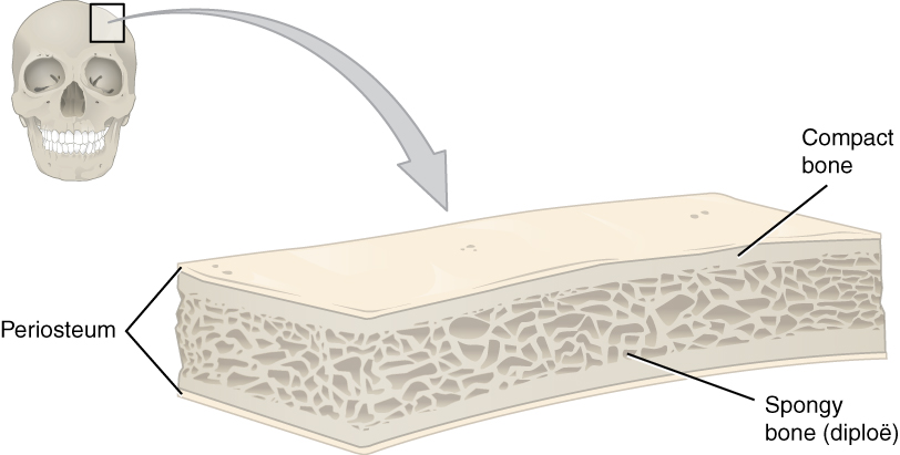
The skull is a bone protective cavity for the brain. The skull is composed of four types of bone i.e., cranial bones, facial bones, ear ossicles and hyoid bone. However two parts are more prominent: the cranium and the mandible. In humans, these two parts are the neurocranium and the viscerocranium ( facial skeleton) that includes the mandible as its largest bone. The skull forms the anterior-most portion of the skeleton and is a product of cephalisation—housing the brain, and several sensory structures such as the eyes, ears, nose, and mouth. In humans these sensory structures are part of the facial skeleton. Functions of the skull include protection of the brain, fixing the distance between the eyes to allow stereoscopic vision, and fixing the position of the ears to enable sound localisation of the direction and distance of sounds. In some animals, such as horned ungulates (mammals with hooves), the skull also has a defensive function by providing the mount (on the front ... [...More Info...] [...Related Items...] OR: [Wikipedia] [Google] [Baidu] |

.jpg)


