|
Middle Meningeal Artery
The middle meningeal artery ('' la, arteria meningea media'') is typically the third branch of the first portion of the maxillary artery. After branching off the maxillary artery in the infratemporal fossa, it runs through the foramen spinosum to supply the dura mater (the outer meningeal layer) and the calvaria. The middle meningeal artery is the largest of the three (paired) arteries that supply the meninges, the others being the anterior meningeal artery and the posterior meningeal artery. The anterior branch of the middle meningeal artery runs beneath the pterion. It is vulnerable to injury at this point, where the skull is thin. Rupture of the artery may give rise to an epidural hematoma. In the dry cranium, the middle meningeal, which runs within the dura mater surrounding the brain, makes a deep groove in the calvarium. The middle meningeal artery is intimately associated with the auriculotemporal nerve, which wraps around the artery making the two easily identifiabl ... [...More Info...] [...Related Items...] OR: [Wikipedia] [Google] [Baidu] |
Maxillary Artery
The maxillary artery supplies deep structures of the face. It branches from the external carotid artery just deep to the neck of the mandible. Structure The maxillary artery, the larger of the two terminal branches of the external carotid artery, arises behind the neck of the mandible, and is at first imbedded in the substance of the parotid gland; it passes forward between the ramus of the mandible and the sphenomandibular ligament, and then runs, either superficial or deep to the lateral pterygoid muscle, to the pterygopalatine fossa. It supplies the deep structures of the face, and may be divided into mandibular, pterygoid, and pterygopalatine portions. First portion The ''first'' or ''mandibular '' or ''bony'' portion passes horizontally forward, between the neck of the mandible and the sphenomandibular ligament, where it lies parallel to and a little below the auriculotemporal nerve; it crosses the inferior alveolar nerve, and runs along the lower border of the lateral ... [...More Info...] [...Related Items...] OR: [Wikipedia] [Google] [Baidu] |
Surgery
Surgery ''cheirourgikē'' (composed of χείρ, "hand", and ἔργον, "work"), via la, chirurgiae, meaning "hand work". is a medical specialty that uses operative manual and instrumental techniques on a person to investigate or treat a pathological condition such as a disease or injury, to help improve bodily function, appearance, or to repair unwanted ruptured areas. The act of performing surgery may be called a surgical procedure, operation, or simply "surgery". In this context, the verb "operate" means to perform surgery. The adjective surgical means pertaining to surgery; e.g. surgical instruments or surgical nurse. The person or subject on which the surgery is performed can be a person or an animal. A surgeon is a person who practices surgery and a surgeon's assistant is a person who practices surgical assistance. A surgical team is made up of the surgeon, the surgeon's assistant, an anaesthetist, a circulating nurse and a surgical technologist. Surgery usually span ... [...More Info...] [...Related Items...] OR: [Wikipedia] [Google] [Baidu] |
Scalp
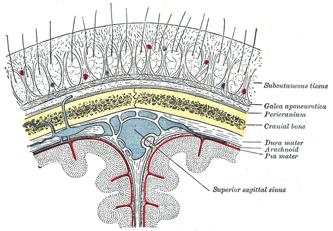
The scalp is the anatomical area bordered by the human face at the front, and by the neck at the sides and back. Structure The scalp is usually described as having five layers, which can conveniently be remembered as a mnemonic: * S: The skin on the head from which head hair grows. It contains numerous sebaceous glands and hair follicles. * C: Connective tissue. A dense subcutaneous layer of fat and fibrous tissue that lies beneath the skin, containing the nerves and vessels of the scalp. * A: The aponeurosis called epicranial aponeurosis (or galea aponeurotica) is the next layer. It is a tough layer of dense fibrous tissue which runs from the frontalis muscle anteriorly to the occipitalis posteriorly. * L: The loose areolar connective tissue layer provides an easy plane of separation between the upper three layers and the pericranium. In scalping the scalp is torn off through this layer. It also provides a plane of access in craniofacial surgery and neurosurgery. Th ... [...More Info...] [...Related Items...] OR: [Wikipedia] [Google] [Baidu] |
Human Skull
The skull is a bone protective cavity for the brain. The skull is composed of four types of bone i.e., cranial bones, facial bones, ear ossicles and hyoid bone. However two parts are more prominent: the cranium and the mandible. In humans, these two parts are the neurocranium and the viscerocranium ( facial skeleton) that includes the mandible as its largest bone. The skull forms the anterior-most portion of the skeleton and is a product of cephalisation—housing the brain, and several sensory structures such as the eyes, ears, nose, and mouth. In humans these sensory structures are part of the facial skeleton. Functions of the skull include protection of the brain, fixing the distance between the eyes to allow stereoscopic vision, and fixing the position of the ears to enable sound localisation of the direction and distance of sounds. In some animals, such as horned ungulates (mammals with hooves), the skull also has a defensive function by providing the mount (on the f ... [...More Info...] [...Related Items...] OR: [Wikipedia] [Google] [Baidu] |
Anastomosis
An anastomosis (, plural anastomoses) is a connection or opening between two things (especially cavities or passages) that are normally diverging or branching, such as between blood vessels, leaf veins, or streams. Such a connection may be normal (such as the foramen ovale in a fetus's heart) or abnormal (such as the patent foramen ovale in an adult's heart); it may be acquired (such as an arteriovenous fistula) or innate (such as the arteriovenous shunt of a metarteriole); and it may be natural (such as the aforementioned examples) or artificial (such as a surgical anastomosis). The reestablishment of an anastomosis that had become blocked is called a reanastomosis. Anastomoses that are abnormal, whether congenital or acquired, are often called fistulas. The term is used in medicine, biology, mycology, geology, and geography. Etymology Anastomosis: medical or Modern Latin, from Greek ἀναστόμωσις, anastomosis, "outlet, opening", Gr ana- "up, on, upon", stoma "mouth", ... [...More Info...] [...Related Items...] OR: [Wikipedia] [Google] [Baidu] |
Posterior Meningeal Arteries
The posterior meningeal artery is the largest vessel supplying the dura region of the posterior fossa. It typically arises from the ascending pharyngeal artery although other origins have been seen, such as the occipital artery. The artery or its branches enter the cranium through jugular foramen, foramen magnum or hypoglossal canal.Diagnostic Cerebral Angiography, 2nd edition, Anne G. Osborn See also * Meninges In anatomy, the meninges (, ''singular:'' meninx ( or ), ) are the three membranes that envelop the brain and spinal cord. In mammals, the meninges are the dura mater, the arachnoid mater, and the pia mater. Cerebrospinal fluid is located in ... References * Diagnostic Cerebral Angiography, 2nd edition, Anne G. Osborn Arteries of the head and neck {{circulatory-stub ... [...More Info...] [...Related Items...] OR: [Wikipedia] [Google] [Baidu] |
Anterior Meningeal Arteries
Standard anatomical terms of location are used to unambiguously describe the anatomy of animals, including humans. The terms, typically derived from Latin or Greek roots, describe something in its standard anatomical position. This position provides a definition of what is at the front ("anterior"), behind ("posterior") and so on. As part of defining and describing terms, the body is described through the use of anatomical planes and anatomical axes. The meaning of terms that are used can change depending on whether an organism is bipedal or quadrupedal. Additionally, for some animals such as invertebrates, some terms may not have any meaning at all; for example, an animal that is radially symmetrical will have no anterior surface, but can still have a description that a part is close to the middle ("proximal") or further from the middle ("distal"). International organisations have determined vocabularies that are often used as standard vocabularies for subdisciplines of anatom ... [...More Info...] [...Related Items...] OR: [Wikipedia] [Google] [Baidu] |
Temporal Bone
The temporal bones are situated at the sides and base of the skull, and lateral to the temporal lobes of the cerebral cortex. The temporal bones are overlaid by the sides of the head known as the temples, and house the structures of the ears. The lower seven cranial nerves and the major vessels to and from the brain traverse the temporal bone. Structure The temporal bone consists of four parts— the squamous, mastoid, petrous and tympanic parts. The squamous part is the largest and most superiorly positioned relative to the rest of the bone. The zygomatic process is a long, arched process projecting from the lower region of the squamous part and it articulates with the zygomatic bone. Posteroinferior to the squamous is the mastoid part. Fused with the squamous and mastoid parts and between the sphenoid and occipital bones lies the petrous part, which is shaped like a pyramid. The tympanic part is relatively small and lies inferior to the squamous part, anterior to t ... [...More Info...] [...Related Items...] OR: [Wikipedia] [Google] [Baidu] |
Squama Temporalis
The squamous part of temporal bone, or temporal squama, forms the front and upper part of the temporal bone, and is scale-like, thin, and translucent. Surfaces Its outer surface is smooth and convex; it affords attachment to the temporal muscle, and forms part of the temporal fossa; on its hinder part is a vertical groove for the middle temporal artery. A curved line, the ''temporal line'', or ''supramastoid crest'', runs backward and upward across its posterior part; it serves for the attachment of the temporal fascia, and limits the origin of the temporalis muscle. The boundary between the squamous part and the mastoid portion of the bone, as indicated by traces of the original suture, lies about 1 cm. below this line. Projecting from the lower part of the squamous part is a long, arched process, the ''zygomatic process''. This process is at first directed lateralward, its two surfaces looking upward and downward; it then appears as if twisted inward upon itself, and ru ... [...More Info...] [...Related Items...] OR: [Wikipedia] [Google] [Baidu] |
Occiput
The occipital bone () is a cranial dermal bone and the main bone of the occiput (back and lower part of the skull). It is trapezoidal in shape and curved on itself like a shallow dish. The occipital bone overlies the occipital lobes of the cerebrum. At the base of skull in the occipital bone, there is a large oval opening called the foramen magnum, which allows the passage of the spinal cord. Like the other cranial bones, it is classed as a flat bone. Due to its many attachments and features, the occipital bone is described in terms of separate parts. From its front to the back is the basilar part, also called the basioccipital, at the sides of the foramen magnum are the lateral parts, also called the exoccipitals, and the back is named as the squamous part. The basilar part is a thick, somewhat quadrilateral piece in front of the foramen magnum and directed towards the pharynx. The squamous part is the curved, expanded plate behind the foramen magnum and is the largest par ... [...More Info...] [...Related Items...] OR: [Wikipedia] [Google] [Baidu] |
Vertex (anatomy)
In arthropod and vertebrate anatomy, the vertex (or ''cranial vertex'') is the highest point of the head. In humans, the vertex is formed by four bones of the skull: the frontal bone, the two parietal bones, and the occipital bone. These bones are connected by the coronal suture between the frontal and parietal bones, the sagittal suture between the two parietal bones, and the lambdoid suture between the parietal and occipital bones. ''Vertex baldness'' refers to a form of male pattern baldness in which the baldness is limited to the vertex, resembling a tonsure. In childbirth, ''vertex birth'' refers to the common head-first presentation of the baby, as opposed to the buttocks-first position of a breech birth. In entomology, the color and shape of an insect's vertex and the structures arising from it are commonly used in identifying species. See also * Calvaria (skull) *Crown (anatomy) The crown is the top portion of the head behind the vertex. The anatomy of the crown v ... [...More Info...] [...Related Items...] OR: [Wikipedia] [Google] [Baidu] |
Parietal Bone
The parietal bones () are two bones in the skull which, when joined at a fibrous joint, form the sides and roof of the cranium. In humans, each bone is roughly quadrilateral in form, and has two surfaces, four borders, and four angles. It is named from the Latin ''paries'' (''-ietis''), wall. Surfaces External The external surface ig. 1is convex, smooth, and marked near the center by an eminence, the parietal eminence (''tuber parietale''), which indicates the point where ossification commenced. Crossing the middle of the bone in an arched direction are two curved lines, the superior and inferior temporal lines; the former gives attachment to the temporal fascia, and the latter indicates the upper limit of the muscular origin of the temporal muscle. Above these lines the bone is covered by a tough layer of fibrous tissue – the epicranial aponeurosis; below them it forms part of the temporal fossa, and affords attachment to the temporal muscle. At the back part and clos ... [...More Info...] [...Related Items...] OR: [Wikipedia] [Google] [Baidu] |
.jpg)


