|
Microtome
A microtome (from the Greek ''mikros'', meaning "small", and ''temnein'', meaning "to cut") is a cutting tool used to produce extremely thin slices of material known as ''sections''. Important in science, microtomes are used in microscopy, allowing for the preparation of samples for observation under transmitted light or electron radiation. Microtomes use steel, glass or diamond blades depending upon the specimen being sliced and the desired thickness of the sections being cut. Steel blades are used to prepare histological sections of animal or plant tissues for light microscopy. Glass knives are used to slice sections for light microscopy and to slice very thin sections for electron microscopy. Industrial grade diamond knives are used to slice hard materials such as bone, teeth and tough plant matter for both light microscopy and for electron microscopy. Gem-quality diamond knives are also used for slicing thin sections for electron microscopy. Microtomy is a method for the p ... [...More Info...] [...Related Items...] OR: [Wikipedia] [Google] [Baidu] |
Histological
Histology, also known as microscopic anatomy or microanatomy, is the branch of biology which studies the microscopic anatomy of biological tissues. Histology is the microscopic counterpart to gross anatomy, which looks at larger structures visible without a microscope. Although one may divide microscopic anatomy into ''organology'', the study of organs, ''histology'', the study of tissues, and ''cytology'', the study of cells, modern usage places all of these topics under the field of histology. In medicine, histopathology is the branch of histology that includes the microscopic identification and study of diseased tissue. In the field of paleontology, the term paleohistology refers to the histology of fossil organisms. Biological tissues Animal tissue classification There are four basic types of animal tissues: muscle tissue, nervous tissue, connective tissue, and epithelial tissue. All animal tissues are considered to be subtypes of these four principal tissue types (fo ... [...More Info...] [...Related Items...] OR: [Wikipedia] [Google] [Baidu] |
Cutting Tool
In the context of machining, a cutting tool or cutter is typically a hardened metal tool that is used to cut, shape, and remove material from a workpiece by means of machining tools as well as abrasive tools by way of shear deformation. The majority of these tools are designed exclusively for metals. There are several different types of single edge cutting tools that are made from a variety of hardened metal alloys that are ground to a specific shape in order to perform a specific part of the turning process resulting in a finished machined part. Single edge cutting tools are used mainly in the turning operations performed by a lathe in which they vary in size as well as alloy composition depending on the size and the type of material being turned. These cutting tools are held stationary by what is known as a tool post which is what manipulates the tools to cut the material into the desired shape. Single edge cutting tools are also the means of cutting material performed by metal s ... [...More Info...] [...Related Items...] OR: [Wikipedia] [Google] [Baidu] |
Focused Ion Beam
Focused ion beam, also known as FIB, is a technique used particularly in the semiconductor industry, materials science and increasingly in the biological field for site-specific analysis, deposition, and ablation of materials. A FIB setup is a scientific instrument that resembles a scanning electron microscope (SEM). However, while the SEM uses a focused beam of electrons to image the sample in the chamber, a FIB setup uses a focused beam of ions instead. FIB can also be incorporated in a system with both electron and ion beam columns, allowing the same feature to be investigated using either of the beams. FIB should not be confused with using a beam of focused ions for direct write lithography (such as in proton beam writing). These are generally quite different systems where the material is modified by other mechanisms. Ion beam source Most widespread instruments are using liquid metal ion sources (LMIS), especially gallium ion sources. Ion sources based on elemental gold an ... [...More Info...] [...Related Items...] OR: [Wikipedia] [Google] [Baidu] |
Fixation (histology)
In the fields of histology, pathology, and cell biology, fixation is the preservation of biological tissues from decay due to autolysis or putrefaction. It terminates any ongoing biochemical reactions and may also increase the treated tissues' mechanical strength or stability. Tissue fixation is a critical step in the preparation of histological sections, its broad objective being to preserve cells and tissue components and to do this in such a way as to allow for the preparation of thin, stained sections. This allows the investigation of the tissues' structure, which is determined by the shapes and sizes of such macromolecules (in and around cells) as proteins and nucleic acids. Purposes In performing their protective role, fixatives denature proteins by coagulation, by forming additive compounds, or by a combination of coagulation and additive processes. A compound that adds chemically to macromolecules stabilizes structure most effectively if it is able to combine with part ... [...More Info...] [...Related Items...] OR: [Wikipedia] [Google] [Baidu] |
Immunohistochemistry
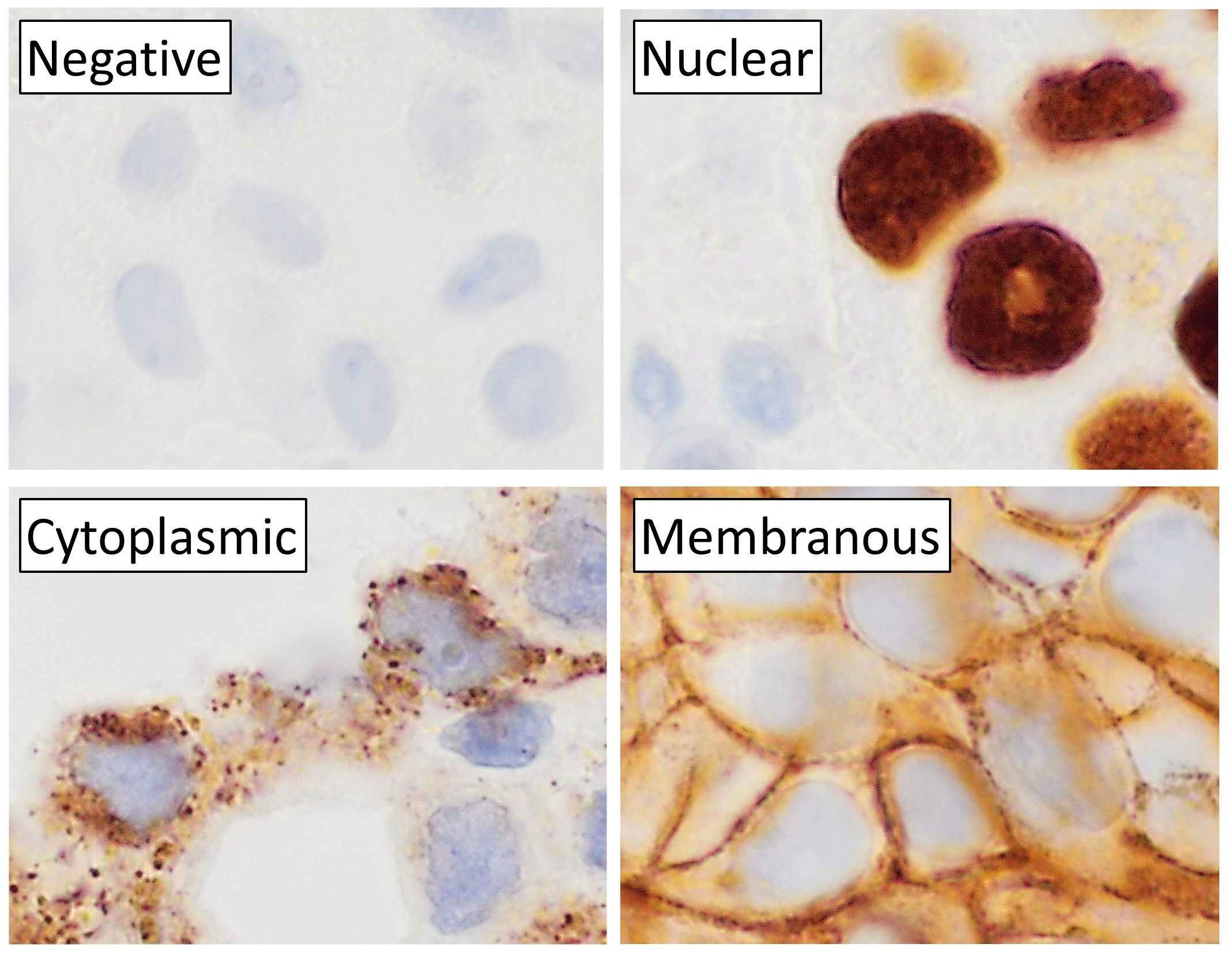
Immunohistochemistry (IHC) is the most common application of immunostaining. It involves the process of selectively identifying antigens (proteins) in cells of a tissue section by exploiting the principle of antibodies binding specifically to antigens in biological tissues. IHC takes its name from the roots "immuno", in reference to antibodies used in the procedure, and "histo", meaning tissue (compare to immunocytochemistry). Albert Coons conceptualized and first implemented the procedure in 1941. Visualising an antibody-antigen interaction can be accomplished in a number of ways, mainly either of the following: * ''Chromogenic immunohistochemistry'' (CIH), wherein an antibody is conjugated to an enzyme, such as peroxidase (the combination being termed immunoperoxidase), that can catalyse a colour-producing reaction. * '' Immunofluorescence'', where the antibody is tagged to a fluorophore, such as fluorescein or rhodamine. Immunohistochemical staining is widely used in the dia ... [...More Info...] [...Related Items...] OR: [Wikipedia] [Google] [Baidu] |
Cryostat
A cryostat (from ''cryo'' meaning cold and ''stat'' meaning stable) is a device used to maintain low cryogenic temperatures of samples or devices mounted within the cryostat. Low temperatures may be maintained within a cryostat by using various refrigeration methods, most commonly using cryogenic fluid bath such as liquid helium. Hence it is usually assembled into a vessel, similar in construction to a vacuum flask or Dewar. Cryostats have numerous applications within science, engineering, and medicine. Types Closed-cycle cryostats Closed-cycle cryostats consist of a chamber through which cold helium vapour is pumped. An external mechanical refrigerator extracts the warmer helium exhaust vapour, which is cooled and recycled. Closed-cycle cryostats consume a relatively large amount of electrical power, but need not be refilled with helium and can run continuously for an indefinite period. Objects may be cooled by attaching them to a metallic coldplate inside a vacuum ch ... [...More Info...] [...Related Items...] OR: [Wikipedia] [Google] [Baidu] |
Frozen Section Procedure
The frozen section procedure is a pathological laboratory procedure to perform rapid microscopic analysis of a specimen. It is used most often in oncological surgery. The technical name for this procedure is cryosection. The microtome device that cold cuts thin blocks of frozen tissue is called a cryotome. The quality of the slides produced by frozen section is of lower quality than formalin fixed paraffin embedded tissue processing. While diagnosis can be rendered in many cases, fixed tissue processing is preferred in many conditions for more accurate diagnosis. The intraoperative consultation is the name given to the whole intervention by the pathologist, which includes not only frozen section but also gross evaluation of the specimen, examination of cytology preparations taken on the specimen (e.g. touch imprints), and aliquoting of the specimen for special studies (e.g. molecular pathology techniques, flow cytometry). The report given by the pathologist is often limited to ... [...More Info...] [...Related Items...] OR: [Wikipedia] [Google] [Baidu] |
Paraffin Wax
Paraffin wax (or petroleum wax) is a soft colorless solid derived from petroleum, coal, or oil shale that consists of a mixture of hydrocarbon molecules containing between 20 and 40 carbon atoms. It is solid at room temperature and begins to melt above approximately , and its boiling point is above . Common applications for paraffin wax include lubrication, electrical insulation, and candles; dyed paraffin wax can be made into crayons. It is distinct from kerosene and other petroleum products that are sometimes called paraffin. Un-dyed, unscented paraffin candles are odorless and bluish-white. Paraffin wax was first created by Carl Reichenbach in Germany in 1830 and marked a major advancement in candlemaking technology, as it burned more cleanly and reliably than tallow candles and was cheaper to produce. In chemistry, ''paraffin'' is used synonymously with ''alkane'', indicating hydrocarbons with the general formula C''n''H2''n''+2. The name is derived from Latin ''parum'' (" ... [...More Info...] [...Related Items...] OR: [Wikipedia] [Google] [Baidu] |
Jan Evangelista Purkyně
Jan Evangelista Purkyně (; also written Johann Evangelist Purkinje) (17 or 18 December 1787 – 28 July 1869) was a Czech anatomist and physiologist. In 1839, he coined the term '' protoplasm'' for the fluid substance of a cell. He was one of the best known scientists of his time. Such was his fame that when people from outside Europe wrote letters to him, all that they needed to put as the address was "Purkyně, Europe". Biography Purkyně was born in the Kingdom of Bohemia (then part of the Austrian monarchy, now Czech Republic). After completing senior high school in 1804, Purkyně joined the Piarists order as a monk but subsequently left "to deal more freely with science." In 1818, he graduated from Charles University in Prague with a degree in medicine, where he was appointed a Professor of Physiology. He discovered the Purkinje effect, the human eye's much reduced sensitivity to dim red light compared to dim blue light, and published in 1823 description of several entopt ... [...More Info...] [...Related Items...] OR: [Wikipedia] [Google] [Baidu] |
Wilhelm His, Sr , the Dutch national anthem
{{Disambiguation ...
Wilhelm may refer to: People and fictional characters * William Charles John Pitcher, costume designer known professionally as "Wilhelm" * Wilhelm (name), a list of people and fictional characters with the given name or surname Other uses * Mount Wilhelm, the highest mountain in Papua New Guinea * Wilhelm Archipelago, Antarctica * Wilhelm (crater), a lunar crater See also * Wilhelm scream, a stock sound effect * SS ''Kaiser Wilhelm II'', or USS ''Agamemnon'', a German steam ship * Wilhelmus "Wilhelmus van Nassouwe", usually known just as "Wilhelmus" ( nl, Het Wilhelmus, italic=no; ; English translation: "The William"), is the national anthem of both the Netherlands and the Kingdom of the Netherlands. It dates back to at least 1572 ... [...More Info...] [...Related Items...] OR: [Wikipedia] [Google] [Baidu] |
Alexander Cummings
Alexander Cumming FRSE (sometimes referred to as Alexander Cummings; 1733 – 8 March 1814) was a Scottish watchmaker and instrument inventor, who was the first to patent a design of the flush toilet in 1775, which had been pioneered by Sir John Harington, but without solving the problem of foul smells. As well as improving the flush mechanism, Cummings included an S-trap (or bend) to retain water permanently within the waste pipe, thus preventing sewer gases from entering buildings. Most modern flush toilets still include a similar trap. Early life Cumming was a mathematician and mechanic as well as a watchmaker. Little is known of his early life. He was born in Edinburgh in 1733, the son of James Cumming of Duthil. He is recorded as having been apprenticed to an Edinburgh watchmaker. Career In the 1750s he was employed by Archibald Campbell, 3rd Duke of Argyll, at Inverary as an organ builder as well as a clockmaker. After his move to England he continued to work in both ... [...More Info...] [...Related Items...] OR: [Wikipedia] [Google] [Baidu] |


