|
Mesothelium
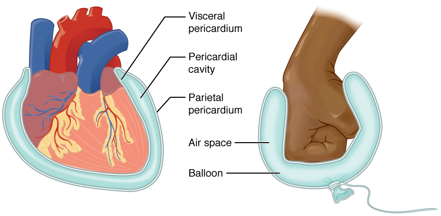
The mesothelium is a membrane composed of simple squamous epithelial cells of mesodermal origin, which forms the lining of several body cavities: the pleura (pleural cavity around the lungs), peritoneum (abdominopelvic cavity including the mesentery, omenta, falciform ligament and the perimetrium) and pericardium (around the heart). Mesothelial tissue also surrounds the male testis (as the tunica vaginalis) and occasionally the spermatic cord (in a patent processus vaginalis). Mesothelium that covers the internal organs is called visceral mesothelium, while one that covers the surrounding body walls is called the parietal mesothelium. The mesothelium that secretes serous fluid as a main function is also known as a serosa. Origin Mesothelium derives from the embryonic mesoderm cell layer, that lines the coelom (body cavity) in the embryo. It develops into the layer of cells that covers and protects most of the internal organs of the body. Structure The mesothelium forms a ... [...More Info...] [...Related Items...] OR: [Wikipedia] [Google] [Baidu] |
Mesoderm
The mesoderm is the middle layer of the three germ layers that develops during gastrulation in the very early development of the embryo of most animals. The outer layer is the ectoderm, and the inner layer is the endoderm.Langman's Medical Embryology, 11th edition. 2010. The mesoderm forms mesenchyme, mesothelium, non-epithelial blood cells and coelomocytes. Mesothelium lines coeloms. Mesoderm forms the muscles in a process known as myogenesis, septa (cross-wise partitions) and mesenteries (length-wise partitions); and forms part of the gonads (the rest being the gametes). Myogenesis is specifically a function of mesenchyme. The mesoderm differentiates from the rest of the embryo through intercellular signaling, after which the mesoderm is polarized by an organizing center. The position of the organizing center is in turn determined by the regions in which beta-catenin is protected from degradation by GSK-3. Beta-catenin acts as a co-factor that alters the activity of ... [...More Info...] [...Related Items...] OR: [Wikipedia] [Google] [Baidu] |
Cytology
Cell biology (also cellular biology or cytology) is a branch of biology that studies the structure, function, and behavior of cells. All living organisms are made of cells. A cell is the basic unit of life that is responsible for the living and functioning of organisms. Cell biology is the study of structural and functional units of cells. Cell biology encompasses both prokaryotic and eukaryotic cells and has many subtopics which may include the study of cell metabolism, cell communication, cell cycle, biochemistry, and cell composition. The study of cells is performed using several microscopy techniques, cell culture, and cell fractionation. These have allowed for and are currently being used for discoveries and research pertaining to how cells function, ultimately giving insight into understanding larger organisms. Knowing the components of cells and how cells work is fundamental to all biological sciences while also being essential for research in biomedical fields such as ca ... [...More Info...] [...Related Items...] OR: [Wikipedia] [Google] [Baidu] |
Falciform Ligament
In human anatomy, the falciform ligament () is a ligament that attaches the liver to the front body wall and divides the liver into the left lobe and right lobe. The falciform ligament is a broad and thin fold of peritoneum, its base being directed downward and backward and its apex upward and forward. It droops down from the hilum of the liver. Structure The falciform ligament stretches obliquely from the front to the back of the abdomen, with one surface in contact with the peritoneum behind the right rectus abdominis muscle and the diaphragm, and the other in contact with the left lobe of the liver. The ligament stretches from the underside of the diaphragm to the posterior surface of the sheath of the right rectus abdominis muscle, as low down as the umbilicus; by its right margin it extends from the notch on the anterior margin of the liver, as far back as the posterior surface. It is composed of two layers of peritoneum closely united together. Its base or free edge ... [...More Info...] [...Related Items...] OR: [Wikipedia] [Google] [Baidu] |
Embryo
An embryo is an initial stage of development of a multicellular organism. In organisms that reproduce sexually, embryonic development is the part of the life cycle that begins just after fertilization of the female egg cell by the male sperm cell. The resulting fusion of these two cells produces a single-celled zygote that undergoes many cell divisions that produce cells known as blastomeres. The blastomeres are arranged as a solid ball that when reaching a certain size, called a morula, takes in fluid to create a cavity called a blastocoel. The structure is then termed a blastula, or a blastocyst in mammals. The mammalian blastocyst hatches before implantating into the endometrial lining of the womb. Once implanted the embryo will continue its development through the next stages of gastrulation, neurulation, and organogenesis. Gastrulation is the formation of the three germ layers that will form all of the different parts of the body. Neurulation forms the nervous ... [...More Info...] [...Related Items...] OR: [Wikipedia] [Google] [Baidu] |
Serosa
The serous membrane (or serosa) is a smooth tissue membrane of mesothelium lining the contents and inner walls of body cavities, which secrete serous fluid to allow lubricated sliding movements between opposing surfaces. The serous membrane that covers internal organs is called a ''visceral'' membrane; while the one that covers the cavity wall is called the ''parietal'' membrane. Between the two opposing serosal surfaces is often a potential space, mostly empty except for the small amount of serous fluid. The Latin anatomical name is '' tunica serosa''. Serous membranes line and enclose several body cavities, also known as serous cavities, where they secrete a lubricating fluid which reduces friction from movements. Serosa is entirely different from the adventitia, a connective tissue layer which binds together structures rather than reducing friction between them. The serous membrane covering the heart and lining the mediastinum is referred to as the pericardium, the se ... [...More Info...] [...Related Items...] OR: [Wikipedia] [Google] [Baidu] |
Serous Fluid
In physiology, serous fluid or serosal fluid (originating from the Medieval Latin word ''serosus'', from Latin ''serum'') is any of various body fluids resembling Serum (blood), serum, that are typically pale yellow or transparent and of a benign nature. The fluid fills the inside of body cavity, body cavities. Serous fluid originates from serous glands, with secretions enriched with proteins and water. Serous fluid may also originate from mixed glands, which contain both mucous cell, mucous and serous cells. A common trait of serous fluids is their role in assisting digestion, excretion, and respiratory system, respiration. In medical fields, especially cytopathology, serous fluid is a synonym for effusion fluids from various body cavities. Examples of effusion fluid are pleural effusion and pericardial effusion. There are many causes of effusions which include involvement of the cavity by cancer. Cancer in a serous cavity is called a serous carcinoma. Cytopathology evaluation is ... [...More Info...] [...Related Items...] OR: [Wikipedia] [Google] [Baidu] |
Visceral
In biology, an organ is a collection of tissues joined in a structural unit to serve a common function. In the hierarchy of life, an organ lies between tissue and an organ system. Tissues are formed from same type cells to act together in a function. Tissues of different types combine to form an organ which has a specific function. The intestinal wall for example is formed by epithelial tissue and smooth muscle tissue. Two or more organs working together in the execution of a specific body function form an organ system, also called a biological system or body system. An organ's tissues can be broadly categorized as parenchyma, the functional tissue, and stroma, the structural tissue with supportive, connective, or ancillary functions. For example, the gland's tissue that makes the hormones is the parenchyma, whereas the stroma includes the nerves that innervate the parenchyma, the blood vessels that oxygenate and nourish it and carry away its metabolic wastes, and the conne ... [...More Info...] [...Related Items...] OR: [Wikipedia] [Google] [Baidu] |
Tunica (biology)
In biology, a tunica (, ; ) is a layer, coat, sheath, or similar covering. The word came to English from the New Latin of science and medicine. Its literal sense is about the same as that of the word ''tunic'', with which it is cognate. In biology one of its senses used to be the taxonomic name of a genus of plants, but the nomenclature has been revised and those plants are now included in the genus ''Petrorhagia''. In modern biology in general, ''tunica'' occurs as a technical or anatomical term mainly in botany and zoology. It usually refers to membranous structures that line or cover particular organs. In many such contexts ''tunica'' is used interchangeably with ''tunic'' according to preference. An organ or organism that has a tunic(a) may be said to be ''tunicate'', as in a ''tunicate bulb''. This adjective ''tunicate'' is not to be confused with the noun ''tunicate'', which refers to a member of the subphylum '' Tunicata''. Botanical and related usages In botany there are s ... [...More Info...] [...Related Items...] OR: [Wikipedia] [Google] [Baidu] |
Processus Vaginalis
The vaginal process (or processus vaginalis) is an embryonic developmental outpouching of the parietal peritoneum. It is present from around the 12th week of gestation, and commences as a peritoneal outpouching. Sex differences In males, it precedes the testes in their descent down within the gubernaculum, and closes. This closure (also called ''fusion'') occurs at any point from a few weeks before birth, to a few weeks after birth. The remaining portion around the testes becomes the tunica vaginalis. If it does not close in females, it forms the canal of Nuck. Clinical significance Failure of closure of the vaginal process leads to the propensity to develop a number of abnormalities. Peritoneal fluid can travel down a patent vaginal process leading to the formation of a hydrocele. Persistent patent processus vaginalis is more common on the right than the left. Accumulation of blood in a persistent processus vaginalis could result in a hematocele. There is the potential for an ... [...More Info...] [...Related Items...] OR: [Wikipedia] [Google] [Baidu] |
Spermatic Cord
The spermatic cord is the cord-like structure in males formed by the vas deferens (''ductus deferens'') and surrounding tissue that runs from the deep inguinal ring down to each testicle. Its serosal covering, the tunica vaginalis, is an extension of the peritoneum that passes through the transversalis fascia. Each testicle develops in the lower thoracic and upper lumbar region and migrates into the scrotum. During its descent it carries along with it the vas deferens, its vessels, nerves etc. There is one on each side. Structure The spermatic cord is ensheathed in three layers of tissue: * ''external spermatic fascia'', an extension of the innominate fascia that overlies the aponeurosis of the external oblique muscle. * ''cremasteric muscle and fascia'', formed from a continuation of the internal oblique muscle and its fascia. * ''internal spermatic fascia'', continuous with the transversalis fascia. The normal diameter of the spermatic cord is about 16 mm (range 11 to 22 mm). It ... [...More Info...] [...Related Items...] OR: [Wikipedia] [Google] [Baidu] |
Tunica Vaginalis
The tunica vaginalis is the pouch of serous membrane that covers the testes. It is derived from the vaginal process of the peritoneum, which in the fetus precedes the descent of the testes from the abdomen into the scrotum. After its descent, that portion of the pouch which extends from the abdominal inguinal ring to near the upper part of the gland becomes obliterated; the lower portion remains as a shut sac, which invests the surface of each testis, and is reflected on to the internal surface of the scrotum; hence it may be described as consisting of a visceral and a parietal lamina. Visceral lamina The visceral lamina (lamina visceralis) covers the greater part of the testis and epididymis, connecting the latter to the testis by means of a distinct fold. From the posterior border of the gland it is reflected on to the internal surface of the scrotum. Parietal lamina The parietal lamina (lamina parietalis) is far more extensive than the visceral, extending upward for some di ... [...More Info...] [...Related Items...] OR: [Wikipedia] [Google] [Baidu] |


