|
Median Antebrachial Vein
The median antebrachial vein is a superficial vein of the (anterior) forearm. It arises from - and drains - the superficial palmar venous arch, ascending superficially along the anterior forearm before terminating by draining into either the basilic vein and/or median cubital vein In human anatomy, the median cubital vein (or median basilic vein) is a superficial vein of the upper limb. It lies in the cubital fossa superficial to the bicipital aponeurosis. It connects the cephalic vein and the basilic vein. It becomes promi ... (it may bifurcate distal to the elbow and proceed to drain into both aforementioned veins). References Veins of the upper limb {{circulatory-stub ... [...More Info...] [...Related Items...] OR: [Wikipedia] [Google] [Baidu] |
Superficial Vein
Superficial veins are veins that are close to the surface of the body, as opposed to deep veins, which are far from the surface. Superficial veins are not paired with an artery, unlike the deep veins, which are typically associated with an artery of the same name. Superficial veins are important physiologically for cooling of the body. When the body is too hot, the body shunts blood from the deep veins to the superficial veins to facilitate heat transfer to the body's surroundings. Superficial veins are often visible underneath the skin. Those below the level of the heart tend to bulge out, which can be readily witnessed in the hand, where the veins bulge significantly less after the arm has been raised above the head for a short time. Veins become more visually prominent when lifting heavy weight, especially after a period of proper strength training. Physiologically, the superficial veins are not as important as the deep veins (as they carry less blood) and are sometimes r ... [...More Info...] [...Related Items...] OR: [Wikipedia] [Google] [Baidu] |
Palmar Venous Plexus
The palmar digital veins on each finger are connected to the dorsal digital veins by oblique intercapitular veins. They drain into a venous plexus which is situated over the thenar and hypothenar eminences and across the front of the wrist In human anatomy, the wrist is variously defined as (1) the Carpal bones, carpus or carpal bones, the complex of eight bones forming the proximal skeletal segment of the hand; "The wrist contains eight bones, roughly aligned in two rows, known .... References Veins of the upper limb {{circulatory-stub ... [...More Info...] [...Related Items...] OR: [Wikipedia] [Google] [Baidu] |
Basilic Vein
The basilic vein is a large superficial vein of the upper limb that helps drain parts of the hand and forearm. It originates on the medial (ulnar) side of the dorsal venous network of the hand and travels up the base of the forearm, where its course is generally visible through the skin as it travels in the subcutaneous fat and fascia lying superficial to the muscles. The basilic vein terminates by uniting with the brachial veins to form the axillary vein. Anatomy Course As it ascends the medial side of the biceps in the arm proper (between the elbow and shoulder), the basilic vein normally perforates the brachial fascia (deep fascia) superior to the medial epicondyle, or even as high as mid-arm. Tributaries and anastomoses Near the region anterior to the cubital fossa (in the bend of the elbow joint), the basilic vein usually communicates with the cephalic vein (the other large superficial vein of the upper extremity) via the median cubital vein. The layout of superfic ... [...More Info...] [...Related Items...] OR: [Wikipedia] [Google] [Baidu] |
Median Cubital Vein
In human anatomy, the median cubital vein (or median basilic vein) is a superficial vein of the upper limb. It lies in the cubital fossa superficial to the bicipital aponeurosis. It connects the cephalic vein and the basilic vein. It becomes prominent when pressure is applied. It is routinely used for venipuncture (taking blood) and as a site for an intravenous cannula. This is due to its particularly wide lumen, and its tendency to remain stationary upon needle insertion. Structure The median cubital vein is a superficial vein of the upper limb. It lies in the cubital fossa superficial to the bicipital aponeurosis. It connects the cephalic vein and the basilic vein. It becomes prominent when pressure is applied to upstream veins, as venous blood builds up. Variations The median cubital vein shows a wide range of variations. More commonly, the vein forms an H-pattern with the cephalic and basilic veins making up the sides. Other forms include an M-pattern, where the vein bran ... [...More Info...] [...Related Items...] OR: [Wikipedia] [Google] [Baidu] |
Superficial Vein
Superficial veins are veins that are close to the surface of the body, as opposed to deep veins, which are far from the surface. Superficial veins are not paired with an artery, unlike the deep veins, which are typically associated with an artery of the same name. Superficial veins are important physiologically for cooling of the body. When the body is too hot, the body shunts blood from the deep veins to the superficial veins to facilitate heat transfer to the body's surroundings. Superficial veins are often visible underneath the skin. Those below the level of the heart tend to bulge out, which can be readily witnessed in the hand, where the veins bulge significantly less after the arm has been raised above the head for a short time. Veins become more visually prominent when lifting heavy weight, especially after a period of proper strength training. Physiologically, the superficial veins are not as important as the deep veins (as they carry less blood) and are sometimes r ... [...More Info...] [...Related Items...] OR: [Wikipedia] [Google] [Baidu] |
Forearm
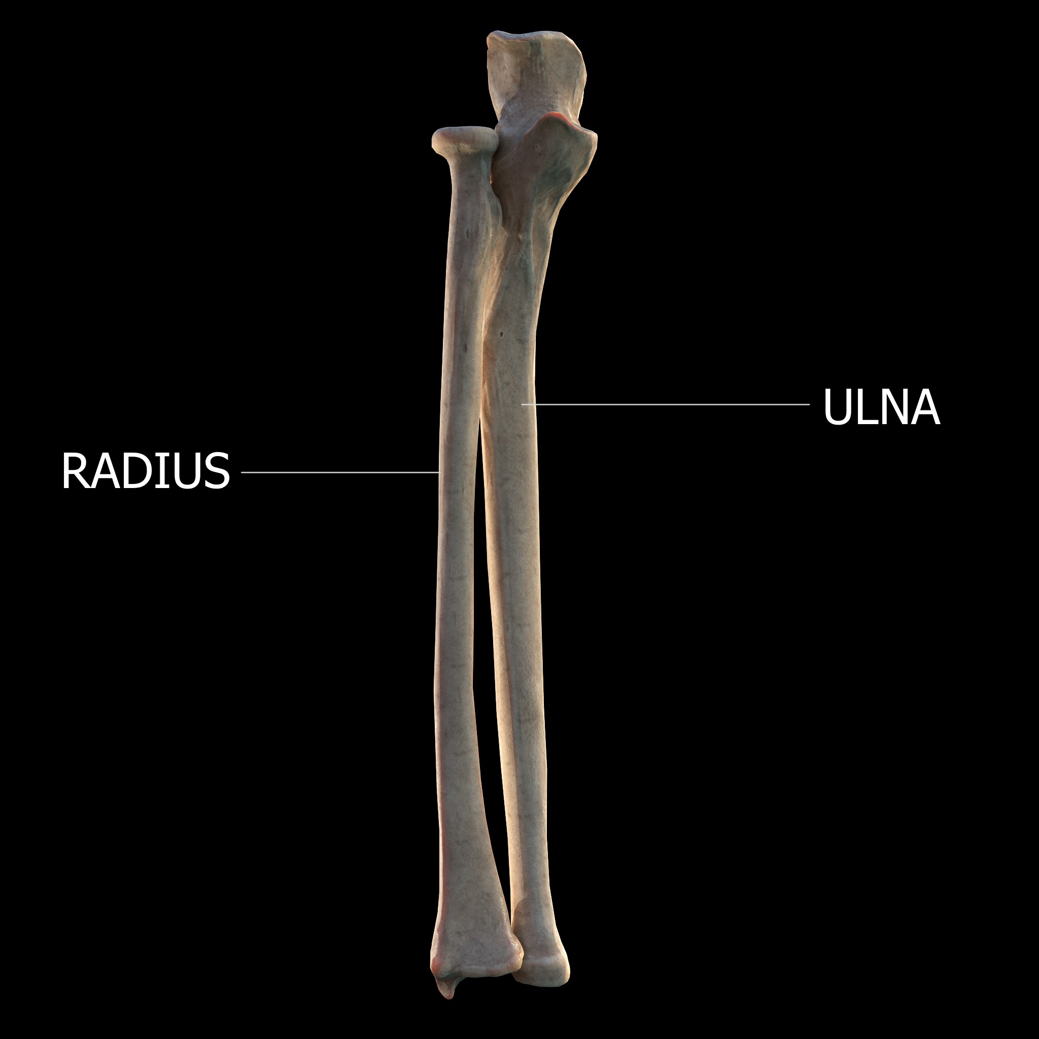
The forearm is the region of the upper limb between the elbow and the wrist. The term forearm is used in anatomy to distinguish it from the arm, a word which is most often used to describe the entire appendage of the upper limb, but which in anatomy, technically, means only the region of the upper arm, whereas the lower "arm" is called the forearm. It is homologous to the region of the leg that lies between the knee and the ankle joints, the crus. The forearm contains two long bones, the radius and the ulna, forming the two radioulnar joints. The interosseous membrane connects these bones. Ultimately, the forearm is covered by skin, the anterior surface usually being less hairy than the posterior surface. The forearm contains many muscles, including the flexors and extensors of the wrist, flexors and extensors of the digits, a flexor of the elbow (brachioradialis), and pronators and supinators that turn the hand to face down or upwards, respectively. In cross-section, the for ... [...More Info...] [...Related Items...] OR: [Wikipedia] [Google] [Baidu] |
Superficial Palmar Arch
The superficial palmar arch is formed predominantly by the ulnar artery, with a contribution from the superficial palmar branch of the radial artery. However, in some individuals the contribution from the radial artery might be absent, and instead anastomoses with either the princeps pollicis artery, the radialis indicis artery, or the median artery, the former two of which are branches from the radial artery. Alternative names for this arterial arch are: superficial volar arch, superficial ulnar arch, arcus palmaris superficialis, or arcus volaris superficialis.Again, ''palmar'' and ''volar'' may be used synonymously, but ''arcus volaris superficialis'' does not occur in the TA, and can therefore be considered deprecated. The arch passes across the palm in a curve (Boeckel's line) with its convexity downward, If one were to fully extend the thumb, the superficial palmar arch would lie approximately 1 cm distal from a line drawn between the first web space to the Hook of H ... [...More Info...] [...Related Items...] OR: [Wikipedia] [Google] [Baidu] |
Vena Mediana Cubiti
In human anatomy, the median cubital vein (or median basilic vein) is a superficial vein of the upper limb. It lies in the cubital fossa superficial to the bicipital aponeurosis. It connects the cephalic vein and the basilic vein. It becomes prominent when pressure is applied. It is routinely used for venipuncture (taking blood) and as a site for an intravenous cannula. This is due to its particularly wide lumen, and its tendency to remain stationary upon needle insertion. Structure The median cubital vein is a superficial vein of the upper limb. It lies in the cubital fossa superficial to the bicipital aponeurosis. It connects the cephalic vein and the basilic vein. It becomes prominent when pressure is applied to upstream veins, as venous blood builds up. Variations The median cubital vein shows a wide range of variations. More commonly, the vein forms an H-pattern with the cephalic and basilic veins making up the sides. Other forms include an M-pattern, where the vein bran ... [...More Info...] [...Related Items...] OR: [Wikipedia] [Google] [Baidu] |
