|
Marginal Sulcus
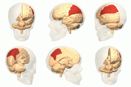
In neuroanatomy, the marginal sulcus (margin of the cingulate sulcus) is a Sulcus (neuroanatomy), sulcus (crevice) that may be considered the termination of the cingulate sulcus. It separates the paracentral lobule anteriorly and the precuneus posteriorly. Additional images File:Marginal sulcus animation small.gif, Position of marginal sulcus (shown in red). File:Cingulate sulcus of Vervet Monkey.png, Transverse plane, Transverse sections of brains of vervet monkey. It showing difference of the relative position of the left and right ascending ramus of the cingulate sulcus. References External links * https://web.archive.org/web/20070416083824/http://www.med.harvard.edu/AANLIB/cases/case3/mr1/033.html Parietal lobe Sulci (neuroanatomy) {{neuroanatomy-stub ... [...More Info...] [...Related Items...] OR: [Wikipedia] [Google] [Baidu] |
Neuroanatomy
Neuroanatomy is the study of the structure and organization of the nervous system. In contrast to animals with radial symmetry, whose nervous system consists of a distributed network of cells, animals with bilateral symmetry have segregated, defined nervous systems. Their neuroanatomy is therefore better understood. In vertebrates, the nervous system is segregated into the internal structure of the brain and spinal cord (together called the central nervous system, or CNS) and the routes of the nerves that connect to the rest of the body (known as the peripheral nervous system, or PNS). The delineation of distinct structures and regions of the nervous system has been critical in investigating how it works. For example, much of what neuroscientists have learned comes from observing how damage or "lesions" to specific brain areas affects behavior or other neural functions. For information about the composition of non-human animal nervous systems, see nervous system. For information ab ... [...More Info...] [...Related Items...] OR: [Wikipedia] [Google] [Baidu] |
Sulcus (neuroanatomy)
In neuroanatomy, a sulcus (Latin: "furrow", pl. ''sulci'') is a depression or groove in the cerebral cortex. It surrounds a gyrus (pl. gyri), creating the characteristic folded appearance of the brain in humans and other mammals. The larger sulci are usually called fissures. Structure Sulci, the grooves, and gyri, the folds or ridges, make up the folded surface of the cerebral cortex. Larger or deeper sulci are termed fissures, and in many cases the two terms are interchangeable. The folded cortex creates a larger surface area for the brain in humans and other mammals. When looking at the human brain, two-thirds of the surface are hidden in the grooves. The sulci and fissures are both grooves in the cortex, but they are differentiated by size. A sulcus is a shallower groove that surrounds a gyrus. A fissure is a large furrow that divides the brain into lobes and also into the two hemispheres as the longitudinal fissure. Importance of expanded surface area As the surfac ... [...More Info...] [...Related Items...] OR: [Wikipedia] [Google] [Baidu] |
Cingulate Sulcus
The cingulate sulcus is a sulcus (brain fold) on the cingulate cortex in the medial wall of the cerebral cortex. The frontal and parietal lobes are separated from the cingulate gyrus by the cingulate sulcus. It terminates as the marginal sulcus of the cingulate sulcus. It sends a ramus to separate the paracentral lobule from the frontal gyri, the paracentral sulcus. Additional images File:Cingulate sulcus animation small.gif, Position of cingulate sulcus (shown in red). File:LobesCaptsMedial1.png, Medial surface of right cerebral hemisphere. Cingulate sulcus (labeled as sulcus cinguli) and brain lobes. File:Slide2ZEN.JPG, Medial surface of cerebral hemisphere.Medial view.Deep dissection. File:Slide3ZEN.JPG, Medial surface of cerebral hemisphere.Medial view.Deep dissection. File:Slide4ZE.JPG, Medial surface of cerebral hemisphere.Medial view.Deep dissection. External links * NIF Search - Cingulate Sulcusvia the Neuroscience Information Framework The Neuroscience Information ... [...More Info...] [...Related Items...] OR: [Wikipedia] [Google] [Baidu] |
Paracentral Lobule
In neuroanatomy, the paracentral lobule is on the medial surface of the cerebral hemisphere and is the continuation of the precentral and postcentral gyri. The paracentral lobule controls motor and sensory innervations of the contralateral lower extremity. It is also responsible for control of defecation and urination. It includes portions of the frontal and parietal lobes: * The anterior portion of the paracentral lobule is part of the frontal lobe and contains a little portion of Brodmann's area 6 (SMA): this is because the paracentral sulcus (branch of the limbic sulcus) does not correspond to the precentral sulcus on the medial plane. * The posterior portion is considered part of the parietal lobe and deals with somatosensory of the distal limbs. While the boundary between the lobes, the central sulcus, is easy to locate on the lateral surface of the cerebral hemispheres, this boundary is often discerned in a cytoarchetectonic manner in cases where the central sulcus i ... [...More Info...] [...Related Items...] OR: [Wikipedia] [Google] [Baidu] |
Precuneus
In neuroanatomy, the precuneus is the portion of the superior parietal lobule on the medial surface of each brain hemisphere. It is located in front of the cuneus (the upper portion of the occipital lobe). The precuneus is bounded in front by the marginal branch of the cingulate sulcus, at the rear by the parieto-occipital sulcus, and underneath by the subparietal sulcus. It is involved with episodic memory, visuospatial processing, reflections upon self, and aspects of consciousness. The location of the precuneus makes it difficult to study. Furthermore, it is rarely subject to isolated injury due to strokes, or trauma such as gunshot wounds. This has resulted in it being "one of the less accurately mapped areas of the whole cortical surface". While originally described as homogeneous by Korbinian Brodmann, it is now appreciated to contain three subdivisions. It is also known after Achille-Louis Foville as the ''quadrate lobule of Foville''. The Latin form of was first used ... [...More Info...] [...Related Items...] OR: [Wikipedia] [Google] [Baidu] |
Transverse Plane
The transverse plane (also known as the horizontal plane, axial plane and transaxial plane) is an anatomical plane that divides the body into Anatomical terms of location#Superior and inferior, superior and inferior sections. It is perpendicular to the coronal plane, coronal and sagittal plane, sagittal planes. List of clinically relevant anatomical planes * Transverse ''thoracic plane'' * ''Xiphosternal plane'' (or xiphosternal junction) * ''Transpyloric plane'' * ''Subcostal plane'' * ''Umbilical plane'' (or transumbilical plane) * ''Supracristal plane'' * ''Intertubercular plane'' (or transtubercular plane) * ''Interspinous plane'' Clinically relevant anatomical planes with associated structures * The transverse ''thoracic plane'' ** Plane through T4 & T5 vertebral junction and Sternal angle, sternal angle of Louis. ** Marks the: *** Attachment of costal cartilage of rib 2 at the sternal angle; *** Aortic arch (beginning and end); *** Upper margin of Superior vena cava, SVC ... [...More Info...] [...Related Items...] OR: [Wikipedia] [Google] [Baidu] |
Vervet Monkey
The vervet monkey (''Chlorocebus pygerythrus''), or simply vervet, is an Old World monkey of the family Cercopithecidae native to Africa. The term "vervet" is also used to refer to all the members of the genus ''Chlorocebus''. The five distinct subspecies can be found mostly throughout Southern Africa, as well as some of the eastern countries. Vervets were introduced to Florida, St. Kitts and Nevis, Barbados, and Cape Verde. These mostly herbivorous monkeys have black faces and grey body hair color, ranging in body length from about for females, to about for males. In addition to behavioral research on natural populations, vervet monkeys serve as a nonhuman primate model for understanding genetic and social behaviors of humans. They have been noted for having human-like characteristics, such as hypertension, anxiety, and social and dependent alcohol (drug), alcohol use. Vervets live in social groups ranging from 10 to 70 individuals, with males moving to other groups at the tim ... [...More Info...] [...Related Items...] OR: [Wikipedia] [Google] [Baidu] |
Parietal Lobe
The parietal lobe is one of the four major lobes of the cerebral cortex in the brain of mammals. The parietal lobe is positioned above the temporal lobe and behind the frontal lobe and central sulcus. The parietal lobe integrates sensory information among various modalities, including spatial sense and navigation (proprioception), the main sensory receptive area for the sense of touch in the somatosensory cortex which is just posterior to the central sulcus in the postcentral gyrus, and the dorsal stream of the visual system. The major sensory inputs from the skin (touch, temperature, and pain receptors), relay through the thalamus to the parietal lobe. Several areas of the parietal lobe are important in language processing. The somatosensory cortex can be illustrated as a distorted figure – the cortical homunculus (Latin: "little man") in which the body parts are rendered according to how much of the somatosensory cortex is devoted to them. The superior parietal lobule and in ... [...More Info...] [...Related Items...] OR: [Wikipedia] [Google] [Baidu] |


