|
Manubrium - Animation
The sternum or breastbone is a long flat bone located in the central part of the chest. It connects to the ribs via cartilage and forms the front of the rib cage, thus helping to protect the heart, human lung, lungs, and major blood vessels from injury. Shaped roughly like a necktie, it is one of the largest and longest flat bones of the body. Its three regions are the manubrium, the body, and the xiphoid process. The word "sternum" originates from the Ancient Greek στέρνον (stérnon), meaning "chest". Structure The sternum is a narrow, flat bone, forming the middle portion of the front of the chest. The top of the sternum supports the clavicles (collarbones) and its edges join with the costal cartilages of the first two pairs of ribs. The inner surface of the sternum is also the attachment of the sternopericardial ligaments. Its top is also connected to the sternocleidomastoid muscle. The sternum consists of three main parts, listed from the top: * Manubrium * Body (glad ... [...More Info...] [...Related Items...] OR: [Wikipedia] [Google] [Baidu] |
Xiphoid Process
The xiphoid process , or xiphisternum or metasternum, is a small cartilaginous process (extension) of the inferior (lower) part of the sternum, which is usually ossified in the adult human. It may also be referred to as the ensiform process. Both the Greek-derived ''xiphoid'' and its Latin equivalent ''ensiform'' mean 'swordlike' or 'sword-shaped' Structure The xiphoid process is considered to be at the level of the 9th thoracic vertebra and the T7 dermatome. Development In newborns and young (especially small) infants, the tip of the xiphoid process may be both seen and felt as a lump just below the sternal notch. At 15 to 29 years old, the xiphoid usually fuses to the body of the sternum with a fibrous joint. Unlike the synovial articulation of major joints, this is non-movable. Ossification of the xiphoid process occurs around age 40. Variation The xiphoid process can be naturally bifurcated or sometimes perforated (xiphoidal foramen). These variances in morphology are inher ... [...More Info...] [...Related Items...] OR: [Wikipedia] [Google] [Baidu] |
Suprasternal Notch
The suprasternal notch, also known as the fossa jugularis sternalis, jugular notch, or Plender gap, is a large, visible dip in between the neck in humans, between the clavicles, and above the manubrium of the sternum. Structure The suprasternal notch is a visible dip in between the neck, between the clavicles, and above the manubrium of the sternum. It is at the level of the T2 and T3 vertebrae. The trachea lies just behind it, rising about 5 cm above it in adults. Clinical significance Intrathoracic pressure is measured by using a transducer held in such a way over the body that an actuator engages the soft tissue that is located above the suprasternal notch. Arcot J. Chandrasekhar, MD of Loyola University, Chicago, is the author of an evaluative test for the aorta using the suprasternal notch. - Evaluative tests using the suprasternal notch The test can help recognize the following conditions: * Aneurysm * Dissecting aneurysm * Atherosclerosis * Hypertension Hypert ... [...More Info...] [...Related Items...] OR: [Wikipedia] [Google] [Baidu] |
Sternocostal Joints
The sternocostal joints, also known as sternochondral joints or costosternal articulations, are synovial plane joints of the costal cartilages of the true ribs with the sternum. The only exception is the first rib, which has a synchondrosis joint since the cartilage is directly united with the sternum. The sternocostal joints are important for thoracic wall mobility. The ligaments connecting them are: * Articular capsules * Interarticular sternocostal ligament * Radiate sternocostal ligaments * Costoxiphoid ligaments Clinical significance Ankylosis, joint stiffness caused by ossification, may occur at the sternocostal joints. See also * Costochondritis Costochondritis, also known as chest wall pain syndrome or costosternal syndrome, is a benign inflammation of the upper costochondral (rib to cartilage) and sternocostal (cartilage to sternum) joints. 90% of patients are affected in multiple ri ... References External links Joints Thorax (human anatomy) {{mu ... [...More Info...] [...Related Items...] OR: [Wikipedia] [Google] [Baidu] |
Articular Facets
A joint or articulation (or articular surface) is the connection made between bones, ossicles, or other hard structures in the body which link an animal's skeletal system into a functional whole.Saladin, Ken. Anatomy & Physiology. 7th ed. McGraw-Hill Connect. Webp.274/ref> They are constructed to allow for different degrees and types of movement. Some joints, such as the knee, elbow, and shoulder, are self-lubricating, almost frictionless, and are able to withstand compression and maintain heavy loads while still executing smooth and precise movements. Other joints such as sutures between the bones of the skull permit very little movement (only during birth) in order to protect the brain and the sense organs. The connection between a tooth and the jawbone is also called a joint, and is described as a fibrous joint known as a gomphosis. Joints are classified both structurally and functionally. Classification The number of joints depends on if sesamoids are included, age of th ... [...More Info...] [...Related Items...] OR: [Wikipedia] [Google] [Baidu] |
Vascular
The blood vessels are the components of the circulatory system that transport blood throughout the human body. These vessels transport blood cells, nutrients, and oxygen to the tissues of the body. They also take waste and carbon dioxide away from the tissues. Blood vessels are needed to sustain life, because all of the body's tissues rely on their functionality. There are five types of blood vessels: the arteries, which carry the blood away from the heart; the arterioles; the capillaries, where the exchange of water and chemicals between the blood and the tissues occurs; the venules; and the veins, which carry blood from the capillaries back towards the heart. The word ''vascular'', meaning relating to the blood vessels, is derived from the Latin ''vas'', meaning vessel. Some structures – such as cartilage, the epithelium, and the lens and cornea of the eye – do not contain blood vessels and are labeled ''avascular''. Etymology * artery: late Middle English; from Latin ' ... [...More Info...] [...Related Items...] OR: [Wikipedia] [Google] [Baidu] |
Cardiopulmonary Resuscitation
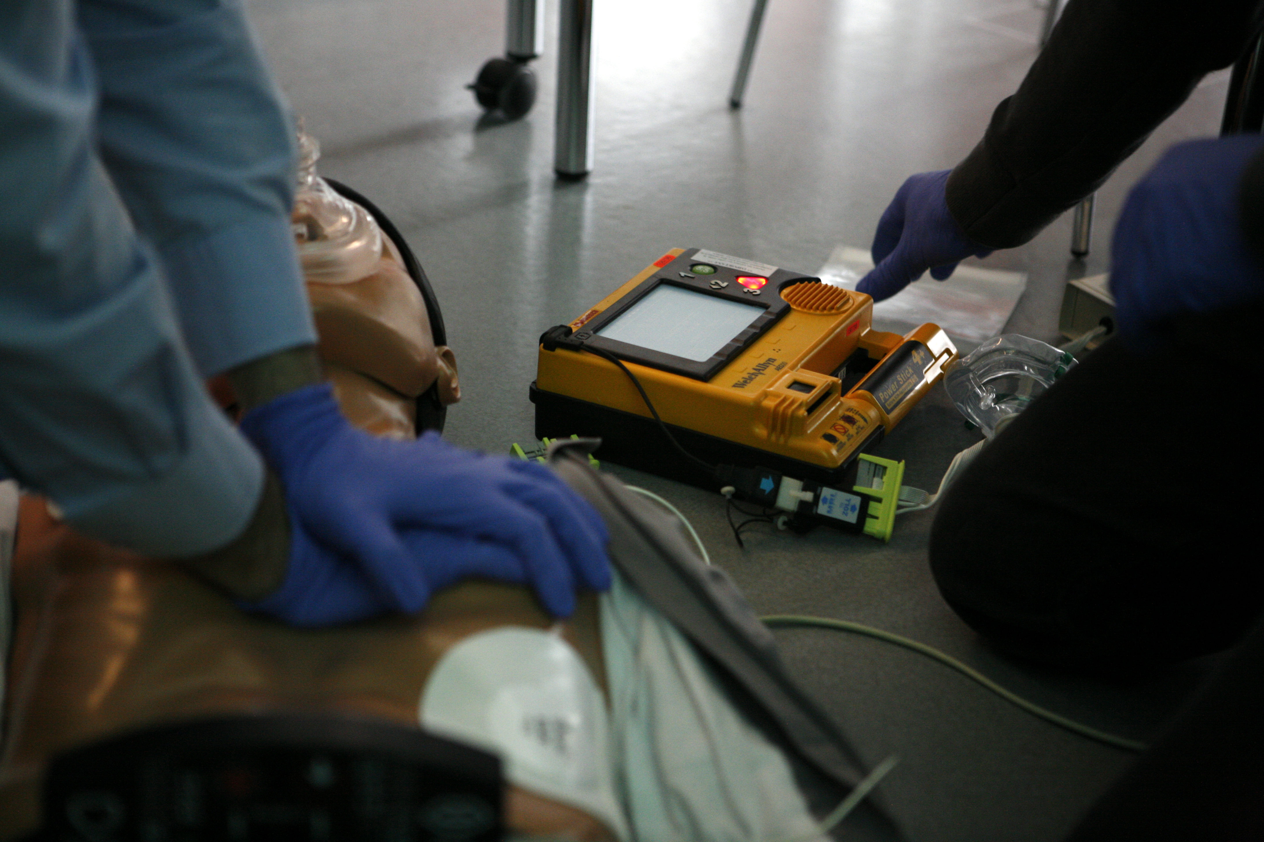
Cardiopulmonary resuscitation (CPR) is an emergency procedure consisting of chest compressions often combined with artificial ventilation in an effort to manually preserve intact brain function until further measures are taken to restore spontaneous blood circulation and breathing in a person who is in cardiac arrest. It is recommended in those who are unresponsive with no breathing or abnormal breathing, for example, agonal respirations. CPR involves chest compressions for adults between and deep and at a rate of at least 100 to 120 per minute. The rescuer may also provide artificial ventilation by either exhaling air into the subject's mouth or nose (mouth-to-mouth resuscitation) or using a device that pushes air into the subject's lungs (mechanical ventilation). Current recommendations place emphasis on early and high-quality chest compressions over artificial ventilation; a simplified CPR method involving only chest compressions is recommended for untrained rescuers. Wit ... [...More Info...] [...Related Items...] OR: [Wikipedia] [Google] [Baidu] |
Xiphoid Process - Close-up - Animation
The xiphoid process , or xiphisternum or metasternum, is a small cartilaginous process (extension) of the inferior (lower) part of the sternum, which is usually ossified in the adult human. It may also be referred to as the ensiform process. Both the Greek-derived ''xiphoid'' and its Latin equivalent ''ensiform'' mean 'swordlike' or 'sword-shaped' Structure The xiphoid process is considered to be at the level of the 9th thoracic vertebra and the T7 dermatome. Development In newborns and young (especially small) infants, the tip of the xiphoid process may be both seen and felt as a lump just below the sternal notch. At 15 to 29 years old, the xiphoid usually fuses to the body of the sternum with a fibrous joint. Unlike the synovial articulation of major joints, this is non-movable. Ossification of the xiphoid process occurs around age 40. Variation The xiphoid process can be naturally bifurcated or sometimes perforated (xiphoidal foramen). These variances in morphology are inher ... [...More Info...] [...Related Items...] OR: [Wikipedia] [Google] [Baidu] |
True Rib
The rib cage, as an enclosure that comprises the ribs, vertebral column and sternum in the thorax of most vertebrates, protects vital organs such as the heart, lungs and great vessels. The sternum, together known as the thoracic cage, is a semi-rigid bony and cartilaginous structure which surrounds the thoracic cavity and supports the shoulder girdle to form the core part of the human skeleton. A typical human thoracic cage consists of 12 pairs of ribs and the adjoining costal cartilages, the sternum (along with the manubrium and xiphoid process), and the 12 thoracic vertebrae articulating with the ribs. Together with the skin and associated fascia and muscles, the thoracic cage makes up the thoracic wall and provides attachments for extrinsic skeletal muscles of the neck, upper limbs, upper abdomen and back. The rib cage intrinsically holds the muscles of respiration ( diaphragm, intercostal muscles, etc.) that are crucial for active inhalation and forced exhalation, and t ... [...More Info...] [...Related Items...] OR: [Wikipedia] [Google] [Baidu] |
Costal Cartilage
The costal cartilages are bars of hyaline cartilage that serve to prolong the ribs forward and contribute to the elasticity of the walls of the thorax. Costal cartilage is only found at the anterior ends of the ribs, providing medial extension. Differences from Ribs 1-12 The first seven pairs are connected with the sternum; the next three are each articulated with the lower border of the cartilage of the preceding rib; the last two have pointed extremities, which end in the wall of the abdomen. Like the ribs, the costal cartilages vary in their length, breadth, and direction. They increase in length from the first to the seventh, then gradually decrease to the twelfth. Their breadth, as well as that of the intervals between them, diminishes from the first to the last. They are broad at their attachments to the ribs, and taper toward their sternal extremities, excepting the first two, which are of the same breadth throughout, and the sixth, seventh, and eighth, which are enlarge ... [...More Info...] [...Related Items...] OR: [Wikipedia] [Google] [Baidu] |
Sternal Angle
The sternal angle (also known as the angle of Louis, angle of Ludovic or manubriosternal junction) is the synarthrotic joint formed by the articulation of the manubrium and the body of the sternum. The sternal angle is a palpable clinical landmark in surface anatomy. Anatomy The sternal angle, which varies around 162 degrees in males, marks the approximate level of the 2nd pair of costal cartilages, which attach to the second ribs, and the level of the intervertebral disc between T4 and T5. In clinical applications, the sternal angle can be palpated at the T4 vertebral level. The sternal angle is used in the definition of the thoracic plane. This marks the level of a number of other anatomical structures: The angle also marks a number of other features: :* Carina of the trachea is deep to the sternal angle :* :*Passage of the thoracic duct from right to left behind esophagus :* :* Ligamentum arteriosum :* :* Loop of left recurrent laryngeal nerve around aortic arch The ang ... [...More Info...] [...Related Items...] OR: [Wikipedia] [Google] [Baidu] |
Transversus Thoracis
The transversus thoracis muscle (), also known as triangularis sterni, lies internal to the thoracic cage, anteriorly. It is usually a thin plane of muscular and tendinous fibers, however on athletic individuals it can be a thick 'slab of meat', situated upon the inner surface of the front wall of the chest. It is in the same layer as the subcostal muscles and the innermost intercostal muscles. Structure It arises on either side from the lower third of the posterior surface of the body of the sternum, from the posterior surface of the xiphoid process, and from the sternal ends of the costal cartilages of the lower three or four true ribs. Its fibers diverge upward and lateralward, to be inserted by slips into the lower borders and inner surfaces of the costal cartilages of the second, third, fourth, fifth, and sixth ribs. The lowest fibers of this muscle are horizontal in their direction, and are continuous with those of the transversus abdominis; the intermediate fibers are ob ... [...More Info...] [...Related Items...] OR: [Wikipedia] [Google] [Baidu] |
Pectoralis Major
The pectoralis major () is a thick, fan-shaped or triangular convergent muscle, situated at the chest of the human body. It makes up the bulk of the chest muscles and lies under the breast. Beneath the pectoralis major is the pectoralis minor, a thin, triangular muscle. The pectoralis major's primary functions are flexion, adduction, and internal rotation of the humerus. The pectoral major may colloquially be referred to as "pecs", "pectoral muscle", or "chest muscle", because it is the largest and most superficial muscle in the chest area. Structure It arises from the anterior surface of the sternal half of the clavicle from breadth of the half of the anterior surface of the sternum, as low down as the attachment of the cartilage of the sixth or seventh rib; from the cartilages of all the true ribs, with the exception, frequently, of the first or seventh, and from the aponeurosis of the abdominal external oblique muscle. From this extensive origin the fibers converge toward the ... [...More Info...] [...Related Items...] OR: [Wikipedia] [Google] [Baidu] |


