|
Mandibular Canal
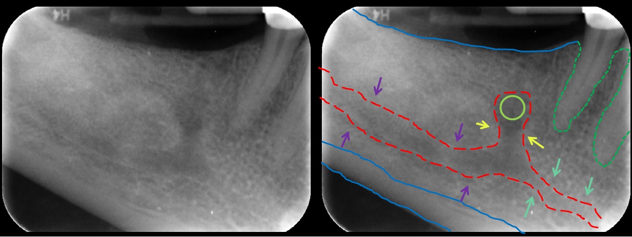
In human anatomy, the mandibular canal is a canal within the mandible that contains the inferior alveolar nerve, inferior alveolar artery, and inferior alveolar vein. It runs obliquely downward and forward in the ramus, and then horizontally forward in the body, where it is placed under the alveoli and communicates with them by small openings. On arriving at the incisor teeth, it turns back to communicate with the mental foramen, giving off a small canal known as the mandibular incisive canal, which run to the cavities containing the incisor teeth. It carries branches of the inferior alveolar nerve and artery. It is continuous with the mental foramen (which opens onto front of mandible) and mandibular foramen (on medial aspect of ramus). Variations The mandibular canal is fairly close to the apices of the second molar in 50% of the radiographs. In 40%, canal is away from the root apices, and in only 10% of the radiographs the root apices appeared to penetrate the canal. ... [...More Info...] [...Related Items...] OR: [Wikipedia] [Google] [Baidu] |
Mandibular Incisive Canal Highlighted
In anatomy, the mandible, lower jaw or jawbone is the largest, strongest and lowest bone in the human facial skeleton. It forms the lower jaw and holds the lower tooth, teeth in place. The mandible sits beneath the maxilla. It is the only movable bone of the skull (discounting the ossicles of the middle ear). It is connected to the temporal bones by the temporomandibular joints. The bone is formed prenatal development, in the fetus from a fusion of the left and right mandibular prominences, and the point where these sides join, the mandibular symphysis, is still visible as a faint ridge in the midline. Like other symphyses in the body, this is a midline articulation where the bones are joined by fibrocartilage, but this articulation fuses together in early childhood.Illustrated Anatomy of the Head and Neck, Fehrenbach and Herring, Elsevier, 2012, p. 59 The word "mandible" derives from the Latin word ''mandibula'', "jawbone" (literally "one used for chewing"), from ''wikt:mandere ... [...More Info...] [...Related Items...] OR: [Wikipedia] [Google] [Baidu] |
Human Mandible
In anatomy, the mandible, lower jaw or jawbone is the largest, strongest and lowest bone in the human facial skeleton. It forms the lower jaw and holds the lower tooth, teeth in place. The mandible sits beneath the maxilla. It is the only movable bone of the skull (discounting the ossicles of the middle ear). It is connected to the temporal bones by the temporomandibular joints. The bone is formed prenatal development, in the fetus from a fusion of the left and right mandibular prominences, and the point where these sides join, the mandibular symphysis, is still visible as a faint ridge in the midline. Like other symphyses in the body, this is a midline articulation where the bones are joined by fibrocartilage, but this articulation fuses together in early childhood.Illustrated Anatomy of the Head and Neck, Fehrenbach and Herring, Elsevier, 2012, p. 59 The word "mandible" derives from the Latin word ''mandibula'', "jawbone" (literally "one used for chewing"), from ''wikt:mandere ... [...More Info...] [...Related Items...] OR: [Wikipedia] [Google] [Baidu] |
Inferior Alveolar Nerve
The inferior alveolar nerve (IAN) (also the inferior dental nerve) is a branch of the mandibular nerve, which is itself the third branch of the trigeminal nerve. The inferior alveolar nerves supply sensation to the lower teeth. Structure The inferior alveolar nerve is a branch of the mandibular nerve. After branching from the mandibular nerve, the inferior alveolar nerve travels behind the lateral pterygoid muscle. It gives off a branch, the mylohyoid nerve, and then enters the mandibular foramen. While in the mandibular canal within the mandible, it supplies the lower teeth (molars and second premolar) with sensory branches that form into the inferior dental plexus and give off small gingival and dental nerves to the teeth. Anteriorly, the nerve gives off the mental nerve at about the level of the mandibular 2nd premolars, which exits the mandible via the mental foramen and supplies sensory branches to the chin and lower lip. The inferior alveolar nerve continues anteriorl ... [...More Info...] [...Related Items...] OR: [Wikipedia] [Google] [Baidu] |
Inferior Alveolar Artery
The inferior alveolar artery (inferior dental artery) is an artery of the face. It is a branch of the first portion of the maxillary artery. Structure It descends with the inferior alveolar nerve to the mandibular foramen on the medial surface of the ramus of the mandible. It runs along the mandibular canal in the substance of the bone, accompanied by the nerve, and opposite the first premolar tooth divides into two branches, incisor and mental. Incisor branch The ''incisor branch'' is continued forward beneath the incisor teeth as far as the middle line, where it anastomoses with the artery of the opposite side The inferior alveolar artery and its incisor branch during their course through the substance of the bone give off a few twigs which are lost in the cancellous tissue, and a series of branches which correspond in number to the roots of the teeth: these enter the minute apertures at the extremities of the roots, and supply the pulp of the teeth. Mental branch The ''m ... [...More Info...] [...Related Items...] OR: [Wikipedia] [Google] [Baidu] |
Inferior Alveolar Vein
{{disambiguation ...
Inferior may refer to: * Inferiority complex * An anatomical term of location * Inferior angle of the scapula, in the human skeleton * ''Inferior'' (book), by Angela Saini * ''The Inferior'', a 2007 novel by Peadar Ó Guilín See also *Junior (other) Junior or Juniors may refer to: Arts and entertainment Music * ''Junior'' (Junior Mance album), 1959 * ''Junior'' (Röyksopp album), 2009 * ''Junior'' (Kaki King album), 2010 * ''Junior'' (LaFontaines album), 2019 Films * ''Junior'' (1994 ... [...More Info...] [...Related Items...] OR: [Wikipedia] [Google] [Baidu] |
Ramus Of The Mandible
In anatomy, the mandible, lower jaw or jawbone is the largest, strongest and lowest bone in the human facial skeleton. It forms the lower jaw and holds the lower teeth in place. The mandible sits beneath the maxilla. It is the only movable bone of the skull (discounting the ossicles of the middle ear). It is connected to the temporal bones by the temporomandibular joints. The bone is formed in the fetus from a fusion of the left and right mandibular prominences, and the point where these sides join, the mandibular symphysis, is still visible as a faint ridge in the midline. Like other symphyses in the body, this is a midline articulation where the bones are joined by fibrocartilage, but this articulation fuses together in early childhood.Illustrated Anatomy of the Head and Neck, Fehrenbach and Herring, Elsevier, 2012, p. 59 The word "mandible" derives from the Latin word ''mandibula'', "jawbone" (literally "one used for chewing"), from '' mandere'' "to chew" and ''-bula'' (i ... [...More Info...] [...Related Items...] OR: [Wikipedia] [Google] [Baidu] |
Dental Alveolus
Dental alveoli (singular ''alveolus'') are sockets in the jaws in which the roots of teeth are held in the alveolar process with the periodontal ligament. The lay term for dental alveoli is tooth sockets. A joint that connects the roots of the teeth and the alveolus is called ''gomphosis'' (plural ''gomphoses''). Alveolar bone is the bone that surrounds the roots of the teeth forming bone sockets. In mammals, tooth sockets are found in the maxilla, the premaxilla, and the mandible. Etymology 1706, "a hollow," especially "the socket of a tooth," from Latin alveolus "a tray, trough, basin; bed of a small river; small hollow or cavity," diminutive of alvus "belly, stomach, paunch, bowels; hold of a ship," from PIE root *aulo- "hole, cavity" (source also of Greek aulos "flute, tube, pipe;" Serbo-Croatian, Polish, Russian ulica "street," originally "narrow opening;" Old Church Slavonic uliji, Lithuanian aulys "beehive" (hollow trunk), Armenian yli "pregnant"). The word was extended in ... [...More Info...] [...Related Items...] OR: [Wikipedia] [Google] [Baidu] |
Mental Foramen
The mental foramen is one of two foramina (openings) located on the anterior surface of the mandible. It is part of the mandibular canal. It transmits the terminal branches of the inferior alveolar nerve and the mental vessels. Structure The mental foramen is located on the anterior surface of the mandible. It is directly below the commisure of the lips, and the tendon of depressor labii inferioris muscle. It is at the end of the mandibular canal, which begins at the mandibular foramen on the posterior surface of the mandible. It transmits the terminal branches of the inferior alveolar nerve (the mental nerve), the mental artery, and the mental vein. Variation The mental foramen descends slightly in toothless individuals. The mental foramen is in line with the longitudinal axis of the 2nd premolar in 63% of people. It generally lies at the level of the vestibular fornix and about a finger's breadth above the inferior border of the mandible. In the general population, 17% ... [...More Info...] [...Related Items...] OR: [Wikipedia] [Google] [Baidu] |
Mandibular Incisive Canal
The mandibular incisive canal is a bony canal within the anterior mandible that runs bilaterally from the mental foramina usually to the region of the ipsilateral lateral incisor teeth. After branching into the mental nerve that exits the foramen of the same name, the inferior alveolar nerve The inferior alveolar nerve (IAN) (also the inferior dental nerve) is a branch of the mandibular nerve, which is itself the third branch of the trigeminal nerve. The inferior alveolar nerves supply sensation to the lower teeth. Structure The in ... continues anteriorly within the mandibular incisive canal as the incisive nerve, providing innervation to the mandibular first premolar, canine and lateral and central incisors.Greenstein, G; Cavallaro, J; Tarnow, D. "Practical Application of Anatomy for the Dental Implant Surgeon," ''J Perio'' 2008;79:1833-1846 The mandibular incisive nerve either terminates as nerve endings within the anterior teeth or adjacent bone, or may join nerve end ... [...More Info...] [...Related Items...] OR: [Wikipedia] [Google] [Baidu] |
Incisor Teeth
Incisors (from Latin ''incidere'', "to cut") are the front teeth present in most mammals. They are located in the premaxilla above and on the mandible below. Humans have a total of eight (two on each side, top and bottom). Opossums have 18, whereas armadillos have none. Structure Adult humans normally have eight incisors, two of each type. The types of incisor are: * maxillary central incisor (upper jaw, closest to the center of the lips) * maxillary lateral incisor (upper jaw, beside the maxillary central incisor) * mandibular central incisor (lower jaw, closest to the center of the lips) * mandibular lateral incisor (lower jaw, beside the mandibular central incisor) Children with a full set of deciduous teeth (primary teeth) also have eight incisors, named the same way as in permanent teeth. Young children may have from zero to eight incisors depending on the stage of their tooth eruption and tooth development. Typically, the mandibular central incisors erupt first, follo ... [...More Info...] [...Related Items...] OR: [Wikipedia] [Google] [Baidu] |
Dennis Tarnow
Dennis Perry Tarnow (b. May 28) is an American dentist specializing in dental implant research. He is director of implant dentistry at Columbia University College of Dental Medicine and a speaker on the subject of implant dentistry.Ohio Dental Association - Dr. Dennis Tarnow to present full-day program at the 142nd Annual Session April 1, 2008 Early years and education Tarnow was born and raised in , to Joseph and Mildred Tarnow. After graduating from[...More Info...] [...Related Items...] OR: [Wikipedia] [Google] [Baidu] |
Mandibular Foramen
The mandibular foramen is an opening on the internal surface of the ramus of the mandible. It allows for divisions of the mandibular nerve and blood vessels to pass through. Structure The mandibular foramen is an opening on the internal surface of the ramus of the mandible. It allows for divisions of the mandibular nerve and blood vessels to pass through. Variation There are two distinct anatomies to its rim. * In the common form the rim is “V” shaped, with a groove separating the anterior and posterior parts. * In the horizontal-oval form there is no groove, and the rim is horizontally oriented and oval in shape, the anterior and posterior parts connected. Rarely, a bifid inferior alveolar nerve may be present, in which case a second mandibular foramen, more inferiorly placed, exists and can be detected by noting a doubled mandibular canal on a radiograph. Function The mandibular nerve is one of three branches of the trigeminal nerve, and the only one having motor ... [...More Info...] [...Related Items...] OR: [Wikipedia] [Google] [Baidu] |


