|
Mycena Semivestipes
''Mycena semivestipes'' is a species of agaric fungus in the family Mycenaceae. It is found in eastern North America. Taxonomy First described in 1895 as ''Omphalina semivestipes'' by Charles Horton Peck, the species was transferred to '' Mycena'' in 1947 by Alexander H. Smith. The type collection was made in Newfoundland, Canada. Description The cap is initially convex to somewhat conical before flattening out in age; it attains a diameter of wide. The cap surface, smooth and slightly lubricous, is deep viscous to dark brown in the center. The margin is somewhat translucent, so that the grooves made by the gills are discernible. The thin flesh has a firm texture similar to cartilage, a distinct strong nitrous odor, and a mild to somewhat bitter taste. The gills have an adnate attachment, but often recede from the stipe as the mushroom matures. Their color is white to dirty pink. They can develop reddish-brown spots in age. Interspersed between the gills are two or three ... [...More Info...] [...Related Items...] OR: [Wikipedia] [Google] [Baidu] |
Charles Horton Peck
Charles Horton Peck (March 30, 1833 – July 11, 1917) was an American mycologist of the 19th and early 20th centuries. He was the New York State Botanist from 1867 to 1915, a period in which he described over 2,700 species of North American fungi. Biography Charles Horton Peck was born on March 30, 1833, in the northeastern part of the town Sand Lake, New York, now called Averill Park. After suffering a light stroke early in November 1912 and then a severe stroke in 1913, he died at his house in Menands, New York, on July 11, 1917. In 1794, Eleazer Peck (his great grandfather) moved from Farmington, Conn. to Sand Lake, NY attracted by oak timber that was manufactured for the Albany market. Later on, Pamelia Horton Peck married Joel B., both from English descent, and became Charles Peck parents (Burnham 1919; Atkinson 1918). Even though his family was rich and locally prominent, his education was provincial (Haines 1986). During his childhood, he used to enjoy fishing and h ... [...More Info...] [...Related Items...] OR: [Wikipedia] [Google] [Baidu] |
Adnation
Adnation in Angiosperms is the fusion of two or more whorls of a flower, e.g. stamens to petals". This is in contrast to connation, the fusion among a single whorl A whorl ( or ) is an individual circle, oval, volution or equivalent in a whorled pattern, which consists of a spiral or multiple concentric objects (including circles, ovals and arcs). Whorls in nature File:Photograph and axial plane floral .... References Plant anatomy {{botany-stub ... [...More Info...] [...Related Items...] OR: [Wikipedia] [Google] [Baidu] |
Fungi Of North America
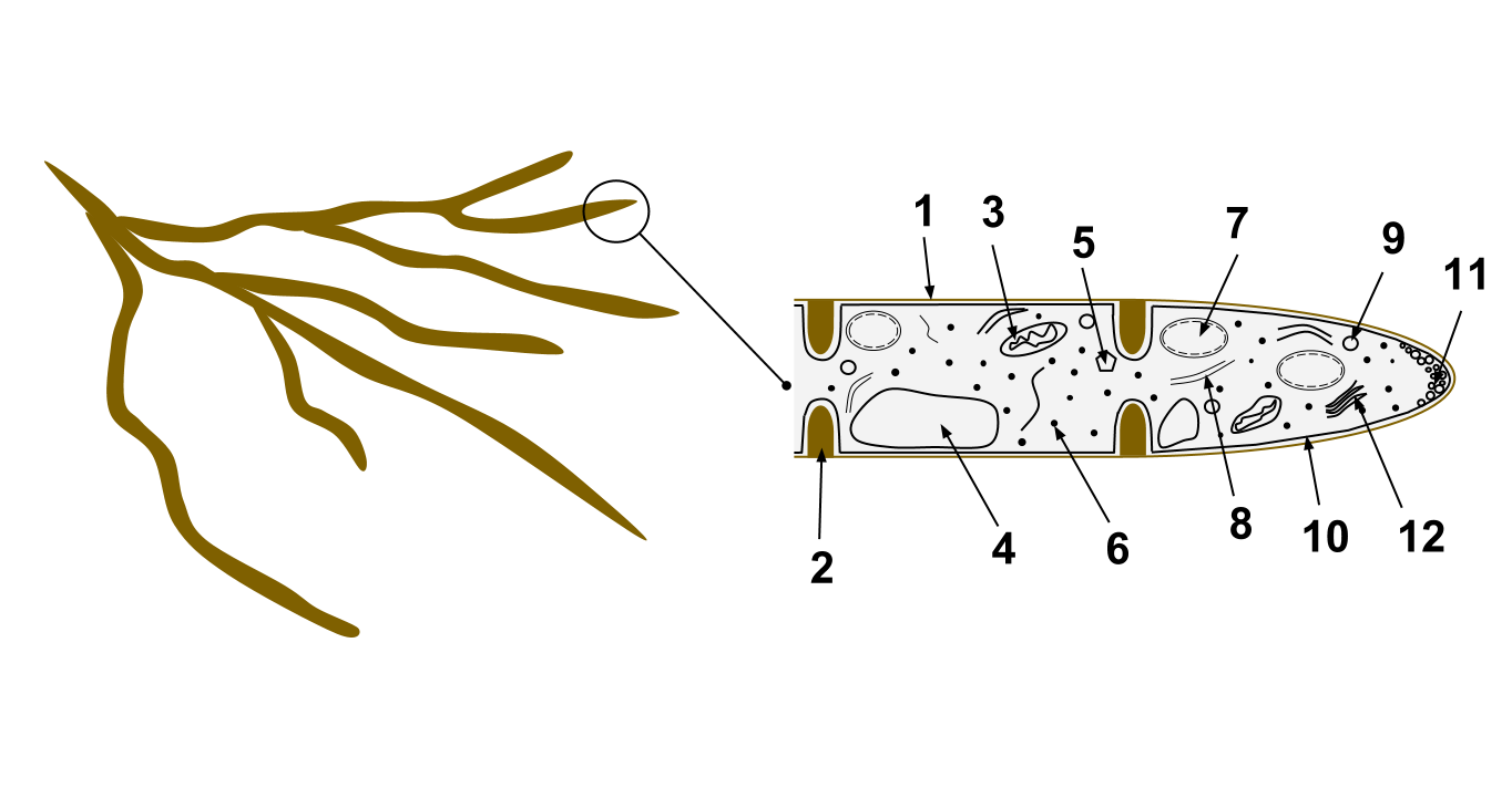
A fungus ( : fungi or funguses) is any member of the group of eukaryotic organisms that includes microorganisms such as yeasts and molds, as well as the more familiar mushrooms. These organisms are classified as a kingdom, separately from the other eukaryotic kingdoms, which by one traditional classification include Plantae, Animalia, Protozoa, and Chromista. A characteristic that places fungi in a different kingdom from plants, bacteria, and some protists is chitin in their cell walls. Fungi, like animals, are heterotrophs; they acquire their food by absorbing dissolved molecules, typically by secreting digestive enzymes into their environment. Fungi do not photosynthesize. Growth is their means of mobility, except for spores (a few of which are flagellated), which may travel through the air or water. Fungi are the principal decomposers in ecological systems. These and other differences place fungi in a single group of related organisms, named the ''Eumycota'' (''true fungi' ... [...More Info...] [...Related Items...] OR: [Wikipedia] [Google] [Baidu] |
MycoBank
MycoBank is an online database, documenting new mycological names and combinations, eventually combined with descriptions and illustrations. It is run by the Westerdijk Fungal Biodiversity Institute in Utrecht. Each novelty, after being screened by nomenclatural experts and found in accordance with the ICN ( International Code of Nomenclature for algae, fungi, and plants), is allocated a unique MycoBank number before the new name has been validly published. This number then can be cited by the naming author in the publication where the new name is being introduced. Only then, this unique number becomes public in the database. By doing so, this system can help solve the problem of knowing which names have been validly published and in which year. MycoBank is linked to other important mycological databases such as ''Index Fungorum'', Life Science Identifiers, Global Biodiversity Information Facility (GBIF) and other databases. MycoBank is one of three nomenclatural repositories r ... [...More Info...] [...Related Items...] OR: [Wikipedia] [Google] [Baidu] |
Saprobic
Saprotrophic nutrition or lysotrophic nutrition is a process of chemoheterotrophic extracellular digestion involved in the processing of decayed (dead or waste) organic matter. It occurs in saprotrophs, and is most often associated with fungi (for example ''Mucor'') and soil bacteria. Saprotrophic microscopic fungi are sometimes called saprobes; saprotrophic plants or bacterial flora are called saprophytes ( sapro- 'rotten material' + -phyte 'plant'), although it is now believed that all plants previously thought to be saprotrophic are in fact parasites of microscopic fungi or other plants. The process is most often facilitated through the active transport of such materials through endocytosis within the internal mycelium and its constituent hyphae. states the purpose of saprotrophs and their internal nutrition, as well as the main two types of fungi that are most often referred to, as well as describes, visually, the process of saprotrophic nutrition through a diagram of hyph ... [...More Info...] [...Related Items...] OR: [Wikipedia] [Google] [Baidu] |
Cystidia
A cystidium (plural cystidia) is a relatively large cell found on the sporocarp of a basidiomycete (for example, on the surface of a mushroom gill), often between clusters of basidia. Since cystidia have highly varied and distinct shapes that are often unique to a particular species or genus, they are a useful micromorphological characteristic in the identification of basidiomycetes. In general, the adaptive significance of cystidia is not well understood. Classification of cystidia By position Cystidia may occur on the edge of a lamella (or analogous hymenophoral structure) (cheilocystidia), on the face of a lamella (pleurocystidia), on the surface of the cap (dermatocystidia or pileocystidia), on the margin of the cap (circumcystidia) or on the stipe (caulocystidia). Especially the pleurocystidia and cheilocystidia are important for identification within many genera. Sometimes the cheilocystidia give the gill edge a distinct colour which is visible to the naked eye or wit ... [...More Info...] [...Related Items...] OR: [Wikipedia] [Google] [Baidu] |
Basidia
A basidium () is a microscopic sporangium (a spore-producing structure) found on the hymenophore of fruiting bodies of basidiomycete fungi which are also called tertiary mycelium, developed from secondary mycelium. Tertiary mycelium is highly-coiled secondary myceliuma dikaryon. The presence of basidia is one of the main characteristic features of the Basidiomycota. A basidium usually bears four sexual spores called basidiospores; occasionally the number may be two or even eight. In a typical basidium, each basidiospore is borne at the tip of a narrow prong or horn called a sterigma (), and is forcibly discharged upon maturity. The word ''basidium'' literally means "little pedestal", from the way in which the basidium supports the spores. However, some biologists suggest that the structure more closely resembles a club. An immature basidium is known as a basidiole. Structure Most basidiomycota have single celled basidia (holobasidia), but in some groups basidia can be multice ... [...More Info...] [...Related Items...] OR: [Wikipedia] [Google] [Baidu] |
Amyloid (mycology)
In mycology a tissue or feature is said to be amyloid if it has a positive amyloid reaction when subjected to a crude chemical test using iodine as an ingredient of either Melzer's reagent or Lugol's solution, producing a blue to blue-black staining. The term "amyloid" is derived from the Latin ''amyloideus'' ("starch-like"). It refers to the fact that starch gives a similar reaction, also called an amyloid reaction. The test can be on microscopic features, such as spore walls or hyphal walls, or the apical apparatus or entire ascus wall of an ascus, or be a macroscopic reaction on tissue where a drop of the reagent is applied. Negative reactions, called inamyloid or nonamyloid, are for structures that remain pale yellow-brown or clear. A reaction producing a deep reddish to reddish-brown staining is either termed a dextrinoid reaction (pseudoamyloid is a synonym) or a hemiamyloid reaction. Melzer's reagent reactions Hemiamyloidity Hemiamyloidity in mycology refers to a special ... [...More Info...] [...Related Items...] OR: [Wikipedia] [Google] [Baidu] |
Micrometre
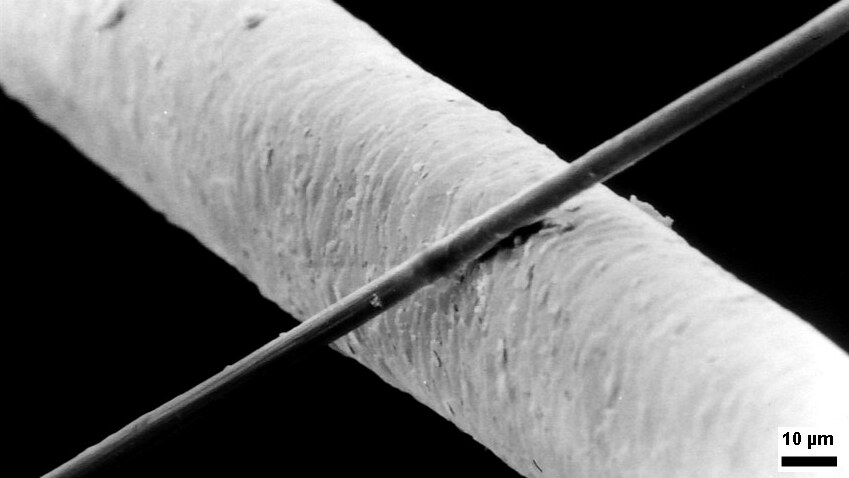
The micrometre ( international spelling as used by the International Bureau of Weights and Measures; SI symbol: μm) or micrometer (American spelling), also commonly known as a micron, is a unit of length in the International System of Units (SI) equalling (SI standard prefix "micro-" = ); that is, one millionth of a metre (or one thousandth of a millimetre, , or about ). The nearest smaller common SI unit is the nanometre, equivalent to one thousandth of a micrometre, one millionth of a millimetre or one billionth of a metre (). The micrometre is a common unit of measurement for wavelengths of infrared radiation as well as sizes of biological cells and bacteria, and for grading wool by the diameter of the fibres. The width of a single human hair ranges from approximately 20 to . The longest human chromosome, chromosome 1, is approximately in length. Examples Between 1 μm and 10 μm: * 1–10 μm – length of a typical bacterium * 3–8 μm – width of ... [...More Info...] [...Related Items...] OR: [Wikipedia] [Google] [Baidu] |
Hyaline
A hyaline substance is one with a glassy appearance. The word is derived from el, ὑάλινος, translit=hyálinos, lit=transparent, and el, ὕαλος, translit=hýalos, lit=crystal, glass, label=none. Histopathology Hyaline cartilage is named after its glassy appearance on fresh gross pathology. On light microscopy of H&E stained slides, the extracellular matrix of hyaline cartilage looks homogeneously pink, and the term "hyaline" is used to describe similarly homogeneously pink material besides the cartilage. Hyaline material is usually acellular and proteinaceous. For example, arterial hyaline is seen in aging, high blood pressure, diabetes mellitus and in association with some drugs (e.g. calcineurin inhibitors). It is bright pink with PAS staining. Ichthyology and entomology In ichthyology and entomology, ''hyaline'' denotes a colorless, transparent substance, such as unpigmented fins of fishes or clear insect wings. Resh, Vincent H. and R. T. Cardé, Eds. Encyclo ... [...More Info...] [...Related Items...] OR: [Wikipedia] [Google] [Baidu] |
Ellipsoid
An ellipsoid is a surface that may be obtained from a sphere by deforming it by means of directional scalings, or more generally, of an affine transformation. An ellipsoid is a quadric surface; that is, a surface that may be defined as the zero set of a polynomial of degree two in three variables. Among quadric surfaces, an ellipsoid is characterized by either of the two following properties. Every planar cross section is either an ellipse, or is empty, or is reduced to a single point (this explains the name, meaning "ellipse-like"). It is bounded, which means that it may be enclosed in a sufficiently large sphere. An ellipsoid has three pairwise perpendicular axes of symmetry which intersect at a center of symmetry, called the center of the ellipsoid. The line segments that are delimited on the axes of symmetry by the ellipsoid are called the ''principal axes'', or simply axes of the ellipsoid. If the three axes have different lengths, the figure is a triaxial ellipsoid (r ... [...More Info...] [...Related Items...] OR: [Wikipedia] [Google] [Baidu] |
Basidiospore
A basidiospore is a reproductive spore produced by Basidiomycete fungi, a grouping that includes mushrooms, shelf fungi, rusts, and smuts. Basidiospores typically each contain one haploid nucleus that is the product of meiosis, and they are produced by specialized fungal cells called basidia. Typically, four basidiospores develop on appendages from each basidium, of which two are of one strain and the other two of its opposite strain. In gills under a cap of one common species, there exist millions of basidia. Some gilled mushrooms in the order Agaricales have the ability to release billions of spores. The puffball fungus ''Calvatia gigantea'' has been calculated to produce about five trillion basidiospores. Most basidiospores are forcibly discharged, and are thus considered ballistospores. These spores serve as the main air dispersal units for the fungi. The spores are released during periods of high humidity and generally have a night-time or pre-dawn peak concentration in the ... [...More Info...] [...Related Items...] OR: [Wikipedia] [Google] [Baidu] |


