|
Mucopolysaccharidosis Type I
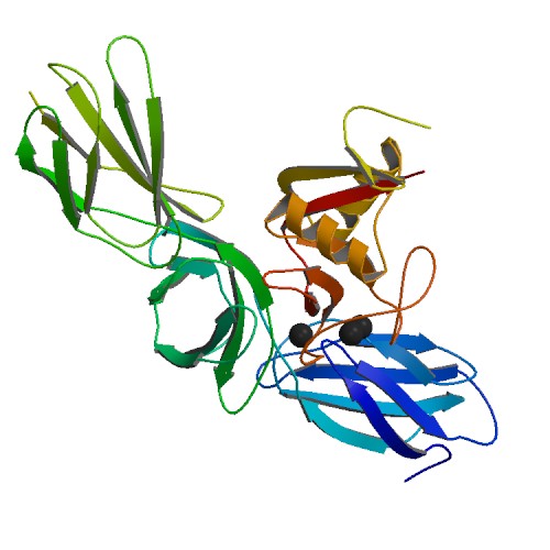
Mucopolysaccharidosis type I is a spectrum of diseases in the mucopolysaccharidosis family. It results in the buildup of glycosaminoglycans (or GAGs, or mucopolysaccharides) due to a deficiency of alpha-L iduronidase, an enzyme responsible for the degradation of GAGs in lysosomes. Without this enzyme, a buildup of dermatan sulfate and heparan sulfate occurs in the body. MPS I may present with a wide spectrum of symptoms, depending on how much functional enzyme is produced. In severe forms, symptoms appear during childhood, and early death can occur due to organ damage. In mild cases, the patient may live into adulthood. Signs and symptoms MPS I affects multiple organ systems. Children with Hurler syndrome (severe MPS I) may appear normal at birth and develop symptoms over the first years of life. Developmental delay may become apparent by age 1–2 years, with a maximum functional age of 2–4 years. Progressive deterioration follows. One of the first abnormalities that may be ... [...More Info...] [...Related Items...] OR: [Wikipedia] [Google] [Baidu] |
Hunter Syndrome
Hunter syndrome, or mucopolysaccharidosis type II (MPS II), is a rare genetic disorder in which large sugar molecules called glycosaminoglycans (or GAGs or mucopolysaccharides) build up in body tissues. It is a form of lysosomal storage disease. Hunter syndrome is caused by a deficiency of the lysosomal enzyme iduronate-2-sulfatase (I2S). The lack of this enzyme causes heparan sulfate and dermatan sulfate to accumulate in all body tissues. Hunter syndrome is the only MPS syndrome to exhibit X-linked recessive inheritance. The symptoms of Hunter syndrome are comparable to those of MPS I. It causes abnormalities in many organs, including the skeleton, heart, and respiratory system. In severe cases, this leads to death during the teenaged years. Unlike MPS I, corneal clouding is not associated with this disease. Signs and symptoms Hunter syndrome may present with a wide variety of phenotypes. It has traditionally been categorized as either "mild" or "severe" depending on the pre ... [...More Info...] [...Related Items...] OR: [Wikipedia] [Google] [Baidu] |
Aortic Valve
The aortic valve is a valve in the heart of humans and most other animals, located between the left ventricle and the aorta. It is one of the four valves of the heart and one of the two semilunar valves, the other being the pulmonary valve. The aortic valve normally has three cusps or leaflets, although in 1–2% of the population it is found to congenitally have two leaflets. The aortic valve is the last structure in the heart the blood travels through before stopping the flow through the systemic circulation. Structure The aortic valve normally has three cusps however there is some discrepancy in their naming. They may be called the left coronary, right coronary and non-coronary cusp. Some sources also advocate they be named as a left, right and posterior cusp. Anatomists have traditionally named them the left posterior (origin of left coronary), anterior (origin of the right coronary) and right posterior. The three cusps, when the valve is closed, contain a sinus called an a ... [...More Info...] [...Related Items...] OR: [Wikipedia] [Google] [Baidu] |
Syndromes With Intellectual Disability
A syndrome is a set of medical signs and symptoms which are correlated with each other and often associated with a particular disease or disorder. The word derives from the Greek σύνδρομον, meaning "concurrence". When a syndrome is paired with a definite cause this becomes a disease. In some instances, a syndrome is so closely linked with a pathogenesis or cause that the words ''syndrome'', ''disease'', and ''disorder'' end up being used interchangeably for them. This substitution of terminology often confuses the reality and meaning of medical diagnoses. This is especially true of inherited syndromes. About one third of all phenotypes that are listed in OMIM are described as dysmorphic, which usually refers to the facial gestalt. For example, Down syndrome, Wolf–Hirschhorn syndrome, and Andersen–Tawil syndrome are disorders with known pathogeneses, so each is more than just a set of signs and symptoms, despite the ''syndrome'' nomenclature. In other instances, a syn ... [...More Info...] [...Related Items...] OR: [Wikipedia] [Google] [Baidu] |
Autosomal Recessive Disorders
An autosome is any chromosome that is not a sex chromosome. The members of an autosome pair in a diploid cell have the same morphology, unlike those in allosomal (sex chromosome) pairs, which may have different structures. The DNA in autosomes is collectively known as atDNA or auDNA. For example, humans have a diploid genome that usually contains 22 pairs of autosomes and one allosome pair (46 chromosomes total). The autosome pairs are labeled with numbers (1–22 in humans) roughly in order of their sizes in base pairs, while allosomes are labelled with their letters. By contrast, the allosome pair consists of two X chromosomes in females or one X and one Y chromosome in males. Unusual combinations of XYY, XXY, XXX, XXXX, XXXXX or XXYY, among other Salome combinations, are known to occur and usually cause developmental abnormalities. Autosomes still contain sexual determination genes even though they are not sex chromosomes. For example, the SRY gene on the Y chromosome e ... [...More Info...] [...Related Items...] OR: [Wikipedia] [Google] [Baidu] |
Proteoglycan Metabolism Disorders
Proteoglycans are proteins that are heavily glycosylated. The basic proteoglycan unit consists of a "core protein" with one or more covalently attached glycosaminoglycan (GAG) chain(s). The point of attachment is a serine (Ser) residue to which the glycosaminoglycan is joined through a tetrasaccharide bridge (e.g. chondroitin sulfate-GlcA- Gal-Gal- Xyl-PROTEIN). The Ser residue is generally in the sequence -Ser- Gly-X-Gly- (where X can be any amino acid residue but proline), although not every protein with this sequence has an attached glycosaminoglycan. The chains are long, linear carbohydrate polymers that are negatively charged under physiological conditions due to the occurrence of sulfate and uronic acid groups. Proteoglycans occur in connective tissue. Types Proteoglycans are categorized by their relative size (large and small) and the nature of their glycosaminoglycan chains. Types include: Certain members are considered members of the "small leucine-rich proteoglycan ... [...More Info...] [...Related Items...] OR: [Wikipedia] [Google] [Baidu] |
Hurler–Scheie Syndrome
Hurler–Scheie syndrome is a genetic disorder caused by the buildup of glycosaminoglycans (GAGs) in various organ tissues. It is a cutaneous condition, also characterized by mild mental retardation and corneal clouding. Respiratory problems, sleep apnea, and heart disease may develop in adolescence. Hurler–Scheie syndrome is classified as a lysosomal storage disease. Patients with Hurler–Scheie syndrome lack the ability to break down GAGs in their lysosomes due a deficiency of the enzyme iduronidase. All forms of mucopolysaccharidosis type I (MPS I) are a spectrum of the same disease. Hurler-Sheie is the subtype of MPS I with intermediate severity. Hurler syndrome is the most severe form, while Scheie syndrome is the least severe form. Some clinicians consider the differences between Hurler, Hurler-Scheie, and Scheie syndromes to be arbitrary. Instead, they classify these patients as having "severe", "intermediate", or "attenuated" MPS I. See also * Mucopolysaccharidosis ... [...More Info...] [...Related Items...] OR: [Wikipedia] [Google] [Baidu] |
Scheie Syndrome
Scheie syndrome is a disease caused by a deficiency in the enzyme iduronidase, leading to the buildup of glycosaminoglycans (GAGs) in the body. It is the most mild subtype of mucopolysaccharidosis type I; the most severe subtype of this disease is called Hurler Syndrome. Scheie syndrome is characterized by corneal clouding, facial dysmorphism, and normal lifespan. People with this condition may have aortic regurgitation. Symptoms The symptoms of Scheie syndrome are variable, but are milder than Hurler Syndrome. Symptoms may begin to appear by age 5, but affected children are often not diagnosed until after age 10. Patients with Scheie Syndrome may have normal intelligence, or they may have mild learning impairments or psychiatric problems. Glaucoma, retinal degeneration, and clouded corneas may cause visual impairments. Aortic valve disease may be present, along with carpal tunnel syndrome, deformed hands and feet, stiff joints, or sleep apnea. People with Scheie syndrome may ... [...More Info...] [...Related Items...] OR: [Wikipedia] [Google] [Baidu] |
Hurler Syndrome
Hurler syndrome, also known as mucopolysaccharidosis Type IH (MPS-IH), Hurler's disease, and formerly gargoylism, is a genetic disorder that results in the buildup of large sugar molecules called glycosaminoglycans (GAGs) in lysosomes. The inability to break down these molecules results in a wide variety of symptoms caused by damage to several different organ systems, including but not limited to the nervous system, skeletal system, eyes, and heart. The underlying mechanism is a deficiency of alpha-L iduronidase, an enzyme responsible for breaking down GAGs. Without this enzyme, a buildup of dermatan sulfate and heparan sulfate occurs in the body. Symptoms appear during childhood, and early death usually occurs. Other, less severe forms of MPS Type I include Hurler-Scheie Syndrome (MPS-IHS) and Scheie Syndrome (MPS-IS). Hurler syndrome is classified as a lysosomal storage disease. It is clinically related to Hunter syndrome (MPS II); however, Hunter syndrome is X-linked, wh ... [...More Info...] [...Related Items...] OR: [Wikipedia] [Google] [Baidu] |
Genetic Carrier
A hereditary carrier (genetic carrier or just carrier), is a person or other organism that has inherited a recessive allele for a genetic trait or mutation but usually does not display that trait or show symptoms of the disease. Carriers are, however, able to pass the allele onto their offspring, who may then express the genetic trait. Carriers in autosomal inheritances Autosomal dominant-recessive inheritance is made possible by the fact that the individuals of most species (including all higher animals and plants) have two alleles of most hereditary predispositions because the chromosomes in the cell nucleus are usually present in pairs (diploid). Carriers can be female or male as the autosomes are homologous independently from the sex. In carriers the expression of a certain characteristic is recessive. The individual has both a genetic predisposition for the dominant trait and a genetic predisposition for the recessive trait, and the dominant expression prevails in the p ... [...More Info...] [...Related Items...] OR: [Wikipedia] [Google] [Baidu] |
Chromosome 4
Chromosome 4 is one of the 23 pairs of chromosomes in humans. People normally have two copies of this chromosome. Chromosome 4 spans more than 186 million base pairs (the building material of DNA) and represents between 6 and 6.5 percent of the total DNA in cells. Genomics The chromosome is ~191 megabases in length. In a 2012 paper, 775 protein-encoding genes were identified on this chromosome.Chen LC, Liu MY, Hsiao YC, Choong WK, Wu HY, Hsu WL, Liao PC, Sung TY, Tsai SF, Yu JS, Chen YJ (2012) Decoding the disease-associated proteins encoded in the human chromosome 4. J Proteome Res 211 (27.9%) of these coding sequences did not have any experimental evidence at the protein level, in 2012. 271 appear to be membrane proteins. 54 have been classified as cancer-associated proteins. Genes Number of genes The following are some of the gene count estimates of human chromosome 4. Because researchers use different approaches to genome annotation their predictions of the number of genes ... [...More Info...] [...Related Items...] OR: [Wikipedia] [Google] [Baidu] |
Autorecessive
In genetics, dominance is the phenomenon of one variant (allele) of a gene on a chromosome masking or overriding the effect of a different variant of the same gene on the other copy of the chromosome. The first variant is termed dominant and the second recessive. This state of having two different variants of the same gene on each chromosome is originally caused by a mutation in one of the genes, either new (''de novo'') or inherited. The terms autosomal dominant or autosomal recessive are used to describe gene variants on non-sex chromosomes ( autosomes) and their associated traits, while those on sex chromosomes (allosomes) are termed X-linked dominant, X-linked recessive or Y-linked; these have an inheritance and presentation pattern that depends on the sex of both the parent and the child (see Sex linkage). Since there is only one copy of the Y chromosome, Y-linked traits cannot be dominant or recessive. Additionally, there are other forms of dominance such as incomplete d ... [...More Info...] [...Related Items...] OR: [Wikipedia] [Google] [Baidu] |
Splenomegaly
Splenomegaly is an enlargement of the spleen. The spleen usually lies in the left upper quadrant (LUQ) of the human abdomen. Splenomegaly is one of the four cardinal signs of ''hypersplenism'' which include: some reduction in number of circulating blood cells affecting granulocytes, erythrocytes or platelets in any combination; a compensatory proliferative response in the bone marrow; and the potential for correction of these abnormalities by splenectomy. Splenomegaly is usually associated with increased workload (such as in hemolytic anemias), which suggests that it is a response to hyperfunction. It is therefore not surprising that splenomegaly is associated with any disease process that involves abnormal red blood cells being destroyed in the spleen. Other common causes include congestion due to portal hypertension and infiltration by leukemias and lymphomas. Thus, the finding of an enlarged spleen, along with caput medusae, is an important sign of portal hypertension. Definiti ... [...More Info...] [...Related Items...] OR: [Wikipedia] [Google] [Baidu] |


.jpg)

