|
Mittendorf's Dot
The hyaloid artery is a branch of the ophthalmic artery, which is itself a branch of the internal carotid artery. It is contained within the optic stalk of the eye and extends from the optic disc through the vitreous humor to the lens. Usually fully regressed before birth, its purpose is to supply nutrients to the developing lens in the growing fetus. During the tenth week of development in humans (time varies depending on species), the lens grows independent of a blood supply and the hyaloid artery usually regresses. Its proximal portion remains as the central artery of the retina. Regression of the hyaloid artery leaves a clear central zone through the vitreous humor, called the hyaloid canal or Cloquet's canal. Cloquet's canal is named after the French physician Jules Germain Cloquet (1790–1883) who first described it. Occasionally the artery may not fully regress, resulting in the condition ''persistent hyaloid artery''. More commonly, small remnants of the artery may re ... [...More Info...] [...Related Items...] OR: [Wikipedia] [Google] [Baidu] |
Central Retinal Artery
The central retinal artery (retinal artery) branches off the ophthalmic artery, running inferior to the optic nerve within its dural sheath to the eyeball. Structure The central retinal artery pierces the eyeball close to the optic nerve, sending branches over the internal surface of the retina, and these terminal branches are the only blood supply to the larger part of it. The central part of the retina where the light rays are focused after passing through the pupil and the lens is a circular area called the macula. The center of this circular area is the fovea. The fovea and a small area surrounding it are not supplied by the central retinal artery or its branches, but instead by the choroid. The central retinal artery is approximately 160 micrometres in diameter. Variation In some cases - approximately 20% of the population - there is a branch of the ciliary circulation called the cilio-retinal artery which supplies the retina between the macula and the optic nerve, includ ... [...More Info...] [...Related Items...] OR: [Wikipedia] [Google] [Baidu] |
Fetus
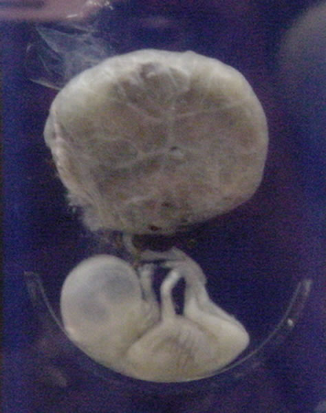
A fetus or foetus (; plural fetuses, feti, foetuses, or foeti) is the unborn offspring that develops from an animal embryo. Following embryonic development the fetal stage of development takes place. In human prenatal development, fetal development begins from the ninth week after fertilization (or eleventh week gestational age) and continues until birth. Prenatal development is a continuum, with no clear defining feature distinguishing an embryo from a fetus. However, a fetus is characterized by the presence of all the major body organs, though they will not yet be fully developed and functional and some not yet situated in their final anatomical location. Etymology The word '' fetus'' (plural '' fetuses'' or '' feti'') is related to the Latin '' fētus'' ("offspring", "bringing forth", "hatching of young") and the Greek "φυτώ" to plant. The word "fetus" was used by Ovid in Metamorphoses, book 1, line 104. The predominant British, Irish, and Commonwealth spelling ... [...More Info...] [...Related Items...] OR: [Wikipedia] [Google] [Baidu] |
Hyaline
A hyaline substance is one with a glassy appearance. The word is derived from el, ὑάλινος, translit=hyálinos, lit=transparent, and el, ὕαλος, translit=hýalos, lit=crystal, glass, label=none. Histopathology Hyaline cartilage is named after its glassy appearance on fresh gross pathology. On light microscopy of H&E stained slides, the extracellular matrix of hyaline cartilage looks homogeneously pink, and the term "hyaline" is used to describe similarly homogeneously pink material besides the cartilage. Hyaline material is usually acellular and proteinaceous. For example, arterial hyaline is seen in aging, high blood pressure, diabetes mellitus Diabetes, also known as diabetes mellitus, is a group of metabolic disorders characterized by a high blood sugar level (hyperglycemia) over a prolonged period of time. Symptoms often include frequent urination, increased thirst and increased ... and in association with some drugs (e.g. calcineurin inhibitors) ... [...More Info...] [...Related Items...] OR: [Wikipedia] [Google] [Baidu] |
Bergmeister's Papilla
Bergmeister's papilla arises from the centre of the optic disc, consists of a small tuft of fibrous tissue and represents a remnant of the fetal hyaloid artery. The hyaloid artery provides nutrition to the lens A lens is a transmissive optical device which focuses or disperses a light beam by means of refraction. A simple lens consists of a single piece of transparent material, while a compound lens consists of several simple lenses (''elements'' ... during development in the fetus, and runs forward to the lens from the optic disc. The optic disc is covered by a plaque of fibrous cells called the central supporting tissue meniscus of Kuhnt. This plaque forms a fibrous sheath around the hyaloid artery where it leaves the optic disc. At birth the hyaloid artery regresses, and is normally completely regressed by the time of eyelid opening. Bergmeister's papilla is a remnant of the hyaloid artery fibrous sheath and is frequently observed as an incidental clinical fin ... [...More Info...] [...Related Items...] OR: [Wikipedia] [Google] [Baidu] |
Floater
Floaters or eye floaters are sometimes visible deposits (e.g., the shadows of tiny structures of protein or other cell debris projected onto the retina) within the eye's vitreous humour ("the vitreous"), which is normally transparent, or between the vitreous and retina.Cline D; Hofstetter HW; Griffin JR. ''Dictionary of Visual Science''. 4th ed. Butterworth-Heinemann, Boston 1997. They can become particularly noticeable when looking at a blank surface or an open monochromatic space, such as blue sky. Each floater can be measured by its size, shape, consistency, refractive index, and motility. They are also called ''muscae volitantes'' (Latin for 'flying flies'), or ''mouches volantes'' (from the same phrase in French). The vitreous usually starts out transparent, but imperfections may gradually develop as one ages. The common type of floater, present in most people's eyes, is due to these degenerative changes of the vitreous. The perception of floaters, which may be annoying or ... [...More Info...] [...Related Items...] OR: [Wikipedia] [Google] [Baidu] |
Jules Germain Cloquet
Jules Germain Cloquet (18 December 1790 – 23 February 1883) was a French physician and surgeon who was born and practiced medicine in Paris. His older brother, Hippolyte Cloquet (1787-1840) and his younger nephew Ernest Cloquet (1818-1855) were also physicians. In 1821 Jules Cloquet became one of the earliest members elected to the Académie Nationale de Médecine in Paris. In 1836, he was elected Honorary Fellow of the Royal College of Surgeons in Ireland. Cloquet was known for his expertise as a surgeon, especially his work with hernial disorders. He was also the first to describe and identify the remnant of the embryonic hyaloid artery. This vestige was to become known as Cloquet's canal. Cloquet's name is associated with three anatomical terms regarding the femoral canal: * "Cloquet's hernia": a hernia of the femoral canal * "Cloquet's septum": a fibrous membrane bounding the annulus femoralis at the base of the femoral canal * " Cloquet's gland": small lymphatic node ... [...More Info...] [...Related Items...] OR: [Wikipedia] [Google] [Baidu] |
Hyaloid Canal
Hyaloid canal (Cloquet's canal and Stilling's canal) is a small transparent canal running through the vitreous body from the optic nerve disc to the lens. It is formed by an invagination of the hyaloid membrane, which encloses the vitreous body. In the fetus, the hyaloid canal contains a prolongation of the central artery of the retina, the hyaloid artery, which supplies blood to the developing lens. Once the lens is fully developed the hyaloid artery retracts and the hyaloid canal contains lymph. The hyaloid canal appears to have no function in the adult eye, though its remnant structure can be seen. Contrary to initial belief, the hyaloid canal does not facilitate changes in the volume of the lens. The lens volume changes by less than 1% over its range of accommodation. Furthermore, lymph, being liquid, is incompressible, so even if the volume of the lens did change, the hyaloid canal could not compensate for it. See also * Hyaloid artery The hyaloid artery is a branch o ... [...More Info...] [...Related Items...] OR: [Wikipedia] [Google] [Baidu] |
Retina
The retina (from la, rete "net") is the innermost, light-sensitive layer of tissue of the eye of most vertebrates and some molluscs. The optics of the eye create a focused two-dimensional image of the visual world on the retina, which then processes that image within the retina and sends nerve impulses along the optic nerve to the visual cortex to create visual perception. The retina serves a function which is in many ways analogous to that of the film or image sensor in a camera. The neural retina consists of several layers of neurons interconnected by synapses and is supported by an outer layer of pigmented epithelial cells. The primary light-sensing cells in the retina are the photoreceptor cells, which are of two types: rods and cones. Rods function mainly in dim light and provide monochromatic vision. Cones function in well-lit conditions and are responsible for the perception of colour through the use of a range of opsins, as well as high-acuity vision used f ... [...More Info...] [...Related Items...] OR: [Wikipedia] [Google] [Baidu] |
Childbirth
Childbirth, also known as labour and delivery, is the ending of pregnancy where one or more babies exits the internal environment of the mother via vaginal delivery or caesarean section. In 2019, there were about 140.11 million births globally. In the developed countries, most deliveries occur in hospitals, while in the developing countries most are home births. The most common childbirth method worldwide is vaginal delivery. It involves four stages of labour: the shortening and opening of the cervix during the first stage, descent and birth of the baby during the second, the delivery of the placenta during the third, and the recovery of the mother and infant during the fourth stage, which is referred to as the postpartum. The first stage is characterized by abdominal cramping or back pain that typically lasts half a minute and occurs every 10 to 30 minutes. Contractions gradually becomes stronger and closer together. Since the pain of childbirth correlates with c ... [...More Info...] [...Related Items...] OR: [Wikipedia] [Google] [Baidu] |
Ophthalmic Artery
The ophthalmic artery (OA) is an artery of the head. It is the first branch of the internal carotid artery distal to the cavernous sinus. Branches of the ophthalmic artery supply all the structures in the orbit around the eye, as well as some structures in the nose, face, and meninges. Occlusion of the ophthalmic artery or its branches can produce sight-threatening conditions. Structure The ophthalmic artery emerges from the internal carotid artery. This is usually just after the internal carotid artery emerges from the cavernous sinus. In some cases, the ophthalmic artery branches just before the internal carotid exits the cavernous sinus. The ophthalmic artery emerges along the medial side of the anterior clinoid process. It runs anteriorly, passing through the optic canal inferolaterally to the optic nerve. It can also pass superiorly to the optic nerve in a minority of cases. In the posterior third of the cone of the orbit, the ophthalmic artery turns sharply and medial ... [...More Info...] [...Related Items...] OR: [Wikipedia] [Google] [Baidu] |
Lens (anatomy)
The lens, or crystalline lens, is a transparent biconvex structure in the eye that, along with the cornea, helps to refract light to be focused on the retina. By changing shape, it functions to change the focal length of the eye so that it can focus on objects at various distances, thus allowing a sharp real image of the object of interest to be formed on the retina. This adjustment of the lens is known as '' accommodation'' (see also below). Accommodation is similar to the focusing of a photographic camera via movement of its lenses. The lens is flatter on its anterior side than on its posterior side. In humans, the refractive power of the lens in its natural environment is approximately 18 dioptres, roughly one-third of the eye's total power. Structure The lens is part of the anterior segment of the human eye. In front of the lens is the iris, which regulates the amount of light entering into the eye. The lens is suspended in place by the suspensory ligament of the l ... [...More Info...] [...Related Items...] OR: [Wikipedia] [Google] [Baidu] |
Vitreous Humor
The vitreous body (''vitreous'' meaning "glass-like"; , ) is the clear gel that fills the space between the lens and the retina of the eyeball (the vitreous chamber) in humans and other vertebrates. It is often referred to as the vitreous humor (also spelled humour, from Latin meaning liquid) or simply "the vitreous". Vitreous fluid or "liquid vitreous" is the liquid component of the vitreous gel, found after a vitreous detachment. It is not to be confused with the aqueous humor, the other fluid in the eye that is found between the cornea and lens. Structure The vitreous humor is a transparent, colorless, gelatinous mass that fills the space in the eye between the lens and the retina. It is surrounded by a layer of collagen called the vitreous membrane (or hyaloid membrane or vitreous cortex) separating it from the rest of the eye. It makes up four-fifths of the volume of the eyeball. The vitreous humour is fluid-like near the centre, and gel-like near the edges. The vitreo ... [...More Info...] [...Related Items...] OR: [Wikipedia] [Google] [Baidu] |


