|
Mitchell's Disease
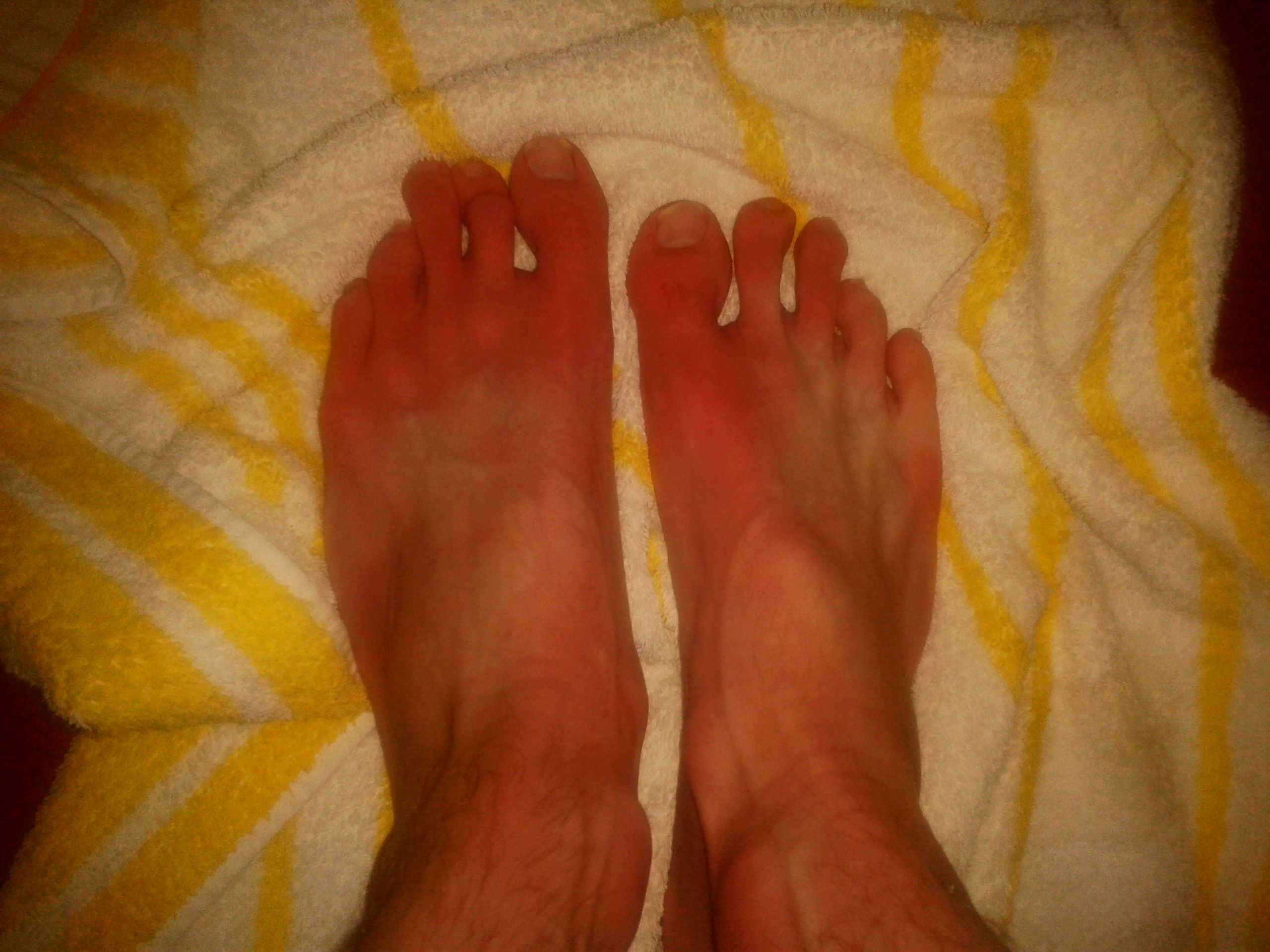
Erythromelalgia or Mitchell's disease (after Silas Weir Mitchell) is a rare vascular peripheral pain disorder in which blood vessels, usually in the lower extremities or hands, are episodically blocked (frequently on and off daily), then become hyperemic and inflamed. There is severe burning pain (in the small fiber sensory nerves) and skin redness. The attacks are periodic and are commonly triggered by heat, pressure, mild activity, exertion, insomnia or stress. Erythromelalgia may occur either as a primary or secondary disorder (i.e. a disorder in and of itself or a symptom of another condition). Secondary erythromelalgia can result from small fiber peripheral neuropathy of any cause, polycythemia vera, essential thrombocytosis, hypercholesterolemia, mushroom or mercury poisoning, and some autoimmune disorders. Primary erythromelalgia is caused by mutation of the voltage-gated sodium channel α-subunit gene '' SCN9A''. In 2004 erythromelalgia became the first human disorde ... [...More Info...] [...Related Items...] OR: [Wikipedia] [Google] [Baidu] |
Silas Weir Mitchell (physician)
Silas Weir Mitchell (February 15, 1829 – January 4, 1914) was an American physician, scientist, novelist, and poet. He is considered the father of medical neurology, and he discovered causalgia (complex regional pain syndrome) and erythromelalgia, and pioneered the rest cure. Biography Silas Weir Mitchell was born on February 15, 1829, in Philadelphia, Pennsylvania, to John Kearsley Mitchell and Sarah Henry Mitchell. He studied at Philadelphia's renowned University of Pennsylvania and later earned the degree of MD at the city's Jefferson Medical College in 1850. During the Civil War, he was director of treatment of nervous injuries and maladies at Turners Lane Hospital, Philadelphia, and at the close of the war became a specialist in neurology. In this field Mitchell pioneered the rest cure for diseases now termed "psychiatric", particularly neurasthenia and hysteria, subsequently taken up by the medical world. The treatment consisted primarily in isolation, confinement to b ... [...More Info...] [...Related Items...] OR: [Wikipedia] [Google] [Baidu] |
Erythema
Erythema (from the Greek , meaning red) is redness of the skin or mucous membranes, caused by hyperemia (increased blood flow) in superficial capillaries. It occurs with any skin injury, infection, or inflammation. Examples of erythema not associated with pathology include nervous blushes. Types * Erythema ab igne * Erythema chronicum migrans * Erythema induratum * Erythema infectiosum (or fifth disease) * Erythema marginatum * Erythema migrans * Erythema multiforme (EM) * Erythema nodosum * Erythema toxicum * Erythema elevatum diutinum * Erythema gyratum repens * Keratolytic winter erythema * Palmar erythema Causes It can be caused by infection, massage, electrical treatment, acne medication, allergies, exercise, solar radiation (sunburn), photosensitization, acute radiation syndrome, mercury toxicity, blister agents, niacin administration, or waxing and tweezing of the hairs—any of which can cause the capillaries to dilate, resulting in redness. Erythema is a common sid ... [...More Info...] [...Related Items...] OR: [Wikipedia] [Google] [Baidu] |
Vascular Smooth Muscle
Vascular smooth muscle is the type of smooth muscle that makes up most of the walls of blood vessels. Structure Vascular smooth muscle refers to the particular type of smooth muscle found within, and composing the majority of the wall of blood vessels. Nerve supply Vascular smooth muscle is innervated primarily by the sympathetic nervous system through adrenergic receptors (adrenoceptors). The three types present are: alpha-1, alpha-2 and beta-2 adrenergic receptors, . The main endogenous agonist of these cell receptors is norepinephrine (NE). The adrenergic receptors exert opposite physiologic effects in the vascular smooth muscle under activation: * alpha-1 receptors. Under NE binding alpha-1 receptors cause vasoconstriction ( contraction of the vascular smooth muscle cells decreasing the diameter of the vessels). Thesea receptors are activated in response to shock or low blood pressure as a defensive reaction trying to restore the normal blood pressure. Antagonists ... [...More Info...] [...Related Items...] OR: [Wikipedia] [Google] [Baidu] |
Cutaneous
Skin is the layer of usually soft, flexible outer tissue covering the body of a vertebrate animal, with three main functions: protection, regulation, and sensation. Other animal coverings, such as the arthropod exoskeleton, have different developmental origin, structure and chemical composition. The adjective cutaneous means "of the skin" (from Latin ''cutis'' 'skin'). In mammals, the skin is an organ of the integumentary system made up of multiple layers of ectodermal tissue and guards the underlying muscles, bones, ligaments, and internal organs. Skin of a different nature exists in amphibians, reptiles, and birds. Skin (including cutaneous and subcutaneous tissues) plays crucial roles in formation, structure, and function of extraskeletal apparatus such as horns of bovids (e.g., cattle) and rhinos, cervids' antlers, giraffids' ossicones, armadillos' osteoderm, and os penis/os clitoris. All mammals have some hair on their skin, even marine mammals like whales, dolphins, a ... [...More Info...] [...Related Items...] OR: [Wikipedia] [Google] [Baidu] |
Sympathetic Nervous System
The sympathetic nervous system (SNS) is one of the three divisions of the autonomic nervous system, the others being the parasympathetic nervous system and the enteric nervous system. The enteric nervous system is sometimes considered part of the autonomic nervous system, and sometimes considered an independent system. The autonomic nervous system functions to regulate the body's unconscious actions. The sympathetic nervous system's primary process is to stimulate the body's fight or flight response. It is, however, constantly active at a basic level to maintain homeostasis. The sympathetic nervous system is described as being antagonistic to the parasympathetic nervous system which stimulates the body to "feed and breed" and to (then) "rest-and-digest". Structure There are two kinds of neurons involved in the transmission of any signal through the sympathetic system: pre-ganglionic and post-ganglionic. The shorter preganglionic neurons originate in the thoracolumbar division o ... [...More Info...] [...Related Items...] OR: [Wikipedia] [Google] [Baidu] |
Nociceptor
A nociceptor ("pain receptor" from Latin ''nocere'' 'to harm or hurt') is a sensory neuron that responds to damaging or potentially damaging stimuli by sending "possible threat" signals to the spinal cord and the brain. The brain creates the sensation of pain to direct attention to the body part, so the threat can be mitigated; this process is called nociception. History Nociceptors were discovered by Charles Scott Sherrington in 1906. In earlier centuries, scientists believed that animals were like mechanical devices that transformed the energy of sensory stimuli into motor responses. Sherrington used many different experiments to demonstrate that different types of stimulation to an afferent nerve fiber's receptive field led to different responses. Some intense stimuli trigger reflex withdrawal, certain autonomic responses, and pain. The specific receptors for these intense stimuli were called nociceptors. Location In mammals, nociceptors are found in any area of the body tha ... [...More Info...] [...Related Items...] OR: [Wikipedia] [Google] [Baidu] |
Dorsal Root Ganglion
A dorsal root ganglion (or spinal ganglion; also known as a posterior root ganglion) is a cluster of neurons (a ganglion) in a dorsal root of a spinal nerve. The cell bodies of sensory neurons known as first-order neurons are located in the dorsal root ganglia. The axons of dorsal root ganglion neurons are known as afferents. In the peripheral nervous system, afferents refer to the axons that relay sensory information into the central nervous system (i.e. the brain and the spinal cord). Structure The neurons comprising the dorsal root ganglion are of the pseudo-unipolar type, meaning they have a cell body (soma) with two branches that act as a single axon, often referred to as a ''distal process'' and a ''proximal process''. Unlike the majority of neurons found in the central nervous system, an action potential in posterior root ganglion neuron may initiate in the ''distal process'' in the periphery, bypass the cell body, and continue to propagate along the ''proximal process ... [...More Info...] [...Related Items...] OR: [Wikipedia] [Google] [Baidu] |
Group C Nerve Fiber
Group C nerve fibers are one of three classes of nerve fiber in the central nervous system (CNS) and peripheral nervous system (PNS). The C group fibers are unmyelinated and have a small diameter and low conduction velocity, whereas Groups A and B are myelinated. Group C fibers include postganglionic fibers in the autonomic nervous system (ANS), and nerve fibers at the dorsal roots (IV fiber). These fibers carry sensory information. Damage or injury to nerve fibers causes neuropathic pain. Capsaicin activates C fibre vanilloid receptors, giving chili peppers a hot sensation. Structure and anatomy Location C fibers are one class of nerve fiber found in the nerves of the somatic sensory system. They are afferent fibers, conveying input signals from the periphery to the central nervous system. Structure C fibers are unmyelinated unlike most other fibers in the nervous system. This lack of myelination is the cause of their slow conduction velocity, which is on the order o ... [...More Info...] [...Related Items...] OR: [Wikipedia] [Google] [Baidu] |
Erythromelalgia
Erythromelalgia or Mitchell's disease (after Silas Weir Mitchell) is a rare vascular peripheral pain disorder in which blood vessels, usually in the lower extremities or hands, are episodically blocked (frequently on and off daily), then become hyperemic and inflamed. There is severe burning pain (in the small fiber sensory nerves) and skin redness. The attacks are periodic and are commonly triggered by heat, pressure, mild activity, exertion, insomnia or stress. Erythromelalgia may occur either as a primary or secondary disorder (i.e. a disorder in and of itself or a symptom of another condition). Secondary erythromelalgia can result from small fiber peripheral neuropathy of any cause, polycythemia vera, essential thrombocytosis, hypercholesterolemia, mushroom or mercury poisoning, and some autoimmune disorders. Primary erythromelalgia is caused by mutation of the voltage-gated sodium channel α-subunit gene ''SCN9A''. In 2004 erythromelalgia became the first human disorder in ... [...More Info...] [...Related Items...] OR: [Wikipedia] [Google] [Baidu] |
Capillary
A capillary is a small blood vessel from 5 to 10 micrometres (μm) in diameter. Capillaries are composed of only the tunica intima, consisting of a thin wall of simple squamous endothelial cells. They are the smallest blood vessels in the body: they convey blood between the arterioles and venules. These microvessels are the site of exchange of many substances with the interstitial fluid surrounding them. Substances which cross capillaries include water, oxygen, carbon dioxide, urea, glucose, uric acid, lactic acid and creatinine. Lymph capillaries connect with larger lymph vessels to drain lymphatic fluid collected in the microcirculation. During early embryonic development, new capillaries are formed through vasculogenesis, the process of blood vessel formation that occurs through a '' de novo'' production of endothelial cells that then form vascular tubes. The term '' angiogenesis'' denotes the formation of new capillaries from pre-existing blood vessels and already present ... [...More Info...] [...Related Items...] OR: [Wikipedia] [Google] [Baidu] |
Neuropathology
Neuropathology is the study of disease of nervous system tissue, usually in the form of either small surgical biopsies or whole-body autopsies. Neuropathologists usually work in a department of anatomic pathology, but work closely with the clinical disciplines of neurology, and neurosurgery, which often depend on neuropathology for a diagnosis. Neuropathology also relates to forensic pathology because brain disease or brain injury can be related to cause of death. Neuropathology should not be confused with neuropathy, which refers to disorders of the nerves themselves (usually in the peripheral nervous system) rather than the tissues. In neuropathology, the branches of the specializations of nervous system as well as the tissues come together into one field of study. Methodology The work of the neuropathologist consists largely of examining autopsy or biopsy tissue from the brain and spinal cord to aid in diagnosis of disease. Tissues are also observed through the eyes, muscles, ... [...More Info...] [...Related Items...] OR: [Wikipedia] [Google] [Baidu] |


