|
Microdeletion Syndrome
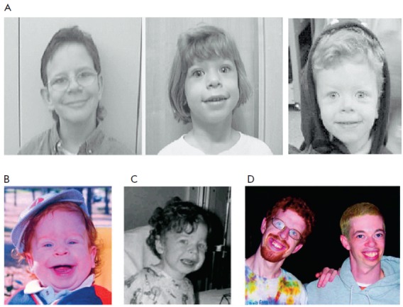
A microdeletion syndrome is a syndrome caused by a chromosomal deletion smaller than 5 million base pairs (5 Mb) spanning several genes that is too small to be detected by conventional cytogenetic methods or high resolution karyotyping (2–5 Mb). Detection is done by fluorescence in situ hybridization (FISH). Larger chromosomal deletion syndromes are detectable using karyotyping techniques. Examples * DiGeorge syndrome or velocardiofacial syndrome – most common microdeletion syndrome * Prader–Willi syndrome * Angelman syndrome * Neurofibromatosis type I * Neurofibromatosis type II * Williams syndrome * Miller–Dieker syndrome * Smith–Magenis syndrome * Rubinstein–Taybi syndrome * Wolf–Hirschhorn syndrome Wolf–Hirschhorn syndrome (WHS) is a chromosomal deletion syndrome resulting from a partial deletion on the short arm of chromosome 4 (del(4p16.3)). Features include a distinct craniofacial phenotype and intellectual disability. Signs and sympt ... References ... [...More Info...] [...Related Items...] OR: [Wikipedia] [Google] [Baidu] |
Deletion (genetics)
In genetics, a deletion (also called gene deletion, deficiency, or deletion mutation) (sign: Δ) is a mutation (a genetic aberration) in which a part of a chromosome or a sequence of DNA is left out during DNA replication. Any number of nucleotides can be deleted, from a single base to an entire piece of chromosome. Some chromosomes have fragile spots where breaks occur which result in the deletion of a part of chromosome. The breaks can be induced by heat, viruses, radiations, chemicals. When a chromosome breaks, a part of it is deleted or lost, the missing piece of chromosome is referred to as deletion or a deficiency. For synapsis to occur between a chromosome with a large intercalary deficiency and a normal complete homolog, the unpaired region of the normal homolog must loop out of the linear structure into a deletion or compensation loop. The smallest single base deletion mutations occur by a single base flipping in the template DNA, followed by template DNA strand sli ... [...More Info...] [...Related Items...] OR: [Wikipedia] [Google] [Baidu] |
Base Pair
A base pair (bp) is a fundamental unit of double-stranded nucleic acids consisting of two nucleobases bound to each other by hydrogen bonds. They form the building blocks of the DNA double helix and contribute to the folded structure of both DNA and RNA. Dictated by specific hydrogen bonding patterns, "Watson–Crick" (or "Watson–Crick–Franklin") base pairs (guanine–cytosine and adenine–thymine) allow the DNA helix to maintain a regular helical structure that is subtly dependent on its nucleotide sequence. The Complementarity (molecular biology), complementary nature of this based-paired structure provides a redundant copy of the genetic information encoded within each strand of DNA. The regular structure and data redundancy provided by the DNA double helix make DNA well suited to the storage of genetic information, while base-pairing between DNA and incoming nucleotides provides the mechanism through which DNA polymerase replicates DNA and RNA polymerase transcribes DNA in ... [...More Info...] [...Related Items...] OR: [Wikipedia] [Google] [Baidu] |
Fluorescence In Situ Hybridization
Fluorescence ''in situ'' hybridization (FISH) is a molecular cytogenetic technique that uses fluorescent probes that bind to only particular parts of a nucleic acid sequence with a high degree of sequence complementarity. It was developed by biomedical researchers in the early 1980s to detect and localize the presence or absence of specific DNA sequences on chromosomes. Fluorescence microscopy can be used to find out where the fluorescent probe is bound to the chromosomes. FISH is often used for finding specific features in DNA for use in genetic counseling, medicine, and species identification. FISH can also be used to detect and localize specific RNA targets (mRNA, lncRNA and miRNA) in cells, circulating tumor cells, and tissue samples. In this context, it can help define the spatial-temporal patterns of gene expression within cells and tissues. Probes – RNA and DNA In biology, a probe is a single strand of DNA or RNA that is complementary to a nucleotide sequence o ... [...More Info...] [...Related Items...] OR: [Wikipedia] [Google] [Baidu] |
Chromosomal Deletion Syndrome
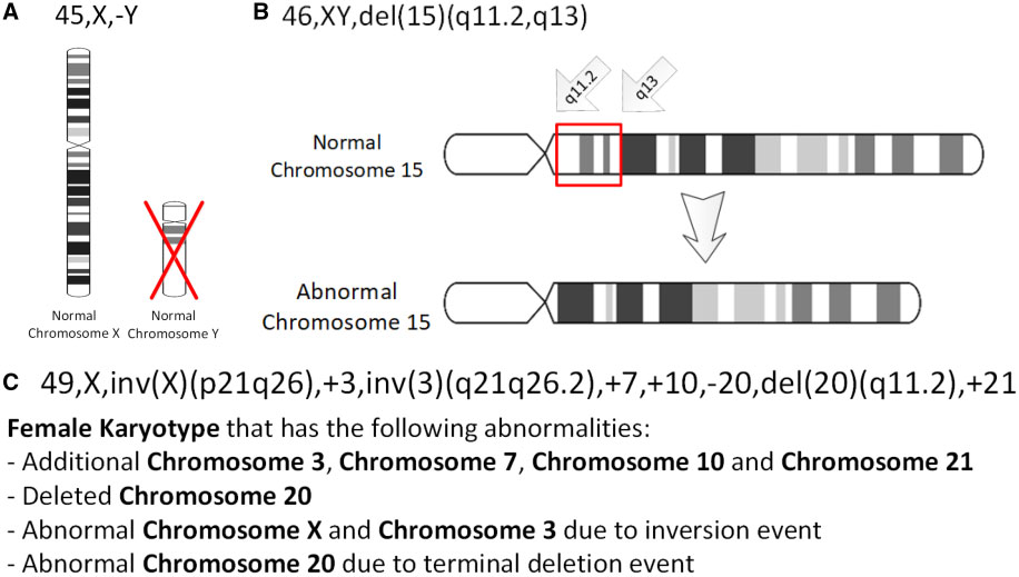
Chromosomal deletion syndromes result from deletion of parts of chromosomes. Depending on the location, size, and whom the deletion is inherited from, there are a few known different variations of chromosome deletions. Chromosomal deletion syndromes typically involve larger deletions that are visible using karyotyping techniques. Smaller deletions result in Microdeletion syndrome, which are detected using fluorescence in situ hybridization (FISH) Examples of chromosomal deletion syndromes include 5p-Deletion (cri du chat syndrome), 4p-Deletion (Wolf–Hirschhorn syndrome), Prader–Willi syndrome, and Angelman syndrome. 5p-Deletion The chromosomal basis of Cri du chat syndrome consists of a deletion of the most terminal portion of the short arm of chromosome 5. 5p deletions, whether terminal or interstitial, occur at different breakpoints; the chromosomal basis generally consists of a deletion on the short arm of chromosome 5. The variability seen among individuals may be att ... [...More Info...] [...Related Items...] OR: [Wikipedia] [Google] [Baidu] |
Karyotyping
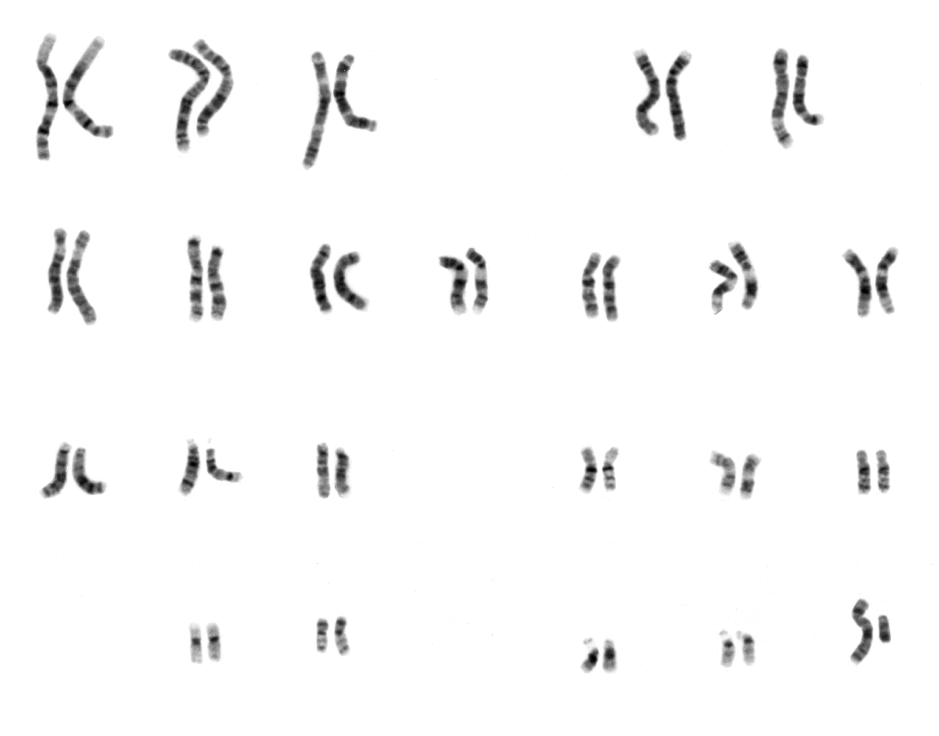
A karyotype is the general appearance of the complete set of metaphase chromosomes in the cells of a species or in an individual organism, mainly including their sizes, numbers, and shapes. Karyotyping is the process by which a karyotype is discerned by determining the chromosome complement of an individual, including the number of chromosomes and any abnormalities. A karyogram or idiogram is a graphical depiction of a karyotype, wherein chromosomes are organized in pairs, ordered by size and position of centromere for chromosomes of the same size. Karyotyping generally combines light microscopy and photography, and results in a photomicrographic (or simply micrographic) karyogram. In contrast, a schematic karyogram is a designed graphic representation of a karyotype. In schematic karyograms, just one of the sister chromatids of each chromosome is generally shown for brevity, and in reality they are generally so close together that they look as one on photomicrographs as well u ... [...More Info...] [...Related Items...] OR: [Wikipedia] [Google] [Baidu] |
DiGeorge Syndrome
DiGeorge syndrome, also known as 22q11.2 deletion syndrome, is a syndrome caused by a microdeletion on the long arm of chromosome 22. While the symptoms can vary, they often include congenital heart problems, specific facial features, frequent infections, developmental delay, learning problems and cleft palate. Associated conditions include kidney problems, schizophrenia, hearing loss and autoimmune disorders such as rheumatoid arthritis or Graves' disease. DiGeorge syndrome is typically due to the deletion of 30 to 40 genes in the middle of chromosome 22 at a location known as ''22q11.2''. About 90% of cases occur due to a new mutation during early development, while 10% are inherited from a person's parents. It is autosomal dominant, meaning that only one affected chromosome is needed for the condition to occur. Diagnosis is suspected based on the symptoms and confirmed by genetic testing. Although there is no cure, treatment can improve symptoms. This often includes a m ... [...More Info...] [...Related Items...] OR: [Wikipedia] [Google] [Baidu] |
Prader–Willi Syndrome
Prader–Willi syndrome (PWS) is a genetic disorder caused by a loss of function of specific genes on chromosome 15. In newborns, symptoms include weak muscles, poor feeding, and slow development. Beginning in childhood, those affected become constantly hungry, which often leads to obesity and type 2 diabetes. Mild to moderate intellectual impairment and behavioral problems are also typical of the disorder. Often, affected individuals have a narrow forehead, small hands and feet, short height, and light skin and hair. Most are unable to have children. About 74% of cases occur when part of the father's chromosome 15 is deleted. In another 25% of cases, the affected person has two copies of the maternal chromosome 15 from the mother and lacks the paternal copy. As parts of the chromosome from the mother are turned off through imprinting, they end up with no working copies of certain genes. PWS is not generally inherited, but rather the genetic changes happen during the format ... [...More Info...] [...Related Items...] OR: [Wikipedia] [Google] [Baidu] |
Angelman Syndrome
Angelman syndrome or Angelman's syndrome (AS) is a genetic disorder that mainly affects the nervous system. Symptoms include a small head and a specific facial appearance, severe intellectual disability, developmental disability, limited to no functional speech, balance and movement problems, seizures, and sleep problems. Children usually have a happy personality and have a particular interest in water. The symptoms generally become noticeable by one year of age. Angelman syndrome is due to a lack of function of part of chromosome 15, typically due to a new mutation rather than one inherited from a person's parents. Most of the time, it is due to a deletion or mutation of the UBE3A gene on that chromosome. Occasionally, it is due to inheriting two copies of chromosome 15 from a person's father and none from their mother ( paternal uniparental disomy). As the father's versions are inactivated by a process known as genomic imprinting, no functional version of the gene remains. D ... [...More Info...] [...Related Items...] OR: [Wikipedia] [Google] [Baidu] |
Neurofibromatosis Type II
Neurofibromatosis type II (also known as MISME syndrome – multiple inherited schwannomas, meningiomas, and ependymomas) is a genetic condition that may be inherited or may arise spontaneously, and causes benign tumors of the brain, spinal cord, and peripheral nerves. The types of tumors frequently associated with NF2 include vestibular schwannomas, meningiomas, and ependymomas. The main manifestation of the condition is the development of bilateral benign brain tumors in the nerve sheath of the cranial nerve VIII, which is the "auditory-vestibular nerve" that transmits sensory information from the inner ear to the brain. Besides, other benign brain and spinal tumors occur. Symptoms depend on the presence, localisation and growth of the tumor(s), in which multiple cranial nerves can be involved. Many people with this condition also experience vision problems. Neurofibromatosis type II (NF2 ''or'' NF II) is caused by mutations of the "Merlin" gene, which seems to influence the for ... [...More Info...] [...Related Items...] OR: [Wikipedia] [Google] [Baidu] |
Williams Syndrome
Williams syndrome (WS) is a genetic disorder that affects many parts of the body. Facial features frequently include a broad forehead, underdeveloped chin, short nose, and full cheeks. Mild to moderate intellectual disability is observed in people with WS, with particular challenges with visual spatial tasks such as drawing. Verbal skills are relatively unaffected. Many people with WS have an outgoing personality, an openness to engaging with other people, and a happy disposition. Medical issues with teeth, heart problems (especially supravalvular aortic stenosis), and periods of high blood calcium are common. Williams syndrome is caused by a genetic abnormality, specifically a deletion of about 27 genes from the long arm of one of the two chromosome 7s. Typically, this occurs as a random event during the formation of the egg or sperm from which a person develops. In a small number of cases, it is inherited from an affected parent in an autosomal dominant manner. The different ... [...More Info...] [...Related Items...] OR: [Wikipedia] [Google] [Baidu] |
Miller–Dieker Syndrome
Miller–Dieker syndrome, Miller–Dieker lissencephaly syndrome (MDLS), and chromosome 17p13.3 deletion syndrome is a micro deletion syndrome characterized by congenital malformations. Congenital malformations are physical defects detectable in an infant at birth which can involve many different parts of the body including the brain, hearts, lungs, liver, bones, or intestinal tract. MDS is a contiguous gene syndrome – a disorder due to the deletion of multiple gene loci adjacent to one another. The disorder arises from the deletion of part of the small arm of chromosome 17p (which includes both the ''LIS1'' and '' 14-3-3 epsilon'' genes), leading to partial monosomy. There may be unbalanced translocations (i.e. 17q:17p or 12q:17p), or the presence of a ring chromosome 17. This syndrome should not be confused with Miller syndrome, an unrelated rare genetic disorder, or Miller Fisher syndrome, a form of Guillain–Barré syndrome. Presentation The brain is abnormally smooth, wit ... [...More Info...] [...Related Items...] OR: [Wikipedia] [Google] [Baidu] |


