|
Massarina Carolinensis
''Massarina carolinensis'' is a species of fungus in the Lophiostomataceae family. The species is found exclusively on the lower parts of the culms of the saltmarsh ''Juncus roemerianus'' on the Atlantic Coast of North Carolina. Taxonomy, classification, and naming The species was first described by mycologists Jan Kohlmeyer, Brigitte Volkmann-Kohlmeyer, and Ove Eriksson in a 1996 ''Mycological Research'' publication. The specific epithet ''carolinensis'' is the Latinized name of the state where it was first discovered. The species was tentatively assigned to ''Massarina'', although this genus is poorly known. Description The roughly spherical fruit bodies are 130–160 μm high by 145–175 μm wide, and immersed in the cortex of the plant. Although initially covered by the plant cuticle, the fungus is later exposed when the cuticle peels off to reveal an ostiolate, leathery ascomata. The ascomata grow singly or in groups, and are clypeate (covered with a shield-lik ... [...More Info...] [...Related Items...] OR: [Wikipedia] [Google] [Baidu] |
Fungus
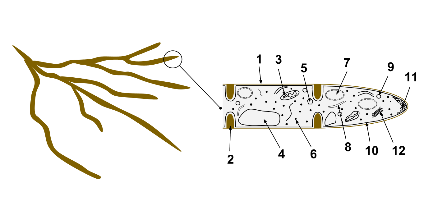
A fungus ( : fungi or funguses) is any member of the group of eukaryotic organisms that includes microorganisms such as yeasts and molds, as well as the more familiar mushrooms. These organisms are classified as a kingdom, separately from the other eukaryotic kingdoms, which by one traditional classification include Plantae, Animalia, Protozoa, and Chromista. A characteristic that places fungi in a different kingdom from plants, bacteria, and some protists is chitin in their cell walls. Fungi, like animals, are heterotrophs; they acquire their food by absorbing dissolved molecules, typically by secreting digestive enzymes into their environment. Fungi do not photosynthesize. Growth is their means of mobility, except for spores (a few of which are flagellated), which may travel through the air or water. Fungi are the principal decomposers in ecological systems. These and other differences place fungi in a single group of related organisms, named the ''Eumycota'' (''true f ... [...More Info...] [...Related Items...] OR: [Wikipedia] [Google] [Baidu] |
Paraphyses
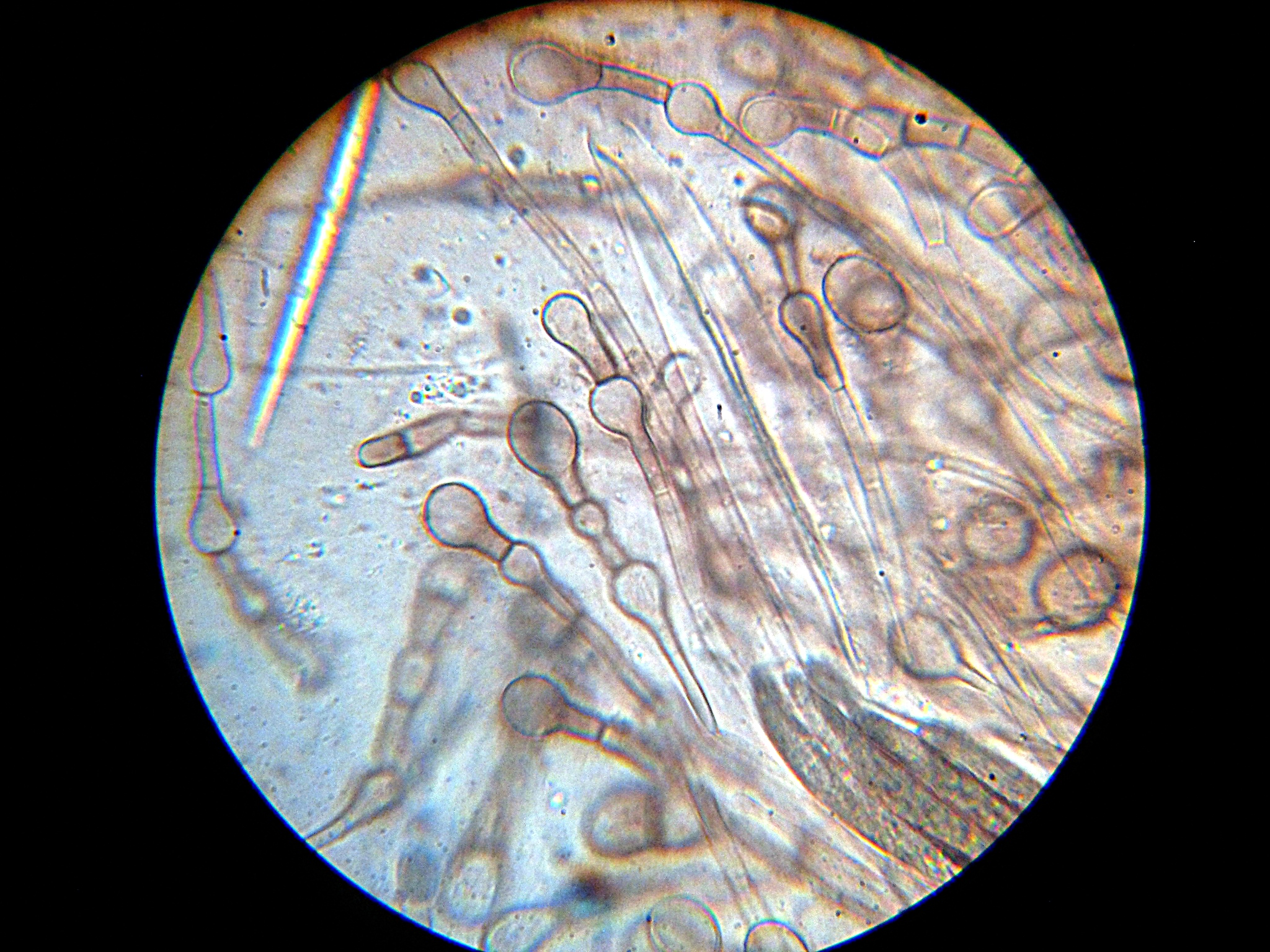
Paraphyses are erect sterile filament-like support structures occurring among the reproductive apparatuses of fungi, ferns, bryophytes and some thallophytes. The singular form of the word is paraphysis. In certain fungi, they are part of the fertile spore-bearing layer. More specifically, paraphyses are sterile filamentous hyphal end cells composing part of the hymenium of Ascomycota and Basidiomycota interspersed among either the asci or basidia respectively, and not sufficiently differentiated to be called cystidia A cystidium (plural cystidia) is a relatively large cell found on the sporocarp of a basidiomycete (for example, on the surface of a mushroom gill), often between clusters of basidia. Since cystidia have highly varied and distinct shapes that ar ..., which are specialized, swollen, often protruding cells. The tips of paraphyses may contain the pigments which colour the hymenium. In ferns and mosses, they are filament-like structures that are found on sporangia ... [...More Info...] [...Related Items...] OR: [Wikipedia] [Google] [Baidu] |
Massarina Ricifera
''Massarina'' is a genus of fungi in the Massarinaceae family. The widespread genus contains about 125 species. Anamorph forms of species in ''Massarina'' include '' Acrocalymma'', '' Ceratophoma'', and '' Tetraploa''. ''Massarina'' was circumscribed by Pier Andrea Saccardo in 1883. The genus name of ''Massarina'' is in honour of Giuseppe Filippo Massara (1792-1839), who was an Italian doctor and botanist, working in Sondrio. Species *''Massarina albocarnis'' *'' Massarina alni'' *''Massarina beaurivagea'' *''Massarina berchemiae'' *''Massarina bipolaris'' *''Massarina brunaudii'' *''Massarina capensis'' *''Massarina carolinensis'' *''Massarina cisti'' *''Massarina clionina'' *''Massarina constricta'' *''Massarina contraria'' *''Massarina cystophorae'' *''Massarina eburnea'' *'' Massarina grandispora'' *'' Massarina igniaria'' *'' Massarina japonica'' *'' Massarina jasminicola'' *'' Massarina lacertensis'' *'' Massarina leucadendri'' *'' Massarina magniarundinacea'' *'' Massarin ... [...More Info...] [...Related Items...] OR: [Wikipedia] [Google] [Baidu] |
Morphology (biology)
Morphology is a branch of biology dealing with the study of the form and structure of organisms and their specific structural features. This includes aspects of the outward appearance (shape, structure, colour, pattern, size), i.e. external morphology (or eidonomy), as well as the form and structure of the internal parts like bones and organs, i.e. internal morphology (or anatomy). This is in contrast to physiology, which deals primarily with function. Morphology is a branch of life science dealing with the study of gross structure of an organism or taxon and its component parts. History The etymology of the word "morphology" is from the Ancient Greek (), meaning "form", and (), meaning "word, study, research". While the concept of form in biology, opposed to function, dates back to Aristotle (see Aristotle's biology), the field of morphology was developed by Johann Wolfgang von Goethe (1790) and independently by the German anatomist and physiologist Karl Friedrich Burdach ... [...More Info...] [...Related Items...] OR: [Wikipedia] [Google] [Baidu] |
Hyaline
A hyaline substance is one with a glassy appearance. The word is derived from el, ὑάλινος, translit=hyálinos, lit=transparent, and el, ὕαλος, translit=hýalos, lit=crystal, glass, label=none. Histopathology Hyaline cartilage is named after its glassy appearance on fresh gross pathology. On light microscopy of H&E stained slides, the extracellular matrix of hyaline cartilage looks homogeneously pink, and the term "hyaline" is used to describe similarly homogeneously pink material besides the cartilage. Hyaline material is usually acellular and proteinaceous. For example, arterial hyaline is seen in aging, high blood pressure, diabetes mellitus and in association with some drugs (e.g. calcineurin inhibitors). It is bright pink with PAS staining. Ichthyology and entomology In ichthyology and entomology, ''hyaline'' denotes a colorless, transparent substance, such as unpigmented fins of fishes or clear insect wings. Resh, Vincent H. and R. T. Cardé, Eds. Encyclo ... [...More Info...] [...Related Items...] OR: [Wikipedia] [Google] [Baidu] |
Locule
A locule (plural locules) or loculus (plural loculi) (meaning "little place" in Latin) is a small cavity or compartment within an organ or part of an organism (animal, plant, or fungus). In angiosperms (flowering plants), the term ''locule'' usually refers to a chamber within an Ovary (plants), ovary (gynoecium or carpel) of the flower and fruits. Depending on the number of locules in the ovary, fruits can be classified as ''uni-locular'' (unilocular), ''bi-locular'', ''tri-locular'' or ''multi-locular''. The number of locules present in a gynoecium may be equal to or less than the number of carpels. The locules contain the ovules or seeds. The term may also refer to chambers within anthers containing pollen. In Ascomycete fungi, locules are chambers within the hymenium in which the perithecium, perithecia develop. References Plant anatomy Plant morphology Fungal morphology and anatomy {{botany-stub ... [...More Info...] [...Related Items...] OR: [Wikipedia] [Google] [Baidu] |
Ascospore
An ascus (; ) is the sexual spore-bearing cell produced in ascomycete fungi. Each ascus usually contains eight ascospores (or octad), produced by meiosis followed, in most species, by a mitotic cell division. However, asci in some genera or species can occur in numbers of one (e.g. ''Monosporascus cannonballus''), two, four, or multiples of four. In a few cases, the ascospores can bud off conidia that may fill the asci (e.g. ''Tympanis'') with hundreds of conidia, or the ascospores may fragment, e.g. some ''Cordyceps'', also filling the asci with smaller cells. Ascospores are nonmotile, usually single celled, but not infrequently may be coenocytic (lacking a septum), and in some cases coenocytic in multiple planes. Mitotic divisions within the developing spores populate each resulting cell in septate ascospores with nuclei. The term ocular chamber, or oculus, refers to the epiplasm (the portion of cytoplasm not used in ascospore formation) that is surrounded by the "bourrelet ... [...More Info...] [...Related Items...] OR: [Wikipedia] [Google] [Baidu] |
Anastomose
An anastomosis (, plural anastomoses) is a connection or opening between two things (especially cavities or passages) that are normally diverging or branching, such as between blood vessels, leaf veins, or streams. Such a connection may be normal (such as the foramen ovale in a fetus's heart) or abnormal (such as the patent foramen ovale in an adult's heart); it may be acquired (such as an arteriovenous fistula) or innate (such as the arteriovenous shunt of a metarteriole); and it may be natural (such as the aforementioned examples) or artificial (such as a surgical anastomosis). The reestablishment of an anastomosis that had become blocked is called a reanastomosis. Anastomoses that are abnormal, whether congenital or acquired, are often called fistulas. The term is used in medicine, biology, mycology, geology, and geography. Etymology Anastomosis: medical or Modern Latin, from Greek ἀναστόμωσις, anastomosis, "outlet, opening", Gr ana- "up, on, upon", stoma "mouth", ... [...More Info...] [...Related Items...] OR: [Wikipedia] [Google] [Baidu] |
Pseudoparaphyses
{{Short pages monitor ... [...More Info...] [...Related Items...] OR: [Wikipedia] [Google] [Baidu] |
Septum
In biology, a septum (Latin for ''something that encloses''; plural septa) is a wall, dividing a cavity or structure into smaller ones. A cavity or structure divided in this way may be referred to as septate. Examples Human anatomy * Interatrial septum, the wall of tissue that is a sectional part of the left and right atria of the heart * Interventricular septum, the wall separating the left and right ventricles of the heart * Lingual septum, a vertical layer of fibrous tissue that separates the halves of the tongue. *Nasal septum: the cartilage wall separating the nostrils of the nose * Alveolar septum: the thin wall which separates the alveoli from each other in the lungs * Orbital septum, a palpebral ligament in the upper and lower eyelids * Septum pellucidum or septum lucidum, a thin structure separating two fluid pockets in the brain * Uterine septum, a malformation of the uterus * Vaginal septum, a lateral or transverse partition inside the vagina * Intermuscular sep ... [...More Info...] [...Related Items...] OR: [Wikipedia] [Google] [Baidu] |
Hymenium
The hymenium is the tissue layer on the hymenophore of a fungal fruiting body where the cells develop into basidia or asci, which produce spores. In some species all of the cells of the hymenium develop into basidia or asci, while in others some cells develop into sterile cells called cystidia (basidiomycetes) or paraphyses (ascomycetes). Cystidia are often important for microscopic identification. The subhymenium consists of the supportive hyphae from which the cells of the hymenium grow, beneath which is the hymenophoral trama, the hyphae that make up the mass of the hymenophore. The position of the hymenium is traditionally the first characteristic used in the classification and identification of mushrooms. Below are some examples of the diverse types which exist among the macroscopic Basidiomycota and Ascomycota. * In agarics, the hymenium is on the vertical faces of the gills. * In boletes and polypores, it is in a spongy mass of downward-pointing tubes. * In puffballs, ... [...More Info...] [...Related Items...] OR: [Wikipedia] [Google] [Baidu] |
Ascus
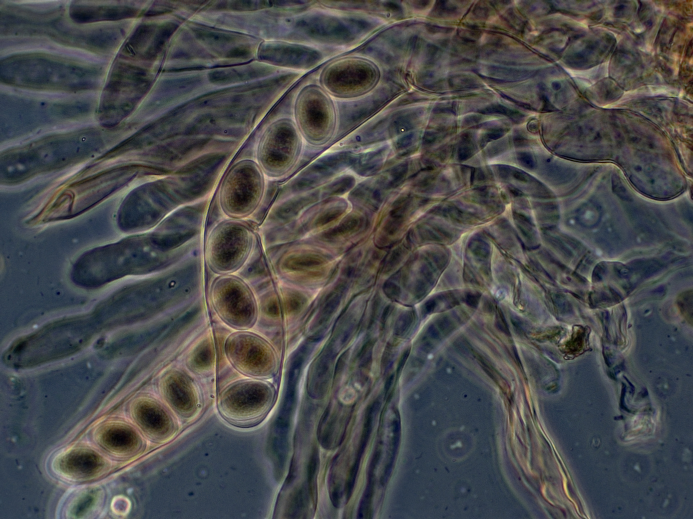
An ascus (; ) is the sexual spore-bearing cell produced in ascomycete fungi. Each ascus usually contains eight ascospores (or octad), produced by meiosis followed, in most species, by a mitotic cell division. However, asci in some genera or species can occur in numbers of one (e.g. ''Monosporascus cannonballus''), two, four, or multiples of four. In a few cases, the ascospores can bud off conidia that may fill the asci (e.g. ''Tympanis'') with hundreds of conidia, or the ascospores may fragment, e.g. some ''Cordyceps'', also filling the asci with smaller cells. Ascospores are nonmotile, usually single celled, but not infrequently may be coenocytic (lacking a septum), and in some cases coenocytic in multiple planes. Mitotic divisions within the developing spores populate each resulting cell in septate ascospores with nuclei. The term ocular chamber, or oculus, refers to the epiplasm (the portion of cytoplasm not used in ascospore formation) that is surrounded by the "bourrelet ... [...More Info...] [...Related Items...] OR: [Wikipedia] [Google] [Baidu] |


