|
Marasmius Sasicola
''Marasmius sasicola'' is a species of Marasmiaceae fungus known from Kanagawa Prefecture, Japan. First collected in 2000, it was described in 2002 by Haruki Takahashi. The species produces small mushrooms with white caps and very short, very thin black stems. Unlike in other, similar species, the stems enter the plant matter on which the mushroom grows. The six to eight white gills are spread out around the cap, and all of them reach the stem. The flesh has no taste or odour. Found in June, the species grows on dead ''Sasa'' leaves, from which it takes its specific epithet. Taxonomy and naming ''Marasmius sasicola'' was first described by Haruki Takahashi (2002) in an article in ''Mycoscience'', based on specimens collected from Ikuta Ryokuchi Park, Kawasaki, Kanagawa Prefecture, Japan in 2000 and 2001. The specific name ''sasicola'' refers to the fact the species grows upon the leaves of ''Sasa'' species. The Japanese common name for the species is ''Sasa-no-houraitake ... [...More Info...] [...Related Items...] OR: [Wikipedia] [Google] [Baidu] |
MycoBank
MycoBank is an online database, documenting new mycological names and combinations, eventually combined with descriptions and illustrations. It is run by the Westerdijk Fungal Biodiversity Institute in Utrecht. Each novelty, after being screened by nomenclatural experts and found in accordance with the ICN ( International Code of Nomenclature for algae, fungi, and plants), is allocated a unique MycoBank number before the new name has been validly published. This number then can be cited by the naming author in the publication where the new name is being introduced. Only then, this unique number becomes public in the database. By doing so, this system can help solve the problem of knowing which names have been validly published and in which year. MycoBank is linked to other important mycological databases such as ''Index Fungorum'', Life Science Identifiers, Global Biodiversity Information Facility (GBIF) and other databases. MycoBank is one of three nomenclatural repositories r ... [...More Info...] [...Related Items...] OR: [Wikipedia] [Google] [Baidu] |
Mycelial Cord
Mycelial cords are linear aggregations of parallel-oriented hypha, hyphae. The mature cords are composed of wide, empty vessel hyphae surrounded by narrower sheathing hyphae. Cords may look similar to plant roots, and also frequently have similar functions; hence they are also called rhizomorphs (literally, "root-forms"). As well as growing underground or on the surface of trees and other plants, some fungi make mycelial cords which hang in the air from vegetation. Mycelial cords are capable of conducting nutrients over long distances. For instance, they can transfer nutrients to a developing fruiting body, or enable wood-rotting fungi to grow through soil from an established food base in search of new food sources. For parasitic fungi, they can help spread infection by growing from established clusters to uninfected parts. The cords of some wood-rotting fungi (like ''Serpula lacrymans'') may be capable of penetrating masonry. The mechanism of the cord formation is not yet precise ... [...More Info...] [...Related Items...] OR: [Wikipedia] [Google] [Baidu] |
Melzer's Reagent
Melzer's reagent (also known as Melzer's iodine reagent, Melzer's solution or informally as Melzer's) is a chemical reagent used by mycologists to assist with the identification of fungi, and by phytopathologists for fungi that are plant pathogens. Composition Melzer's reagent is an aqueous solution of chloral hydrate, potassium iodide, and iodine. Depending on the formulation, it consists of approximately 2.50-3.75% potassium iodide and 0.75–1.25% iodine, with the remainder of the solution being 50% water and 50% chloral hydrate. Melzer's is toxic to humans if ingested due to the presence of iodine and chloral hydrate. Due to the legal status of chloral hydrate, Melzer's reagent is difficult to obtain in the United States. In response to difficulties obtaining chloral hydrate, scientists at Rutgers formulated Visikol (compatible with Lugol's iodine) as a replacement. In 2019, research showed that Visikol behaves differently to Melzer’s reagent in several key situations, notin ... [...More Info...] [...Related Items...] OR: [Wikipedia] [Google] [Baidu] |
Hyphae
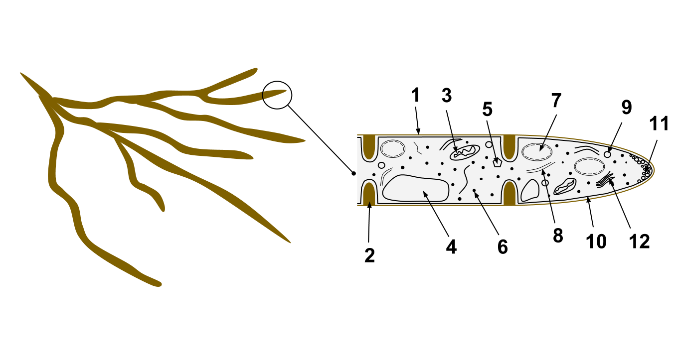
A hypha (; ) is a long, branching, filamentous structure of a fungus, oomycete, or actinobacterium. In most fungi, hyphae are the main mode of vegetative growth, and are collectively called a mycelium. Structure A hypha consists of one or more cells surrounded by a tubular cell wall. In most fungi, hyphae are divided into cells by internal cross-walls called "septa" (singular septum). Septa are usually perforated by pores large enough for ribosomes, mitochondria, and sometimes nuclei to flow between cells. The major structural polymer in fungal cell walls is typically chitin, in contrast to plants and oomycetes that have cellulosic cell walls. Some fungi have aseptate hyphae, meaning their hyphae are not partitioned by septa. Hyphae have an average diameter of 4–6 µm. Growth Hyphae grow at their tips. During tip growth, cell walls are extended by the external assembly and polymerization of cell wall components, and the internal production of new cell membrane. ... [...More Info...] [...Related Items...] OR: [Wikipedia] [Google] [Baidu] |
Clamp Connection
A clamp connection is a hook-like structure formed by growing hyphal cells of certain fungi. It is a characteristic feature of Basidiomycetes fungi. It is created to ensure that each cell, or segment of hypha separated by septa (cross walls), receives a set of differing nuclei, which are obtained through mating of hyphae of differing sexual types. It is used to maintain genetic variation within the hypha much like the mechanisms found in crozier (hook) during sexual reproduction. Formation Clamp connections are formed by the terminal hypha during elongation. Before the clamp connection is formed this terminal segment contains two nuclei. Once the terminal segment is long enough it begins to form the clamp connection. At the same time, each nucleus undergoes mitotic division to produce two daughter nuclei. As the clamp continues to develop it uptakes one of the daughter (green circle) nuclei and separates it from its sister nucleus. While this is occurring the remaining nuclei ... [...More Info...] [...Related Items...] OR: [Wikipedia] [Google] [Baidu] |
Hymenium
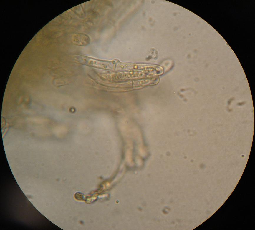
The hymenium is the tissue layer on the hymenophore of a fungal fruiting body where the cells develop into basidia or asci, which produce spores. In some species all of the cells of the hymenium develop into basidia or asci, while in others some cells develop into sterile cells called cystidia (basidiomycetes) or paraphyses (ascomycetes). Cystidia are often important for microscopic identification. The subhymenium consists of the supportive hyphae from which the cells of the hymenium grow, beneath which is the hymenophoral trama, the hyphae that make up the mass of the hymenophore. The position of the hymenium is traditionally the first characteristic used in the classification and identification of mushrooms. Below are some examples of the diverse types which exist among the macroscopic Basidiomycota and Ascomycota. * In agarics, the hymenium is on the vertical faces of the gills. * In boletes and polypores, it is in a spongy mass of downward-pointing tubes. * In puffballs, ... [...More Info...] [...Related Items...] OR: [Wikipedia] [Google] [Baidu] |
Pileipellis
The pileipellis is the uppermost layer of hyphae in the pileus of a fungal fruit body In botany, a fruit is the seed-bearing structure in flowering plants that is formed from the ovary after flowering. Fruits are the means by which flowering plants (also known as angiosperms) disseminate their seeds. Edible fruits in particu .... It covers the trama, the fleshy tissue of the fruit body. The pileipellis is more or less synonymous with the cuticle, but the cuticle generally describes this layer as a macroscopic feature, while pileipellis refers to this structure as a microscopic layer. Pileipellis type is an important character in the identification of fungi. Pileipellis types include the cutis, trichoderm, epithelium, and hymeniderm types. Types Cutis A cutis is a type of pileipellis characterized by hyphae that are repent, that is, that run parallel to the pileus surface. In an ixocutis, the hyphae are gelatinous. Trichoderm In a trichoderm, the outermost hyphae emer ... [...More Info...] [...Related Items...] OR: [Wikipedia] [Google] [Baidu] |
Cystidia
A cystidium (plural cystidia) is a relatively large cell found on the sporocarp of a basidiomycete (for example, on the surface of a mushroom gill), often between clusters of basidia. Since cystidia have highly varied and distinct shapes that are often unique to a particular species or genus, they are a useful micromorphological characteristic in the identification of basidiomycetes. In general, the adaptive significance of cystidia is not well understood. Classification of cystidia By position Cystidia may occur on the edge of a lamella (or analogous hymenophoral structure) (cheilocystidia), on the face of a lamella (pleurocystidia), on the surface of the cap (dermatocystidia or pileocystidia), on the margin of the cap (circumcystidia) or on the stipe (caulocystidia). Especially the pleurocystidia and cheilocystidia are important for identification within many genera. Sometimes the cheilocystidia give the gill edge a distinct colour which is visible to the naked eye or wit ... [...More Info...] [...Related Items...] OR: [Wikipedia] [Google] [Baidu] |
Basidia
A basidium () is a microscopic sporangium (a spore-producing structure) found on the hymenophore of fruiting bodies of basidiomycete fungi which are also called tertiary mycelium, developed from secondary mycelium. Tertiary mycelium is highly-coiled secondary myceliuma dikaryon. The presence of basidia is one of the main characteristic features of the Basidiomycota. A basidium usually bears four sexual spores called basidiospores; occasionally the number may be two or even eight. In a typical basidium, each basidiospore is borne at the tip of a narrow prong or horn called a sterigma (), and is forcibly discharged upon maturity. The word ''basidium'' literally means "little pedestal", from the way in which the basidium supports the spores. However, some biologists suggest that the structure more closely resembles a club. An immature basidium is known as a basidiole. Structure Most basidiomycota have single celled basidia (holobasidia), but in some groups basidia can be multice ... [...More Info...] [...Related Items...] OR: [Wikipedia] [Google] [Baidu] |
Cell Wall
A cell wall is a structural layer surrounding some types of cells, just outside the cell membrane. It can be tough, flexible, and sometimes rigid. It provides the cell with both structural support and protection, and also acts as a filtering mechanism. Cell walls are absent in many eukaryotes, including animals, but are present in some other ones like fungi, algae and plants, and in most prokaryotes (except mollicute bacteria). A major function is to act as pressure vessels, preventing over-expansion of the cell when water enters. The composition of cell walls varies between taxonomic group and species and may depend on cell type and developmental stage. The primary cell wall of land plants is composed of the polysaccharides cellulose, hemicelluloses and pectin. Often, other polymers such as lignin, suberin or cutin are anchored to or embedded in plant cell walls. Algae possess cell walls made of glycoproteins and polysaccharides such as carrageenan and agar that are absent ... [...More Info...] [...Related Items...] OR: [Wikipedia] [Google] [Baidu] |
Amyloid (mycology)
In mycology a tissue or feature is said to be amyloid if it has a positive amyloid reaction when subjected to a crude chemical test using iodine as an ingredient of either Melzer's reagent or Lugol's solution, producing a blue to blue-black staining. The term "amyloid" is derived from the Latin ''amyloideus'' ("starch-like"). It refers to the fact that starch gives a similar reaction, also called an amyloid reaction. The test can be on microscopic features, such as spore walls or hyphal walls, or the apical apparatus or entire ascus wall of an ascus, or be a macroscopic reaction on tissue where a drop of the reagent is applied. Negative reactions, called inamyloid or nonamyloid, are for structures that remain pale yellow-brown or clear. A reaction producing a deep reddish to reddish-brown staining is either termed a dextrinoid reaction (pseudoamyloid is a synonym) or a hemiamyloid reaction. Melzer's reagent reactions Hemiamyloidity Hemiamyloidity in mycology refers to a special ... [...More Info...] [...Related Items...] OR: [Wikipedia] [Google] [Baidu] |


