|
Mammillothalamic Tract
The mammillothalamic tract (mammillothalamic fasciculus, thalamomammillary fasciculus, bundle of Vicq d’Azyr) arises from cells in both the medial and lateral nuclei of the mammillary body and by fibers that are directly continued from the fornix. The mammillothalamic tract then connects the mammillary body to the dorsal tegmental nuclei, the ventral tegmental nuclei, and the anterior thalamic nuclei. Structure The mammillothalamic tract was first described by the French physician, Félix Vicq d'Azyr, from whom it takes its alternate name (bundle of Vicq d'Azyr). There, axons divide within the gray matter; the coarser branches pass into the anterior nucleus of the thalamus as the ''bundle of Vicq d’Azyr''. The finer branches pass downward as the mammillotegmental ''bundle of Gudden''. The bundle of Vicq d’Azyr spreads fan-like as it terminates in the medial dorsal nucleus. Some fibers pass through the dorsal nucleus to the angular nucleus of the thalamus. ("The term 'a ... [...More Info...] [...Related Items...] OR: [Wikipedia] [Google] [Baidu] |
Mammillary Body
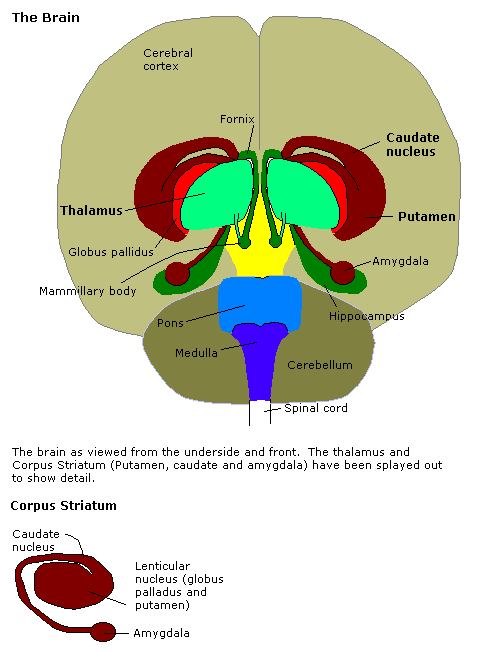
The mammillary bodies are a pair of small round bodies, located on the undersurface of the brain that, as part of the diencephalon, form part of the limbic system. They are located at the ends of the anterior arches of the fornix. They consist of two groups of nuclei, the medial mammillary nuclei and the lateral mammillary nuclei. Neuroanatomists have often categorized the mammillary bodies as part of the posterior part of hypothalamus. Structure Connections They are connected to other parts of the brain (as shown in the schematic, below left), and act as a relay for impulses coming from the amygdalae and hippocampi, via the mamillo-thalamic tract to the thalamus. Function File:Slide5dd.JPG, Mammillary body Mammillary bodies, and their projections to the anterior thalamus via the mammillothalamic tract, are important for recollective memory. The damage of medial mammillary nucleus leads to spatial memory deficit, according to observations in rats with mammillary ... [...More Info...] [...Related Items...] OR: [Wikipedia] [Google] [Baidu] |
Hippocampus
The hippocampus (via Latin from Greek , 'seahorse') is a major component of the brain of humans and other vertebrates. Humans and other mammals have two hippocampi, one in each side of the brain. The hippocampus is part of the limbic system, and plays important roles in the consolidation of information from short-term memory to long-term memory, and in spatial memory that enables navigation. The hippocampus is located in the allocortex, with neural projections into the neocortex in humans, as well as primates. The hippocampus, as the medial pallium, is a structure found in all vertebrates. In humans, it contains two main interlocking parts: the hippocampus proper (also called ''Ammon's horn''), and the dentate gyrus. In Alzheimer's disease (and other forms of dementia), the hippocampus is one of the first regions of the brain to suffer damage; short-term memory loss and disorientation are included among the early symptoms. Damage to the hippocampus can also result from ... [...More Info...] [...Related Items...] OR: [Wikipedia] [Google] [Baidu] |
Hypothalamotegmental Tract
In human neuroanatomy, the hypothalamotegmental tract is a pathway from the hypothalamus to the reticular formation. Axons from the posterior hypothalamus descend through the mesencephalic and pontine reticular formations. They connect with reticular neurons important in visceral and autonomic activity. The tract is a continuation of the medial forebrain bundle in the lateral portion of the tegmentum. It is not visible without special stains. See also *Mammillothalamic tract *Medial forebrain bundle *Midbrain reticular formation The midbrain reticular formation (MRF), also known as reticular formation of midbrain, mesencephalic reticular formation, tegmental reticular formation, and formatio reticularis (tegmenti) mesencephali, is a structure in the midbrain consisting of ... References Hypothalamus {{neuroanatomy-stub ... [...More Info...] [...Related Items...] OR: [Wikipedia] [Google] [Baidu] |
Alcoholic Korsakoff Syndrome
Korsakoff syndrome (KS) is a disorder of the central nervous system characterized by amnesia, deficits in explicit memory, and confabulation. This neurological disorder is caused by a deficiency of thiamine (vitamin B1) in the brain, and it is typically associated with and exacerbated by the prolonged, excessive ingestion of alcohol. Korsakoff syndrome is often accompanied by Wernicke encephalopathy; this combination is called Wernicke–Korsakoff syndrome. Korsakoff syndrome is named after Sergei Korsakoff, the Russian neuropsychiatrist who described it during the late 19th century. Signs and symptoms There are seven major symptoms of Korsakoff syndrome, an amnestic- confabulatory syndrome: # anterograde amnesia, memory loss for events after the onset of the syndrome # retrograde amnesia, memory loss extends back for some time before the onset of the syndrome # amnesia of fixation, also known as fixation amnesia (loss of immediate memory, a person being unable to remember even ... [...More Info...] [...Related Items...] OR: [Wikipedia] [Google] [Baidu] |
Infarction
Infarction is tissue death (necrosis) due to inadequate blood supply to the affected area. It may be caused by artery blockages, rupture, mechanical compression, or vasoconstriction. The resulting lesion is referred to as an infarct (from the Latin ''infarctus'', "stuffed into"). Causes Infarction occurs as a result of prolonged ischemia, which is the insufficient supply of oxygen and nutrition to an area of tissue due to a disruption in blood supply. The blood vessel supplying the affected area of tissue may be blocked due to an obstruction in the vessel (e.g., an arterial embolus, thrombus, or atherosclerotic plaque), compressed by something outside of the vessel causing it to narrow (e.g., tumor, volvulus, or hernia), ruptured by trauma causing a loss of blood pressure downstream of the rupture, or vasoconstricted, which is the narrowing of the blood vessel by contraction of the muscle wall rather than an external force (e.g., cocaine vasoconstriction leading ... [...More Info...] [...Related Items...] OR: [Wikipedia] [Google] [Baidu] |
Spatial Memory
In cognitive psychology and neuroscience, spatial memory is a form of memory responsible for the recording and recovery of information needed to plan a course to a location and to recall the location of an object or the occurrence of an event. Spatial memory is necessary for orientation in space. Spatial memory can also be divided into egocentric and allocentric spatial memory. A person's spatial memory is required to navigate around a familiar city. A rat's spatial memory is needed to learn the location of food at the end of a maze. In both humans and animals, spatial memories are summarized as a cognitive map. Spatial memory has representations within working, short-term memory and long-term memory. Research indicates that there are specific areas of the brain associated with spatial memory. Many methods are used for measuring spatial memory in children, adults, and animals. Short-term spatial memory Short-term memory (STM) can be described as a system allowing one to tempora ... [...More Info...] [...Related Items...] OR: [Wikipedia] [Google] [Baidu] |
Limbic System
The limbic system, also known as the paleomammalian cortex, is a set of brain structures located on both sides of the thalamus, immediately beneath the medial temporal lobe of the cerebrum primarily in the forebrain.Schacter, Daniel L. 2012. ''Psychology''.sec. 3.20 It supports a variety of functions including emotion, behavior, long-term memory, and olfaction. Emotional life is largely housed in the limbic system, and it critically aids the formation of memories. With a primordial structure, the limbic system is involved in lower order emotional processing of input from sensory systems and consists of the amygdaloid nuclear complex (amygdala), mammillary bodies, stria medullaris, central gray and dorsal and ventral nuclei of Gudden. This processed information is often relayed to a collection of structures from the telencephalon, diencephalon, and mesencephalon, including the prefrontal cortex, cingulate gyrus, limbic thalamus, hippocampus including the parahippocampal gyrus an ... [...More Info...] [...Related Items...] OR: [Wikipedia] [Google] [Baidu] |
Thalamus
The thalamus (from Greek θάλαμος, "chamber") is a large mass of gray matter located in the dorsal part of the diencephalon (a division of the forebrain). Nerve fibers project out of the thalamus to the cerebral cortex in all directions, allowing hub-like exchanges of information. It has several functions, such as the relaying of sensory signals, including motor signals to the cerebral cortex and the regulation of consciousness, sleep, and alertness. Anatomically, it is a paramedian symmetrical structure of two halves (left and right), within the vertebrate brain, situated between the cerebral cortex and the midbrain. It forms during embryonic development as the main product of the diencephalon, as first recognized by the Swiss embryologist and anatomist Wilhelm His Sr. in 1893. Anatomy The thalamus is a paired structure of gray matter located in the forebrain which is superior to the midbrain, near the center of the brain, with nerve fibers projecting out to the ... [...More Info...] [...Related Items...] OR: [Wikipedia] [Google] [Baidu] |
Amygdala
The amygdala (; plural: amygdalae or amygdalas; also '; Latin from Greek, , ', 'almond', 'tonsil') is one of two almond-shaped clusters of nuclei located deep and medially within the temporal lobes of the brain's cerebrum in complex vertebrates, including humans. Shown to perform a primary role in the processing of memory, decision making, and emotional responses (including fear, anxiety, and aggression), the amygdalae are considered part of the limbic system. The term "amygdala" was first introduced by Karl Friedrich Burdach in 1822. Structure The regions described as amygdala nuclei encompass several structures of the cerebrum with distinct connectional and functional characteristics in humans and other animals. Among these nuclei are the basolateral complex, the cortical nucleus, the medial nucleus, the central nucleus, and the intercalated cell clusters. The basolateral complex can be further subdivided into the lateral, the basal, and the accessory basal nucle ... [...More Info...] [...Related Items...] OR: [Wikipedia] [Google] [Baidu] |
Fornix Of The Brain
The fornix (from lat, fornix, lit=arch) is a C-shaped bundle of nerve fibers in the brain that acts as the major output tract of the hippocampus. The fornix also carries some afferent fibers to the hippocampus from structures in the diencephalon and basal forebrain. The fornix is part of the limbic system. While its exact function and importance in the physiology of the brain are still not entirely clear, it has been demonstrated in humans that surgical transection—the cutting of the fornix along its body—can cause memory loss. There is some debate over what type of memory is affected by this damage, but it has been found to most closely correlate with recall memory rather than recognition memory. This means that damage to the fornix can cause difficulty in recalling long-term information such as details of past events, but it has little effect on the ability to recognize objects or familiar situations. Structure The fibers begin in the hippocampus on each side of t ... [...More Info...] [...Related Items...] OR: [Wikipedia] [Google] [Baidu] |
Thalamocortical Radiations
In neuroanatomy, thalamocortical radiations are the fibers between the thalamus and the cerebral cortex. Structure Thalamocortical (TC) fibers have been referred to as one of the two constituents of the isothalamus, the other being micro neurons. Thalamocortical fibers have a bush or tree-like appearance as they extend into the internal capsule and project to the layers of the cortex. The main thalamocortical fibers extend from different nuclei of the thalamus and project to the visual cortex, somatosensory (and associated sensori-motor) cortex, and the auditory cortex in the brain. Thalamocortical radiations also innervate gustatory and olfactory pathways, as well as pre-frontal motor areas. Visual input from the optic tract is processed by the lateral geniculate nucleus of the thalamus, auditory input in the medial geniculate nucleus, and somatosensory input in the ventral posterior nucleus of the thalamus. Thalamic nuclei project to cortical areas of distinct architectural or ... [...More Info...] [...Related Items...] OR: [Wikipedia] [Google] [Baidu] |


