|
Mammillotegmental Fasciculus
The mammillotegmental fasciculus (or mammillotegmental tract, mammillo-tegmental bundle of Gudden, or ''Fasciculus mammillotegmentalis'') is a small bundle of efferent fibers from the hypothalamus running from the mammillary body to the tegmentum. Its functions are not well defined for humans, but based on animal studies it seems to be related to regulating visceral function and processing spatial information. The mammillotegmental fasciculus was first described by the German neuroanatomist, Bernhard von Gudden, from which it takes its alternate name, mammillo-tegmental bundle of Gudden. The mammillotegmental fasciculus emerges from the principal mammillary fasciculus of the mammillary body and travels dorsally together with the mammillothalamic tract before splitting off and turning caudally to enter the spinal column. There, it terminates in the tegmentum of the midbrain The midbrain or mesencephalon is the forward-most portion of the brainstem and is associated with visio ... [...More Info...] [...Related Items...] OR: [Wikipedia] [Google] [Baidu] |
Midbrain
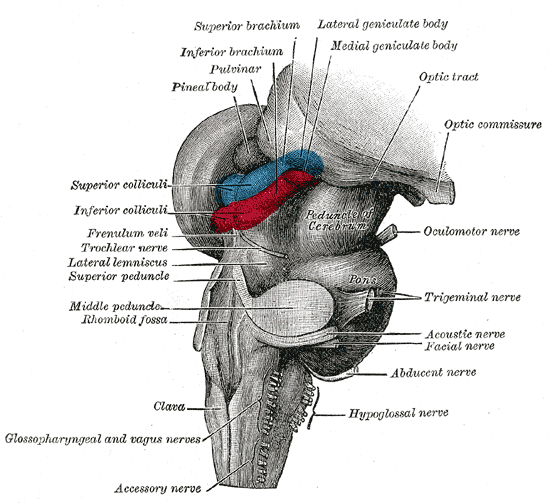
The midbrain or mesencephalon is the forward-most portion of the brainstem and is associated with vision, hearing, motor control, sleep and wakefulness, arousal (alertness), and temperature regulation. The name comes from the Greek ''mesos'', "middle", and ''enkephalos'', "brain". Structure The principal regions of the midbrain are the tectum, the cerebral aqueduct, tegmentum, and the cerebral peduncles. Rostrally the midbrain adjoins the diencephalon (thalamus, hypothalamus, etc.), while caudally it adjoins the hindbrain (pons, medulla and cerebellum). In the rostral direction, the midbrain noticeably splays laterally. Sectioning of the midbrain is usually performed axially, at one of two levels – that of the superior colliculi, or that of the inferior colliculi. One common technique for remembering the structures of the midbrain involves visualizing these cross-sections (especially at the level of the superior colliculi) as the upside-down face of a be ... [...More Info...] [...Related Items...] OR: [Wikipedia] [Google] [Baidu] |
Limbic System
The limbic system, also known as the paleomammalian cortex, is a set of brain structures located on both sides of the thalamus, immediately beneath the medial temporal lobe of the cerebrum primarily in the forebrain.Schacter, Daniel L. 2012. ''Psychology''.sec. 3.20 It supports a variety of functions including emotion, behavior, long-term memory, and olfaction. Emotional life is largely housed in the limbic system, and it critically aids the formation of memories. With a primordial structure, the limbic system is involved in lower order emotional processing of input from sensory systems and consists of the amygdaloid nuclear complex (amygdala), mammillary bodies, stria medullaris, central gray and dorsal and ventral nuclei of Gudden. This processed information is often relayed to a collection of structures from the telencephalon, diencephalon, and mesencephalon, including the prefrontal cortex, cingulate gyrus, limbic thalamus, hippocampus including the parahippocampal gyrus an ... [...More Info...] [...Related Items...] OR: [Wikipedia] [Google] [Baidu] |
Mammillary Body
The mammillary bodies are a pair of small round bodies, located on the undersurface of the brain that, as part of the diencephalon, form part of the limbic system. They are located at the ends of the anterior arches of the fornix. They consist of two groups of nuclei, the medial mammillary nuclei and the lateral mammillary nuclei. Neuroanatomists have often categorized the mammillary bodies as part of the posterior part of hypothalamus. Structure Connections They are connected to other parts of the brain (as shown in the schematic, below left), and act as a relay for impulses coming from the amygdalae and hippocampi, via the mamillo-thalamic tract to the thalamus. Function File:Slide5dd.JPG, Mammillary body Mammillary bodies, and their projections to the anterior thalamus via the mammillothalamic tract, are important for recollective memory. The damage of medial mammillary nucleus leads to spatial memory deficit, according to observations in rats with mammillary ... [...More Info...] [...Related Items...] OR: [Wikipedia] [Google] [Baidu] |
Tegmentum
The tegmentum (from Latin for "covering") is a general area within the brainstem. The tegmentum is the ventral part of the midbrain and the tectum is the dorsal part of the midbrain. It is located between the ventricular system and distinctive basal or ventral structures at each level. It forms the floor of the midbrain (mesencephalon) whereas the tectum forms the ceiling. It is a multisynaptic network of neurons that is involved in many subconscious homeostatic and reflexive pathways. It is a motor center that relays inhibitory signals to the thalamus and basal nuclei preventing unwanted body movement. The tegmentum area includes various different structures, such as the rostral end of the reticular formation, several nuclei controlling eye movements, the periaqueductal gray matter, the red nucleus, the substantia nigra, and the ventral tegmental area. The tegmentum is the location of several cranial nerve (CN) nuclei. The nuclei of CN III and IV are located in the tegmentu ... [...More Info...] [...Related Items...] OR: [Wikipedia] [Google] [Baidu] |
Bernhard Von Gudden
Johann Bernhard Aloys von Gudden (7 June 1824 – 13 June 1886) was a German neuroanatomist and psychiatrist born in Kleve. Career In 1848, von Gudden earned his doctorate from the University of Halle and became an intern at the asylum in Siegburg under Carl Wigand Maximilian Jacobi (1775–1858). From 1851 to 1855 he worked as a psychiatrist under Christian Friedrich Wilhelm Roller (1802–1878) in the mental asylum at Illenau in Baden, then from 1855 to 1869, served as director of the mental institution (''Unterfränkische Landes-Irrenanstalt'') in Werneck. In 1869 he was appointed director of the Burghölzli Hospital, as well as professor of psychiatry at the University of Zürich. In 1872 he was appointed ''Obermedicinalrath'' and director of the Upper Bavarian Kreis-Irrenanstalt (district mental asylum), located in Munich. Shortly afterwards, he became a professor of psychiatry at the University of Munich. Gudden made many contributions in the field of neuroanatomy, especi ... [...More Info...] [...Related Items...] OR: [Wikipedia] [Google] [Baidu] |
Mammillothalamic Tract
The mammillothalamic tract (mammillothalamic fasciculus, thalamomammillary fasciculus, bundle of Vicq d’Azyr) arises from cells in both the medial and lateral nuclei of the mammillary body and by fibers that are directly continued from the fornix. The mammillothalamic tract then connects the mammillary body to the dorsal tegmental nuclei, the ventral tegmental nuclei, and the anterior thalamic nuclei. Structure The mammillothalamic tract was first described by the French physician, Félix Vicq d'Azyr, from whom it takes its alternate name (bundle of Vicq d'Azyr). There, axons divide within the gray matter; the coarser branches pass into the anterior nucleus of the thalamus as the ''bundle of Vicq d’Azyr''. The finer branches pass downward as the mammillotegmental ''bundle of Gudden''. The bundle of Vicq d’Azyr spreads fan-like as it terminates in the medial dorsal nucleus. Some fibers pass through the dorsal nucleus to the angular nucleus of the thalamus. ("The term 'a ... [...More Info...] [...Related Items...] OR: [Wikipedia] [Google] [Baidu] |
Midbrain
The midbrain or mesencephalon is the forward-most portion of the brainstem and is associated with vision, hearing, motor control, sleep and wakefulness, arousal (alertness), and temperature regulation. The name comes from the Greek ''mesos'', "middle", and ''enkephalos'', "brain". Structure The principal regions of the midbrain are the tectum, the cerebral aqueduct, tegmentum, and the cerebral peduncles. Rostrally the midbrain adjoins the diencephalon (thalamus, hypothalamus, etc.), while caudally it adjoins the hindbrain (pons, medulla and cerebellum). In the rostral direction, the midbrain noticeably splays laterally. Sectioning of the midbrain is usually performed axially, at one of two levels – that of the superior colliculi, or that of the inferior colliculi. One common technique for remembering the structures of the midbrain involves visualizing these cross-sections (especially at the level of the superior colliculi) as the upside-down face of a be ... [...More Info...] [...Related Items...] OR: [Wikipedia] [Google] [Baidu] |
Ventral Tegmental Area
The ventral tegmental area (VTA) (tegmentum is Latin for ''covering''), also known as the ventral tegmental area of Tsai, or simply ventral tegmentum, is a group of neurons located close to the midline on the floor of the midbrain. The VTA is the origin of the dopaminergic cell bodies of the mesocorticolimbic dopamine system and other dopamine pathways; it is widely implicated in the drug and natural reward circuitry of the brain. The VTA plays an important role in a number of processes, including reward cognition ( motivational salience, associative learning, and positively-valenced emotions) and orgasm, among others, as well as several psychiatric disorders. Neurons in the VTA project to numerous areas of the brain, ranging from the prefrontal cortex to the caudal brainstem and several regions in between. Structure Neurobiologists have often had great difficulty distinguishing the VTA in humans and other primate brains from the substantia nigra (SN) and surrounding nucl ... [...More Info...] [...Related Items...] OR: [Wikipedia] [Google] [Baidu] |
Tegmental Pontine Reticular Nucleus
The reticulotegmental nucleus, tegmental pontine reticular nucleus (or pontine reticular nucleus of the tegmentum) is an area within the floor of the pons, in the brain stem. This area is known to affect the cerebellum with its axonal projections. These afferent connections have been proven to project not only ipsilaterally, but also to decussate and project to the contralateral side of the vermis. It has also been shown that the projections from the pontine tegmentum to the cerebellar lobes are only crossed fibers. The reticulotegmental nucleus also receives efferent axons from the cerebellum. This nucleus is known for its large number of multipolar cells and its particularly reticular structure. The reticulotegmental nucleus is topographically related to pontine nuclei (non-reticular), being just dorsal to them. The reticulotegmental nucleus has been known to mediate eye movements, otherwise known as saccadic movement. This makes sense concerning their connections, as it w ... [...More Info...] [...Related Items...] OR: [Wikipedia] [Google] [Baidu] |
Hypothalamus
The hypothalamus () is a part of the brain that contains a number of small nuclei with a variety of functions. One of the most important functions is to link the nervous system to the endocrine system via the pituitary gland. The hypothalamus is located below the thalamus and is part of the limbic system. In the terminology of neuroanatomy, it forms the ventral part of the diencephalon. All vertebrate brains contain a hypothalamus. In humans, it is the size of an almond. The hypothalamus is responsible for regulating certain metabolic processes and other activities of the autonomic nervous system. It synthesizes and secretes certain neurohormones, called releasing hormones or hypothalamic hormones, and these in turn stimulate or inhibit the secretion of hormones from the pituitary gland. The hypothalamus controls body temperature, hunger, important aspects of parenting and maternal attachment behaviours, thirst, fatigue, sleep, and circadian rhythms. Structure T ... [...More Info...] [...Related Items...] OR: [Wikipedia] [Google] [Baidu] |


