|
Mammalian Eye
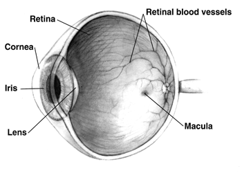
Mammals normally have a pair of eyes. Although mammalian vision is not so excellent as bird vision, it is at least dichromatic for most of mammalian species, with certain families (such as Hominidae) possessing a trichromatic color perception. The dimensions of the eyeball vary only 1–2 mm among humans. The vertical axis is 24 mm; the transverse being larger. At birth it is generally 16–17 mm, enlarging to 22.5–23 mm by three years of age. Between then and age 13 the eye attains its mature size. It weighs 7.5 grams and its volume is roughly 6.5 ml. Along a line through the nodal (central) point of the eye is the optic axis, which is slightly five degrees toward the nose from the visual axis (i.e., that going towards the focused point to the fovea). Three layers The structure of the mammalian eye has a laminar organization that can be divided into three main layers or ''tunics'' whose names reflect their basic functions: the fibrous tunic, t ... [...More Info...] [...Related Items...] OR: [Wikipedia] [Google] [Baidu] |
Plural
The plural (sometimes abbreviated pl., pl, or ), in many languages, is one of the values of the grammatical category of number. The plural of a noun typically denotes a quantity greater than the default quantity represented by that noun. This default quantity is most commonly one (a form that represents this default quantity of one is said to be of ''singular'' number). Therefore, plurals most typically denote two or more of something, although they may also denote fractional, zero or negative amounts. An example of a plural is the English word ''cats'', which corresponds to the singular ''cat''. Words of other types, such as verbs, adjectives and pronouns, also frequently have distinct plural forms, which are used in agreement with the number of their associated nouns. Some languages also have a dual (denoting exactly two of something) or other systems of number categories. However, in English and many other languages, singular and plural are the only grammatical numbers, exce ... [...More Info...] [...Related Items...] OR: [Wikipedia] [Google] [Baidu] |
Ciliary Body
The ciliary body is a part of the eye that includes the ciliary muscle, which controls the shape of the lens, and the ciliary epithelium, which produces the aqueous humor. The aqueous humor is produced in the non-pigmented portion of the ciliary body. The ciliary body is part of the uvea, the layer of tissue that delivers oxygen and nutrients to the eye tissues. The ciliary body joins the ora serrata of the choroid to the root of the iris.Cassin, B. and Solomon, S. ''Dictionary of Eye Terminology''. Gainesville, Florida: Triad Publishing Company, 1990. Structure The ciliary body is a ring-shaped thickening of tissue inside the eye that divides the posterior chamber from the vitreous body. It contains the ciliary muscle, vessels, and fibrous connective tissue. Folds on the inner ciliary epithelium are called ciliary processes, and these secrete aqueous humor into the posterior chamber. The aqueous humor then flows through the pupil into the anterior chamber. The ciliary bo ... [...More Info...] [...Related Items...] OR: [Wikipedia] [Google] [Baidu] |
Color Vision
Color vision, a feature of visual perception, is an ability to perceive differences between light composed of different wavelengths (i.e., different spectral power distributions) independently of light intensity. Color perception is a part of the larger visual system and is mediated by a complex process between neurons that begins with differential stimulation of different types of photoreceptors by light entering the eye. Those photoreceptors then emit outputs that are propagated through many layers of neurons and then ultimately to the brain. Color vision is found in many animals and is mediated by similar underlying mechanisms with common types of biological molecules and a complex history of evolution in different animal taxa. In primates, color vision may have evolved under selective pressure for a variety of visual tasks including the foraging for nutritious young leaves, ripe fruit, and flowers, as well as detecting predator camouflage and emotional states in other pr ... [...More Info...] [...Related Items...] OR: [Wikipedia] [Google] [Baidu] |
Optic Disc
The optic disc or optic nerve head is the point of exit for ganglion cell axons leaving the eye. Because there are no rods or cones overlying the optic disc, it corresponds to a small blind spot in each eye. The ganglion cell axons form the optic nerve after they leave the eye. The optic disc represents the beginning of the optic nerve and is the point where the axons of retinal ganglion cells come together. The optic disc is also the entry point for the major blood vessels that supply the retina. The optic disc in a normal human eye carries 1–1.2 million afferent nerve fibers from the eye towards the brain. Structure The optic disc is placed 3 to 4 mm to the nasal side of the fovea. It is a vertical oval, with average dimensions of 1.76mm horizontally by 1.92mm vertically. There is a central depression, of variable size, called the optic cup. This depression can be a variety of shapes from a shallow indentation to a bean pot—this shape can be significant for diagn ... [...More Info...] [...Related Items...] OR: [Wikipedia] [Google] [Baidu] |
Fovea Centralis
The fovea centralis is a small, central pit composed of closely packed cones in the eye. It is located in the center of the macula lutea of the retina. The fovea is responsible for sharp central vision (also called foveal vision), which is necessary in humans for activities for which visual detail is of primary importance, such as reading and driving. The fovea is surrounded by the ''parafovea'' belt and the ''perifovea'' outer region. The parafovea is the intermediate belt, where the ganglion cell layer is composed of more than five layers of cells, as well as the highest density of cones; the perifovea is the outermost region where the ganglion cell layer contains two to four layers of cells, and is where visual acuity is below the optimum. The perifovea contains an even more diminished density of cones, having 12 per 100 micrometres versus 50 per 100 micrometres in the most central fovea. That, in turn, is surrounded by a larger peripheral area, which delivers highly compres ... [...More Info...] [...Related Items...] OR: [Wikipedia] [Google] [Baidu] |
Cone Cell
Cone cells, or cones, are photoreceptor cells in the retinas of vertebrate eyes including the human eye. They respond differently to light of different wavelengths, and the combination of their responses is responsible for color vision. Cones function best in relatively bright light, called the photopic region, as opposed to rod cells, which work better in dim light, or the scotopic region. Cone cells are densely packed in the fovea centralis, a 0.3 mm diameter rod-free area with very thin, densely packed cones which quickly reduce in number towards the periphery of the retina. Conversely, they are absent from the optic disc, contributing to the blind spot. There are about six to seven million cones in a human eye (vs ~92 million rods), with the highest concentration being towards the macula. Cones are less sensitive to light than the rod cells in the retina (which support vision at low light levels), but allow the perception of color. They are also able to perceive ... [...More Info...] [...Related Items...] OR: [Wikipedia] [Google] [Baidu] |
Rod Cell
Rod cells are photoreceptor cells in the retina of the eye that can function in lower light better than the other type of visual photoreceptor, cone cells. Rods are usually found concentrated at the outer edges of the retina and are used in peripheral vision. On average, there are approximately 92 million rod cells (vs ~6 million cones) in the human retina. Rod cells are more sensitive than cone cells and are almost entirely responsible for night vision. However, rods have little role in color vision, which is the main reason why colors are much less apparent in dim light. Structure Rods are a little longer and leaner than cones but have the same basic structure. Opsin-containing disks lie at the end of the cell adjacent to the retinal pigment epithelium, which in turn is attached to the inside of the eye. The stacked-disc structure of the detector portion of the cell allows for very high efficiency. Rods are much more common than cones, with about 120 million rod cells compar ... [...More Info...] [...Related Items...] OR: [Wikipedia] [Google] [Baidu] |
Retina
The retina (from la, rete "net") is the innermost, light-sensitive layer of tissue of the eye of most vertebrates and some molluscs. The optics of the eye create a focused two-dimensional image of the visual world on the retina, which then processes that image within the retina and sends nerve impulses along the optic nerve to the visual cortex to create visual perception. The retina serves a function which is in many ways analogous to that of the film or image sensor in a camera. The neural retina consists of several layers of neurons interconnected by synapses and is supported by an outer layer of pigmented epithelial cells. The primary light-sensing cells in the retina are the photoreceptor cells, which are of two types: rods and cones. Rods function mainly in dim light and provide monochromatic vision. Cones function in well-lit conditions and are responsible for the perception of colour through the use of a range of opsins, as well as high-acuity vision used for task ... [...More Info...] [...Related Items...] OR: [Wikipedia] [Google] [Baidu] |
Encyclopædia Britannica 2006 Ultimate Reference Suite DVD
An encyclopedia (American English) or encyclopædia (British English) is a reference work or compendium providing summaries of knowledge either general or special to a particular field or discipline. Encyclopedias are divided into articles or entries that are arranged alphabetically by article name or by thematic categories, or else are hyperlinked and searchable. Encyclopedia entries are longer and more detailed than those in most dictionaries. Generally speaking, encyclopedia articles focus on '' factual information'' concerning the subject named in the article's title; this is unlike dictionary entries, which focus on linguistic information about words, such as their etymology, meaning, pronunciation, use, and grammatical forms.Béjoint, Henri (2000)''Modern Lexicography'', pp. 30–31. Oxford University Press. Encyclopedias have existed for around 2,000 years and have evolved considerably during that time as regards language (written in a major international or a verna ... [...More Info...] [...Related Items...] OR: [Wikipedia] [Google] [Baidu] |
Fundus (eye)
The fundus of the eye is the interior surface of the eye opposite the lens and includes the retina, optic disc, macula, fovea, and posterior pole.Cassin, B. and Solomon, S. ''Dictionary of Eye Terminology''. Gainesville, Florida: Triad Publishing Company, 1990. The fundus can be examined by ophthalmoscopy and/or fundus photography. Variation The color of the fundus varies both between and within species. In one study of primates the retina is blue, green, yellow, orange, and red; only the human fundus (from a lightly pigmented blond person) is red. The major differences noted among the "higher" primate species were size and regularity of the border of macular area, size and shape of the optic disc, apparent 'texturing' of retina, and pigmentation of retina. Clinical significance Medical signs that can be detected from observation of eye fundus (generally by funduscopy) include hemorrhages, exudates, cotton wool spots, blood vessel abnormalities (tortuosity, pulsation and n ... [...More Info...] [...Related Items...] OR: [Wikipedia] [Google] [Baidu] |
Pupil
The pupil is a black hole located in the center of the iris of the eye that allows light to strike the retina.Cassin, B. and Solomon, S. (1990) ''Dictionary of Eye Terminology''. Gainesville, Florida: Triad Publishing Company. It appears black because light rays entering the pupil are either absorbed by the tissues inside the eye directly, or absorbed after diffuse reflections within the eye that mostly miss exiting the narrow pupil. The term "pupil" was coined by Gerard of Cremona. In humans, the pupil is round, but its shape varies between species; some cats, reptiles, and foxes have vertical slit pupils, goats have horizontally oriented pupils, and some catfish have annular types. In optical terms, the anatomical pupil is the eye's aperture and the iris is the aperture stop. The image of the pupil as seen from outside the eye is the entrance pupil, which does not exactly correspond to the location and size of the physical pupil because it is magnified by the cornea. On the ... [...More Info...] [...Related Items...] OR: [Wikipedia] [Google] [Baidu] |
Cellular Respiration
Cellular respiration is the process by which biological fuels are oxidised in the presence of an inorganic electron acceptor such as oxygen to produce large amounts of energy, to drive the bulk production of ATP. Cellular respiration may be described as a set of metabolic reactions and processes that take place in the cells of organisms to convert chemical energy from nutrients into adenosine triphosphate (ATP), and then release waste products. The reactions involved in respiration are catabolic reactions, which break large molecules into smaller ones, releasing energy. Respiration is one of the key ways a cell releases chemical energy to fuel cellular activity. The overall reaction occurs in a series of biochemical steps, some of which are redox reactions. Although cellular respiration is technically a combustion reaction, it is an unusual one because of the slow, controlled release of energy from the series of reactions. Nutrients that are commonly used by animal and plan ... [...More Info...] [...Related Items...] OR: [Wikipedia] [Google] [Baidu] |


