|
Myl3
Myosin essential light chain (ELC), ventricular/cardiac isoform is a protein that in humans is encoded by the ''MYL3'' gene. This cardiac ventricular/slow skeletal ELC isoform is distinct from that expressed in fast skeletal muscle (MYL1) and cardiac atrial muscle (MYL4). Ventricular ELC is part of the myosin molecule and is important in modulating cardiac muscle contraction. Structure Cardiac, ventricular ELC is 21.9 kDa and composed of 195 amino acidsSee human MYL3 sequences features here. Cardiac ELC and the second light chain, regulatory light chain (RLC, MYL2), are non-covalently bound to IQXXXRGXXXR motifs in the 9 nm S1-S2 lever arm of the myosin head, both alpha (MYH6) and beta (MYH7) isoforms. Both light chains are members of the EF-hand superfamily of proteins, which possess helix-loop-helix motifs in two globular domains connected by an alpha-helical linker. Though EF hand motifs are specialized to bind divalent ions such as calcium, cardiac ELC does not bind calci ... [...More Info...] [...Related Items...] OR: [Wikipedia] [Google] [Baidu] |
MYL2
Myosin regulatory light chain 2, ventricular/cardiac muscle isoform (MLC-2) also known as the regulatory light chain of myosin (RLC) is a protein that in humans is encoded by the ''MYL2'' gene. This cardiac ventricular RLC isoform is distinct from that expressed in skeletal muscle ( MYLPF), smooth muscle ( MYL12B) and cardiac atrial muscle (MYL7). Ventricular myosin light chain-2 (MLC-2v) refers to the ventricular cardiac muscle form of myosin light chain 2 (Myl2). MLC-2v is a 19-KDa protein composed of 166 amino acids, that belongs to the EF-hand Ca2+ binding superfamily. MLC-2v interacts with the neck/tail region of the muscle thick filament protein myosin to regulate myosin motility and function. Structure Cardiac, ventricular RLC is an 18.8 kDa protein composed of 166 amino acids. RLC and the second ventricular light chain, essential light chain (ELC, MYL3), are non-covalently bound to IQXXXRGXXXR motifs in the 9 nm S1-S2 lever arm of the myosin head, both alpha (MYH6) ... [...More Info...] [...Related Items...] OR: [Wikipedia] [Google] [Baidu] |
MYH7
MYH7 is a gene encoding a myosin heavy chain beta (MHC-β) isoform (slow twitch) expressed primarily in the heart, but also in skeletal muscles (type I fibers). This isoform is distinct from the fast isoform of cardiac myosin heavy chain, MYH6, referred to as MHC-α. MHC-β is the major protein comprising the thick filament in cardiac muscle and plays a major role in cardiac muscle contraction. Structure MHC-β is a 223 kDa protein composed of 1935 amino acids. MHC-β is a hexameric, asymmetric motor forming the bulk of the thick filament in cardiac muscle. MHC-β is composed of N-terminal globular heads (20 nm) that project laterally, and alpha helical tails (130 nm) that dimerize and multimerize into a coiled-coil motif to form the light meromyosin (LMM), thick filament rod. The 9 nm alpha-helical neck region of each MHC-β head non-covalently binds two light chains, essential light chain (MYL3) and regulatory light chain (MYL2). Approximately 300 myosin molecu ... [...More Info...] [...Related Items...] OR: [Wikipedia] [Google] [Baidu] |
Myosin
Myosins () are a superfamily of motor proteins best known for their roles in muscle contraction and in a wide range of other motility processes in eukaryotes. They are ATP-dependent and responsible for actin-based motility. The first myosin (M2) to be discovered was in 1864 by Wilhelm Kühne. Kühne had extracted a viscous protein from skeletal muscle that he held responsible for keeping the tension state in muscle. He called this protein ''myosin''. The term has been extended to include a group of similar ATPases found in the cells of both striated muscle tissue and smooth muscle tissue. Following the discovery in 1973 of enzymes with myosin-like function in '' Acanthamoeba castellanii'', a global range of divergent myosin genes have been discovered throughout the realm of eukaryotes. Although myosin was originally thought to be restricted to muscle cells (hence '' myo-''(s) + '' -in''), there is no single "myosin"; rather it is a very large superfamily of genes whose p ... [...More Info...] [...Related Items...] OR: [Wikipedia] [Google] [Baidu] |
Hypertrophic Cardiomyopathy
Hypertrophic cardiomyopathy (HCM, or HOCM when obstructive) is a condition in which the heart becomes thickened without an obvious cause. The parts of the heart most commonly affected are the interventricular septum and the ventricles. This results in the heart being less able to pump blood effectively and also may cause electrical conduction problems. People who have HCM may have a range of symptoms. People may be asymptomatic, or may have fatigue, leg swelling, and shortness of breath. It may also result in chest pain or fainting. Symptoms may be worse when the person is dehydrated. Complications may include heart failure, an irregular heartbeat, and sudden cardiac death. HCM is most commonly inherited from a person's parents in an autosomal dominant pattern. It is often due to mutations in certain genes involved with making heart muscle proteins. Other inherited causes of left ventricular hypertrophy may include Fabry disease, Friedreich's ataxia, and certain medica ... [...More Info...] [...Related Items...] OR: [Wikipedia] [Google] [Baidu] |
Protein
Proteins are large biomolecules and macromolecules that comprise one or more long chains of amino acid residues. Proteins perform a vast array of functions within organisms, including catalysing metabolic reactions, DNA replication, responding to stimuli, providing structure to cells and organisms, and transporting molecules from one location to another. Proteins differ from one another primarily in their sequence of amino acids, which is dictated by the nucleotide sequence of their genes, and which usually results in protein folding into a specific 3D structure that determines its activity. A linear chain of amino acid residues is called a polypeptide. A protein contains at least one long polypeptide. Short polypeptides, containing less than 20–30 residues, are rarely considered to be proteins and are commonly called peptides. The individual amino acid residues are bonded together by peptide bonds and adjacent amino acid residues. The sequence of amino acid residue ... [...More Info...] [...Related Items...] OR: [Wikipedia] [Google] [Baidu] |
Gene
In biology, the word gene (from , ; "...Wilhelm Johannsen coined the word gene to describe the Mendelian units of heredity..." meaning ''generation'' or ''birth'' or ''gender'') can have several different meanings. The Mendelian gene is a basic unit of heredity and the molecular gene is a sequence of nucleotides in DNA that is transcribed to produce a functional RNA. There are two types of molecular genes: protein-coding genes and noncoding genes. During gene expression, the DNA is first copied into RNA. The RNA can be directly functional or be the intermediate template for a protein that performs a function. The transmission of genes to an organism's offspring is the basis of the inheritance of phenotypic traits. These genes make up different DNA sequences called genotypes. Genotypes along with environmental and developmental factors determine what the phenotypes will be. Most biological traits are under the influence of polygenes (many different genes) as well as gen ... [...More Info...] [...Related Items...] OR: [Wikipedia] [Google] [Baidu] |
MYL1
Myosin light chain 3, skeletal muscle isoform is a protein that in humans is encoded by the ''MYL1'' gene In biology, the word gene (from , ; "...Wilhelm Johannsen coined the word gene to describe the Mendelian units of heredity..." meaning ''generation'' or ''birth'' or ''gender'') can have several different meanings. The Mendelian gene is a ba .... Myosin is a hexameric ATPase cellular motor protein. It is composed of two heavy chains, two nonphosphorylatable alkali light chains, and two phosphorylatable regulatory light chains. This gene encodes a myosin alkali light chain expressed in fast skeletal muscle. Two transcript variants have been identified for this gene. References Further reading * * * * * * * * * * {{gene-2-stub EF-hand-containing proteins ... [...More Info...] [...Related Items...] OR: [Wikipedia] [Google] [Baidu] |
MYL4
Atrial Light Chain-1 (ALC-1), also known as Essential Light Chain, Atrial is a protein that in humans is encoded by the ''MYL4'' gene. ALC-1 is expressed in fetal cardiac ventricular and fetal skeletal muscle, as well as fetal and adult cardiac atrial tissue. ALC-1 expression is reactivated in human ventricular myocardium in various cardiac muscle diseases, including hypertrophic cardiomyopathy, dilated cardiomyopathy, ischemic cardiomyopathy and congenital heart diseases. Structure ALC-1 is a 21.6 kDa protein composed of 197 amino acids. ALC-1 is expressed in fetal cardiac ventricular and fetal skeletal muscle, as well as fetal and adult cardiac atrial tissue. ALC-1 binds the neck region of muscle myosin in adult atria. Two alternatively spliced transcript variants encoding the same protein have been found for this gene. Relative to ventricular essential light chain VLC-1, ALC-1 has an additional ~40 amino-acid N-terminal region that contains four to eleven residues ... [...More Info...] [...Related Items...] OR: [Wikipedia] [Google] [Baidu] |
Muscle Contraction
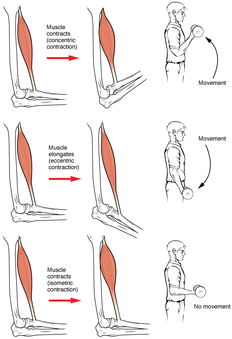
Muscle contraction is the activation of tension-generating sites within muscle cells. In physiology, muscle contraction does not necessarily mean muscle shortening because muscle tension can be produced without changes in muscle length, such as when holding something heavy in the same position. The termination of muscle contraction is followed by muscle relaxation, which is a return of the muscle fibers to their low tension-generating state. For the contractions to happen, the muscle cells must rely on the interaction of two types of filaments which are the thin and thick filaments. Thin filaments are two strands of actin coiled around each, and thick filaments consist of mostly elongated proteins called myosin. Together, these two filaments form myofibrils which are important organelles in the skeletal muscle system. Muscle contraction can also be described based on two variables: length and tension. A muscle contraction is described as isometric if the muscle tension changes ... [...More Info...] [...Related Items...] OR: [Wikipedia] [Google] [Baidu] |
MYH6
Myosin heavy chain, α isoform (MHC-α) is a protein that in humans is encoded by the ''MYH6'' gene. This isoform is distinct from the ventricular/slow myosin heavy chain isoform, MYH7, referred to as MHC-β. MHC-α isoform is expressed predominantly in human cardiac atria, exhibiting only minor expression in human cardiac ventricles. It is the major protein comprising the cardiac muscle thick filament, and functions in cardiac muscle contraction. Mutations in ''MYH6'' have been associated with late-onset hypertrophic cardiomyopathy, atrial septal defects and sick sinus syndrome. Structure MHC-α is a 224 kDa protein composed of 1939 amino acids. The ''MYH6'' gene is located on chromosome 14q12, approximately ~4kb downstream of the ''MYH7'' gene encoding the other major cardiac muscle isoform of myosin heavy chain, MHC-β. MHC-α is a hexameric, asymmetric motor forming the bulk of the thick filament in cardiac muscle; it is the predominant isoform expressed in human cardiac ... [...More Info...] [...Related Items...] OR: [Wikipedia] [Google] [Baidu] |
EF-hand
The EF hand is a helix–loop–helix structural domain or ''motif'' found in a large family of calcium-binding proteins. The EF-hand motif contains a helix–loop–helix topology, much like the spread thumb and forefinger of the human hand, in which the Ca2+ ions are coordinated by ligands within the loop. The motif takes its name from traditional nomenclature used in describing the protein parvalbumin, which contains three such motifs and is probably involved in muscle relaxation via its calcium-binding activity. The EF-hand consists of two alpha helices linked by a short loop region (usually about 12 amino acids) that usually binds calcium ions. EF-hands also appear in each structural domain of the signaling protein calmodulin and in the muscle protein troponin-C. Calcium ion binding site The calcium ion is coordinated in a pentagonal bipyramidal configuration. The six residues involved in the binding are in positions 1, 3, 5, 7, 9 and 12; these residues are denoted by ... [...More Info...] [...Related Items...] OR: [Wikipedia] [Google] [Baidu] |
Lysine
Lysine (symbol Lys or K) is an α-amino acid that is a precursor to many proteins. It contains an α-amino group (which is in the protonated form under biological conditions), an α-carboxylic acid group (which is in the deprotonated −COO− form under biological conditions), and a side chain lysyl ((CH2)4NH2), classifying it as a basic, charged (at physiological pH), aliphatic amino acid. It is encoded by the codons AAA and AAG. Like almost all other amino acids, the α-carbon is chiral and lysine may refer to either enantiomer or a racemic mixture of both. For the purpose of this article, lysine will refer to the biologically active enantiomer L-lysine, where the α-carbon is in the ''S'' configuration. The human body cannot synthesize lysine. It is essential in humans and must therefore be obtained from the diet. In organisms that synthesise lysine, two main biosynthetic pathways exist, the diaminopimelate and α-aminoadipate pathways, which employ distinct e ... [...More Info...] [...Related Items...] OR: [Wikipedia] [Google] [Baidu] |


