|
MRC (file Format)
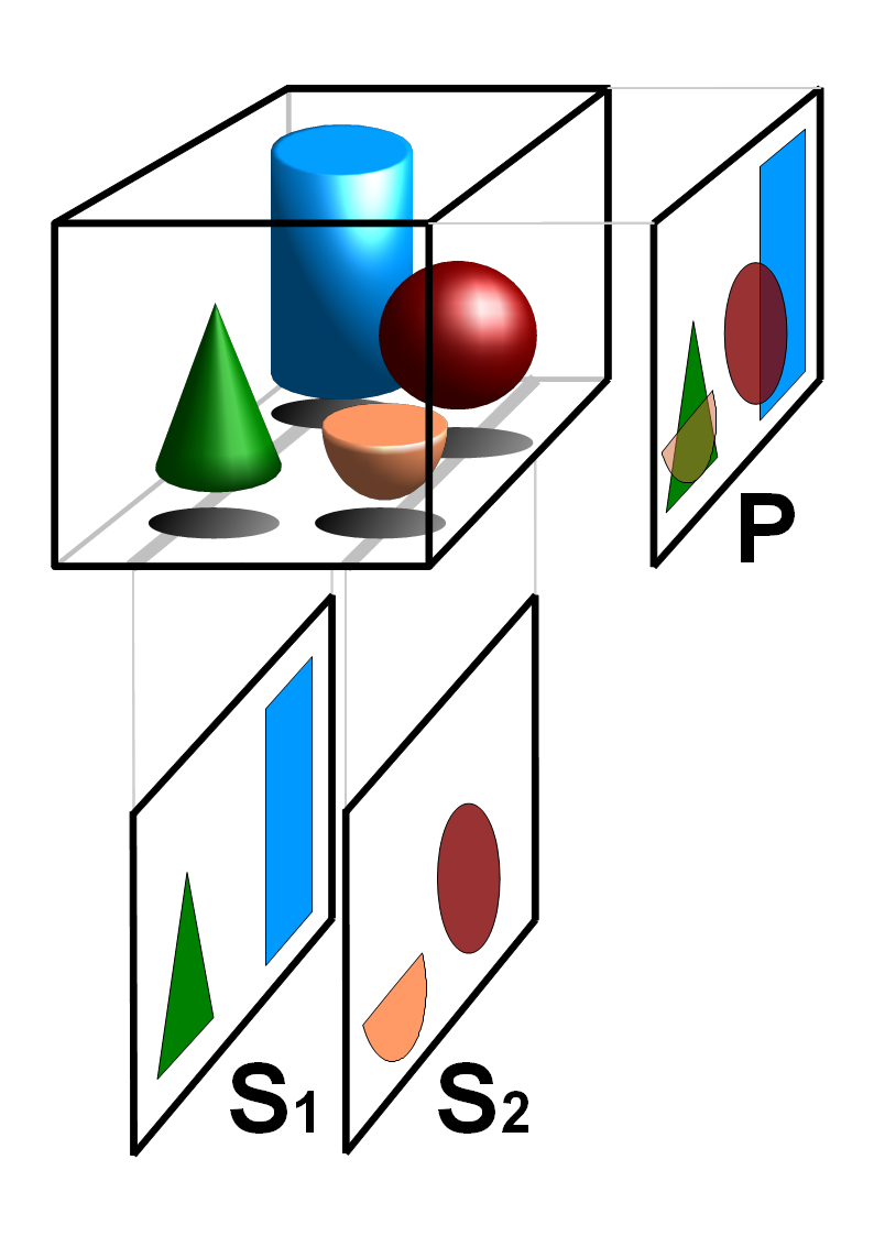
MRC is a file format that has become an industry standard in cryo-electron microscopy (cryoEM) and electron tomography (ET), where the result of the technique is a three-dimensional grid of voxels each with a value corresponding to electron density or electric potential. It was developed by the MRC ( Medical Research Council, UK) Laboratory of Molecular Biology. In 2014, the format was standardised. The format specification is available on thCCP-EM website The MRC format is supported by many of the software packages listed in b:Software Tools For Molecular Microscopy. See also *CCP4 (file format) The CCP4 file format is file generated by the Collaborative Computational Project Number 4 in 1979. The file format for electron density has become industry standard in X-ray crystallography and Cryo-electron microscopy where the result of the ... References External links MRC specification Computational chemistry Structural biology Computer file formats {{Biophy ... [...More Info...] [...Related Items...] OR: [Wikipedia] [Google] [Baidu] |
Electron Density
In quantum chemistry, electron density or electronic density is the measure of the probability of an electron being present at an infinitesimal element of space surrounding any given point. It is a scalar quantity depending upon three spatial variables and is typically denoted as either \rho(\textbf r) or n(\textbf r). The density is determined, through definition, by the normalised N-electron wavefunction which itself depends upon 4N variables (3N spatial and N spin coordinates). Conversely, the density determines the wave function modulo up to a phase factor, providing the formal foundation of density functional theory. According to quantum mechanics, due to the uncertainty principle on an atomic scale the exact location of an electron cannot be predicted, only the probability of its being at a given position; therefore electrons in atoms and molecules act as if they are "smeared out" in space. For one-electron systems, the electron density at any point is proportional to th ... [...More Info...] [...Related Items...] OR: [Wikipedia] [Google] [Baidu] |
Cryo-electron Microscopy
Cryogenic electron microscopy (cryo-EM) is a cryomicroscopy technique applied on samples cooled to cryogenic temperatures. For biological specimens, the structure is preserved by embedding in an environment of vitreous ice. An aqueous sample solution is applied to a grid-mesh and plunge-frozen in liquid ethane or a mixture of liquid ethane and propane. While development of the technique began in the 1970s, recent advances in detector technology and software algorithms have allowed for the determination of biomolecular structures at near-atomic resolution. This has attracted wide attention to the approach as an alternative to X-ray crystallography or NMR spectroscopy for macromolecular structure determination without the need for crystallization. In 2017, the Nobel Prize in Chemistry was awarded to Jacques Dubochet, Joachim Frank, and Richard Henderson "for developing cryo-electron microscopy for the high-resolution structure determination of biomolecules in solution." ''Nature M ... [...More Info...] [...Related Items...] OR: [Wikipedia] [Google] [Baidu] |
Electron Tomography
Electron tomography (ET) is a tomography technique for obtaining detailed 3D structures of sub-cellular, macro-molecular, or materials specimens. Electron tomography is an extension of traditional transmission electron microscopy and uses a transmission electron microscope to collect the data. In the process, a beam of electrons is passed through the sample at incremental degrees of rotation around the center of the target sample. This information is collected and used to assemble a three-dimensional image of the target. For biological applications, the typical resolution of ET systems are in the 5–20 nm range, suitable for examining supra-molecular multi-protein structures, although not the secondary and tertiary structure of an individual protein or polypeptide. Recently, atomic resolution in 3D electron tomography reconstructions has been demonstrated. BF-TEM and ADF-STEM tomography In the field of biology, bright-field transmission electron microscopy (BF-TEM) and high-res ... [...More Info...] [...Related Items...] OR: [Wikipedia] [Google] [Baidu] |
Voxel
In 3D computer graphics, a voxel represents a value on a regular grid in three-dimensional space. As with pixels in a 2D bitmap, voxels themselves do not typically have their position (i.e. coordinates) explicitly encoded with their values. Instead, rendering systems infer the position of a voxel based upon its position relative to other voxels (i.e., its position in the data structure that makes up a single volumetric image). In contrast to pixels and voxels, polygons are often explicitly represented by the coordinates of their vertices (as points). A direct consequence of this difference is that polygons can efficiently represent simple 3D structures with much empty or homogeneously filled space, while voxels excel at representing regularly sampled spaces that are non-homogeneously filled. Voxels are frequently used in the visualization and analysis of medical and scientific data (e.g. geographic information systems (GIS)). Some volumetric displays use voxels to describe ... [...More Info...] [...Related Items...] OR: [Wikipedia] [Google] [Baidu] |
Electric Potential
The electric potential (also called the ''electric field potential'', potential drop, the electrostatic potential) is defined as the amount of work energy needed to move a unit of electric charge from a reference point to the specific point in an electric field. More precisely, it is the energy per unit charge for a test charge that is so small that the disturbance of the field under consideration is negligible. Furthermore, the motion across the field is supposed to proceed with negligible acceleration, so as to avoid the test charge acquiring kinetic energy or producing radiation. By definition, the electric potential at the reference point is zero units. Typically, the reference point is earth or a point at infinity, although any point can be used. In classical electrostatics, the electrostatic field is a vector quantity expressed as the gradient of the electrostatic potential, which is a scalar quantity denoted by or occasionally , equal to the electric potential energy o ... [...More Info...] [...Related Items...] OR: [Wikipedia] [Google] [Baidu] |
Medical Research Council (United Kingdom)
The Medical Research Council (MRC) is responsible for co-coordinating and funding medical research in the United Kingdom. It is part of United Kingdom Research and Innovation (UKRI), which came into operation 1 April 2018, and brings together the UK's seven research councils, Innovate UK and Research England. UK Research and Innovation is answerable to, although politically independent from, the Department for Business, Energy and Industrial Strategy. The MRC focuses on high-impact research and has provided the financial support and scientific expertise behind a number of medical breakthroughs, including the development of penicillin and the discovery of the structure of DNA. Research funded by the MRC has produced 32 Nobel Prize winners to date. History The MRC was founded as the Medical Research Committee and Advisory Council in 1913, with its prime role being the distribution of medical research funds under the terms of the National Insurance Act 1911. This was a consequen ... [...More Info...] [...Related Items...] OR: [Wikipedia] [Google] [Baidu] |
MRC Laboratory Of Molecular Biology
The Medical Research Council (MRC) Laboratory of Molecular Biology (LMB) is a research institute in Cambridge, England, involved in the revolution in molecular biology which occurred in the 1950–60s. Since then it has remained a major medical research laboratory at the forefront of scientific discovery, dedicated to improving the understanding of key biological processes at atomic, molecular and cellular levels using multidisciplinary methods, with a focus on using this knowledge to address key issues in human health. A new replacement building constructed close by to the original site on the Cambridge Biomedical Campus was opened by Her Majesty the Queen in May 2013. The road outside the new building is named Francis Crick Avenue after the 1962 joint Nobel Prize winner and LMB alumnus, who co-discovered the helical structure of DNA in 1953. History Origins: 1947-61 Max Perutz, following undergraduate training in organic chemistry, left Austria in 1936 and came to the Univers ... [...More Info...] [...Related Items...] OR: [Wikipedia] [Google] [Baidu] |
Software Tools For Molecular Microscopy
Transmission electron cryomicroscopy (CryoTEM), commonly known as cryo-EM, is a form of cryogenic electron microscopy, more specifically a type of transmission electron microscopy (TEM) where the sample is studied at cryogenic temperatures (generally liquid-nitrogen temperatures). Cryo-EM is gaining popularity in structural biology. The utility of transmission electron cryomicroscopy stems from the fact that it allows the observation of specimens that have not been stained or fixed in any way, showing them in their native environment. This is in contrast to X-ray crystallography, which requires crystallizing the specimen, which can be difficult, and placing them in non-physiological environments, which can occasionally lead to functionally irrelevant conformational changes. Advances in electron detector technology, particularly DED (Direct Electron Detectors) as well as more powerful software imaging algorithms have allowed for the determination of macromolecular structures at n ... [...More Info...] [...Related Items...] OR: [Wikipedia] [Google] [Baidu] |
CCP4 (file Format)
The CCP4 file format is file generated by the Collaborative Computational Project Number 4 in 1979. The file format for electron density has become industry standard in X-ray crystallography and Cryo-electron microscopy where the result of the technique is a three-dimensional grid of voxels each with a value corresponding to density of electrons (see wave function) The CCP4 format is supported by almost every molecular graphics suite that supports volumetric data. The major packages include: *Visual molecular dynamics *PyMOL *UCSF Chimera *Bsoft *Coot *MOE See also *MTZ (file format) *MRC (file format) * EZD (file format) *Chemical file format **Protein Data Bank (file format) *Voxel In 3D computer graphics, a voxel represents a value on a regular grid in three-dimensional space. As with pixels in a 2D bitmap, voxels themselves do not typically have their position (i.e. coordinates) explicitly encoded with their values. Ins ... - one way of presenting 3D densities Externa ... [...More Info...] [...Related Items...] OR: [Wikipedia] [Google] [Baidu] |
Computational Chemistry
Computational chemistry is a branch of chemistry that uses computer simulation to assist in solving chemical problems. It uses methods of theoretical chemistry, incorporated into computer programs, to calculate the structures and properties of molecules, groups of molecules, and solids. It is essential because, apart from relatively recent results concerning the hydrogen molecular ion (dihydrogen cation, see references therein for more details), the quantum many-body problem cannot be solved analytically, much less in closed form. While computational results normally complement the information obtained by chemical experiments, it can in some cases predict hitherto unobserved chemical phenomena. It is widely used in the design of new drugs and materials. Examples of such properties are structure (i.e., the expected positions of the constituent atoms), absolute and relative (interaction) energies, electronic charge density distributions, dipoles and higher multipole moments, vi ... [...More Info...] [...Related Items...] OR: [Wikipedia] [Google] [Baidu] |
Structural Biology
Structural biology is a field that is many centuries old which, and as defined by the Journal of Structural Biology, deals with structural analysis of living material (formed, composed of, and/or maintained and refined by living cells) at every level of organization. Early structural biologists throughout the 19th and early 20th centuries were primarily only able to study structures to the limit of the naked eye's visual acuity and through magnifying glasses and light microscopes. In the 20th century, a variety of experimental techniques were developed to examine the 3D structures of biological molecules. The most prominent techniques are X-ray crystallography, nuclear magnetic resonance, and electron microscopy. Through the discovery of X-rays and its applications to protein crystals, structural biology was revolutionized, as now scientists could obtain the three-dimensional structures of biological molecules in atomic detail. Likewise, NMR spectroscopy allowed information about p ... [...More Info...] [...Related Items...] OR: [Wikipedia] [Google] [Baidu] |


