|
Low Pressure Receptor Zones
Low pressure baroreceptors are baroreceptors that relay information derived from blood pressure within the autonomic nervous system. They are stimulated by stretching of the vessel wall. They are located in large systemic veins and in the walls of the atria of the heart, and pulmonary vasculature. Low pressure baroreceptors are also referred to as volume receptors and cardiopulmonary baroreceptors.Armstrong, Maggie, et al. ''Physiology, Baroreceptors - Statpearls - NCBI Bookshelf''. 9 Mar. 2022, https://www.ncbi.nlm.nih.gov/books/NBK538172/. Structure There are two types of cardiopulmonary baroreceptors. Type A receptors and Type B receptors are both within the atria of the heart. Type A receptors are activated by wall tension, which develops by atrial contraction during ventricular diastole. Type B receptors are activated by wall stretch, which develops by atrial filling during ventricular systole. In the right atrium, the stretch receptors occur at the junction of the ve ... [...More Info...] [...Related Items...] OR: [Wikipedia] [Google] [Baidu] |
Baroreceptor
Baroreceptors (or archaically, pressoreceptors) are sensors located in the carotid sinus (at the bifurcation of external and internal carotids) and in the aortic arch. They sense the blood pressure and relay the information to the brain, so that a proper blood pressure can be maintained. Baroreceptors are a type of mechanoreceptor sensory neuron that are excited by a stretch of the blood vessel. Thus, increases in the pressure of blood vessel triggers increased action potential generation rates and provides information to the central nervous system. This sensory information is used primarily in autonomic reflexes that in turn influence the heart cardiac output and vascular smooth muscle to influence vascular resistance. Baroreceptors act immediately as part of a negative feedback system called the baroreflex, as soon as there is a change from the usual mean arterial blood pressure, returning the pressure toward a normal level. These reflexes help regulate short-term blood pressure ... [...More Info...] [...Related Items...] OR: [Wikipedia] [Google] [Baidu] |
Kidney
The kidneys are two reddish-brown bean-shaped organs found in vertebrates. They are located on the left and right in the retroperitoneal space, and in adult humans are about in length. They receive blood from the paired renal arteries; blood exits into the paired renal veins. Each kidney is attached to a ureter, a tube that carries excreted urine to the bladder. The kidney participates in the control of the volume of various body fluids, fluid osmolality, acid–base balance, various electrolyte concentrations, and removal of toxins. Filtration occurs in the glomerulus: one-fifth of the blood volume that enters the kidneys is filtered. Examples of substances reabsorbed are solute-free water, sodium, bicarbonate, glucose, and amino acids. Examples of substances secreted are hydrogen, ammonium, potassium and uric acid. The nephron is the structural and functional unit of the kidney. Each adult human kidney contains around 1 million nephrons, while a mouse kidney contains on ... [...More Info...] [...Related Items...] OR: [Wikipedia] [Google] [Baidu] |
High Pressure Receptors
High pressure receptors are the baroreceptors found within the aortic arch and carotid sinus. They are only sensitive to blood pressures above 60 mmHg. When these receptors are activated they elicit a depressor response; which decreases the heart rate and causes a general vasodilation. An increase in arterial blood pressure reflexively elicits an increase in vagal neuronal activity to the heart (i.e. the resulting decreased heart rate). The afferent nerves from the baroreceptors are called buffer nerves. See also * Low pressure receptors * Bainbridge reflex The Bainbridge reflex or Bainbridge effect, also called the atrial reflex, is an increase in heart rate due to an increase in central venous pressure. Increased blood volume is detected by stretch receptors (Cardiac Receptors) located in both sides ... References *"Principles of medical physiology" by A Fonyo page 577 {{neuroanatomy-stub Sensory receptors ... [...More Info...] [...Related Items...] OR: [Wikipedia] [Google] [Baidu] |
Bainbridge Reflex
The Bainbridge reflex or Bainbridge effect, also called the atrial reflex, is an increase in heart rate due to an increase in central venous pressure. Increased blood volume is detected by stretch receptors (Cardiac Receptors) located in both sides of atria at the venoatrial junctions. History Francis Arthur Bainbridge described this as a reflex in 1918 when he was experimenting on dogs. Bainbridge found that infusing blood or saline into the animal increased heart rate. This phenomenon occurred even if arterial blood pressure did not increase. He further observed that heart rate increased when venous pressure rose high enough to distend the right atrium, but denervation of the vagi to the heart eliminated these effects. Subsequent work demonstrated that a stretch-induced increase in cardiac beating rate can also be observed in isolated hearts or even the fully separated SAN. Thus, the positive chronotropic (from Χρόνος, Greek for 'time', and τρέπειν, Greek for 'to ben ... [...More Info...] [...Related Items...] OR: [Wikipedia] [Google] [Baidu] |
Vasopressin
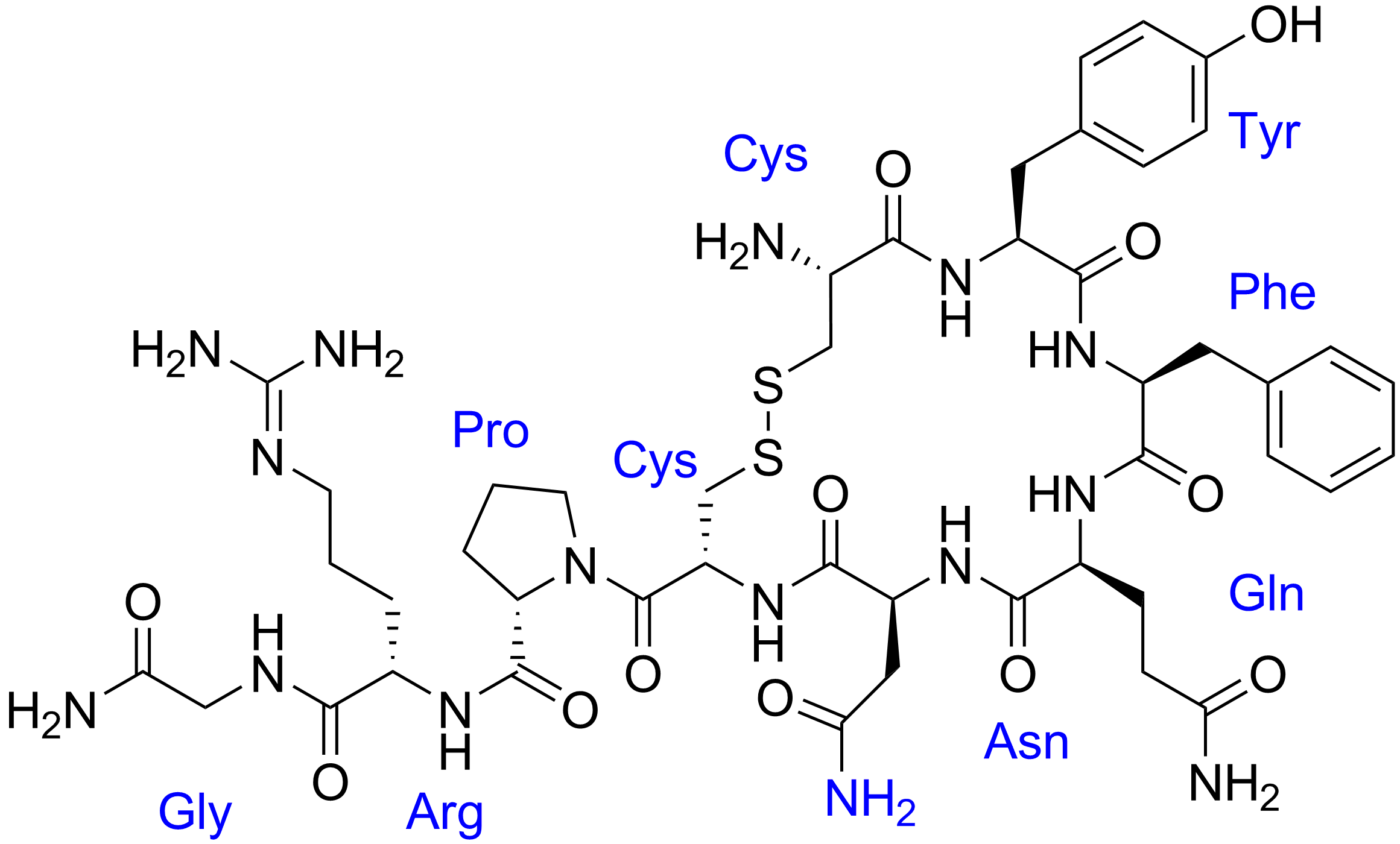
Human vasopressin, also called antidiuretic hormone (ADH), arginine vasopressin (AVP) or argipressin, is a hormone synthesized from the AVP gene as a peptide prohormone in neurons in the hypothalamus, and is converted to AVP. It then travels down the axon terminating in the posterior pituitary, and is released from vesicles into the circulation in response to extracellular fluid hypertonicity (hyperosmolality). AVP has two primary functions. First, it increases the amount of solute-free water reabsorbed back into the circulation from the filtrate in the kidney tubules of the nephrons. Second, AVP constricts arterioles, which increases peripheral vascular resistance and raises arterial blood pressure. A third function is possible. Some AVP may be released directly into the brain from the hypothalamus, and may play an important role in social behavior, sexual motivation and pair bonding, and maternal responses to stress. Vasopressin induces differentiation of stem cells in ... [...More Info...] [...Related Items...] OR: [Wikipedia] [Google] [Baidu] |
Aldosterone
Aldosterone is the main mineralocorticoid steroid hormone produced by the zona glomerulosa of the adrenal cortex in the adrenal gland. It is essential for sodium conservation in the kidney, salivary glands, sweat glands, and colon. It plays a central role in the homeostatic regulation of blood pressure, plasma sodium (Na+), and potassium (K+) levels. It does so primarily by acting on the mineralocorticoid receptors in the distal tubules and collecting ducts of the nephron. It influences the reabsorption of sodium and excretion of potassium (from and into the tubular fluids, respectively) of the kidney, thereby indirectly influencing water retention or loss, blood pressure, and blood volume.Marieb Human Anatomy & Physiology 9th edition, chapter:16, page:629, question number:14 When dysregulated, aldosterone is pathogenic and contributes to the development and progression of cardiovascular and kidney disease. Aldosterone has exactly the opposite function of the atrial natriure ... [...More Info...] [...Related Items...] OR: [Wikipedia] [Google] [Baidu] |
Angiotensin
Angiotensin is a peptide hormone that causes vasoconstriction and an increase in blood pressure. It is part of the renin–angiotensin system, which regulates blood pressure. Angiotensin also stimulates the release of aldosterone from the adrenal cortex to promote sodium retention by the kidneys. An oligopeptide, angiotensin is a hormone and a dipsogen. It is derived from the precursor molecule angiotensinogen, a serum globulin produced in the liver. Angiotensin was isolated in the late 1930s (first named 'angiotonin' or 'hypertensin') and subsequently characterized and synthesized by groups at the Cleveland Clinic and Ciba laboratories. Precursor and types Angiotensinogen Angiotensinogen is an α-2-globulin synthesized in the liver and is a precursor for angiotensin, but has also been indicated as having many other roles not related to angiotensin peptides. It is a member of the serpin family of proteins, leading to another name: Serpin A8, although it is not known to ... [...More Info...] [...Related Items...] OR: [Wikipedia] [Google] [Baidu] |
Sinus Node
The sinoatrial node (also known as the sinuatrial node, SA node or sinus node) is an oval shaped region of special cardiac muscle in the upper back wall of the right atrium made up of cells known as pacemaker cells. The sinus node is approximately fifteen mm long, three mm wide, and one mm thick, located directly below and to the side of the superior vena cava. These cells can produce an electrical impulse an action potential known as a cardiac action potential that travels through the electrical conduction system of the heart, causing it to contract. In a healthy heart, the SA node continuously produces action potentials, setting the rhythm of the heart (sinus rhythm), and so is known as the heart's natural pacemaker. The rate of action potentials produced (and therefore the heart rate) is influenced by the nerves that supply it. Structure The sinoatrial node is a oval-shaped structure that is approximately fifteen mm long, three mm wide, and one mm thick, located directly ... [...More Info...] [...Related Items...] OR: [Wikipedia] [Google] [Baidu] |
Medulla Oblongata
The medulla oblongata or simply medulla is a long stem-like structure which makes up the lower part of the brainstem. It is anterior and partially inferior to the cerebellum. It is a cone-shaped neuronal mass responsible for autonomic (involuntary) functions, ranging from vomiting to sneezing. The medulla contains the cardiac, respiratory, vomiting and vasomotor centers, and therefore deals with the autonomic functions of breathing, heart rate and blood pressure as well as the sleep–wake cycle. During embryonic development, the medulla oblongata develops from the myelencephalon. The myelencephalon is a secondary vesicle which forms during the maturation of the rhombencephalon, also referred to as the hindbrain. The bulb is an archaic term for the medulla oblongata. In modern clinical usage, the word bulbar (as in bulbar palsy) is retained for terms that relate to the medulla oblongata, particularly in reference to medical conditions. The word bulbar can refer to the nerves ... [...More Info...] [...Related Items...] OR: [Wikipedia] [Google] [Baidu] |
Vagus Nerve Stimulation
Vagus nerve stimulation (VNS) is a medical treatment that involves delivering electrical impulses to the vagus nerve. It is used as an add-on treatment for certain types of intractable epilepsy and treatment-resistant depression. Medical use VNS devices are used to treat drug-resistant epilepsy and treatment-resistant major depressive disorder (TR-MDD). In the United States, VNS is approved as adjunctive therapy for those 4 years of age or older with refractory focal onset seizures. In the European Union, VNS is approved as an adjunctive therapy for patients with either generalized or focal onset seizures without any age restrictions. It is recommended that VNS is only pursued following an adequate trial of at least 2 appropriately chosen anti-seizure medications and that the patient is ineligible for epilepsy surgery. This is because epilepsy surgery is associated with a higher probability of resulting in seizure freedom. Patients who have poor adherence or tolerance of ant ... [...More Info...] [...Related Items...] OR: [Wikipedia] [Google] [Baidu] |
Pulmonary Vein
The pulmonary veins are the veins that transfer oxygenated blood from the lungs to the heart. The largest pulmonary veins are the four ''main pulmonary veins'', two from each lung that drain into the left atrium of the heart. The pulmonary veins are part of the pulmonary circulation. Structure There are four main pulmonary veins, two from each lung – an inferior and a superior main vein, emerging from each hilum. The main pulmonary veins receive blood from three or four feeding veins in each lung, and drain into the left atrium. The peripheral feeding veins do not follow the bronchial tree. They run between the pulmonary segments from which they drain the blood. At the root of the lung, the right superior pulmonary vein lies in front of and a little below the pulmonary artery; the inferior is situated at the lowest part of the lung hilum. Behind the pulmonary artery is the bronchus. The right main pulmonary veins (contains oxygenated blood) pass behind the right atrium and ... [...More Info...] [...Related Items...] OR: [Wikipedia] [Google] [Baidu] |
Autonomic Nervous System
The autonomic nervous system (ANS), formerly referred to as the vegetative nervous system, is a division of the peripheral nervous system that supplies viscera, internal organs, smooth muscle and glands. The autonomic nervous system is a control system that acts largely unconsciously and regulates bodily functions, such as the heart rate, its force of contraction, digestion, respiratory rate, pupillary dilation, pupillary response, Micturition, urination, and sexual arousal. This system is the primary mechanism in control of the fight-or-flight response. The autonomic nervous system is regulated by integrated reflexes through the brainstem to the spinal cord and organ (anatomy), organs. Autonomic functions include control of respiration, heart rate, cardiac regulation (the cardiac control center), vasomotor activity (the vasomotor center), and certain reflex, reflex actions such as coughing, sneezing, swallowing and vomiting. Those are then subdivided into other areas and are also ... [...More Info...] [...Related Items...] OR: [Wikipedia] [Google] [Baidu] |


