|
Loeys–Dietz Syndrome
Loeys–Dietz syndrome (LDS) is an autosomal dominant genetic connective tissue disorder. It has features similar to Marfan syndrome and Ehlers–Danlos syndrome. The disorder is marked by aneurysms in the aorta, often in children, and the aorta may also undergo sudden dissection in the weakened layers of the wall of the aorta. Aneurysms and dissections also can occur in arteries other than the aorta. Because aneurysms in children tend to rupture early, children are at greater risk for dying if the syndrome is not identified. Surgery to repair aortic aneurysms is essential for treatment. There are five types of the syndrome, labelled types I through V, which are distinguished by their genetic cause. Type 1, Type 2, Type 3, Type 4 and Type 5 are caused by mutations in '' TGFBR1'', ''TGFBR2'', '' SMAD3'', ''TGFB2'', and ''TGFB3'' respectively. These five genes encoding transforming growth factors play a role in cell signaling that promotes growth and development of the body's tissu ... [...More Info...] [...Related Items...] OR: [Wikipedia] [Google] [Baidu] |
Autosomal Dominant
In genetics, dominance is the phenomenon of one variant (allele) of a gene on a chromosome masking or overriding the effect of a different variant of the same gene on the other copy of the chromosome. The first variant is termed dominant and the second recessive. This state of having two different variants of the same gene on each chromosome is originally caused by a mutation in one of the genes, either new (''de novo'') or inherited. The terms autosomal dominant or autosomal recessive are used to describe gene variants on non-sex chromosomes ( autosomes) and their associated traits, while those on sex chromosomes (allosomes) are termed X-linked dominant, X-linked recessive or Y-linked; these have an inheritance and presentation pattern that depends on the sex of both the parent and the child (see Sex linkage). Since there is only one copy of the Y chromosome, Y-linked traits cannot be dominant or recessive. Additionally, there are other forms of dominance such as incomplete d ... [...More Info...] [...Related Items...] OR: [Wikipedia] [Google] [Baidu] |
Cell Signaling
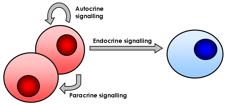
In biology, cell signaling (cell signalling in British English) or cell communication is the ability of a cell to receive, process, and transmit signals with its environment and with itself. Cell signaling is a fundamental property of all cellular life in prokaryotes and eukaryotes. Signals that originate from outside a cell (or extracellular signals) can be physical agents like mechanical pressure, voltage, temperature, light, or chemical signals (e.g., small molecules, peptides, or gas). Cell signaling can occur over short or long distances, and as a result can be classified as autocrine, juxtacrine, intracrine, paracrine, or endocrine. Signaling molecules can be synthesized from various biosynthetic pathways and released through passive or active transports, or even from cell damage. Receptors play a key role in cell signaling as they are able to detect chemical signals or physical stimuli. Receptors are generally proteins located on the cell surface or within the interio ... [...More Info...] [...Related Items...] OR: [Wikipedia] [Google] [Baidu] |
Scar
A scar (or scar tissue) is an area of fibrous tissue that replaces normal skin after an injury. Scars result from the biological process of wound repair in the skin, as well as in other organs, and tissues of the body. Thus, scarring is a natural part of the healing process. With the exception of very minor lesions, every wound (e.g., after accident, disease, or surgery) results in some degree of scarring. An exception to this are animals with complete regeneration, which regrow tissue without scar formation. Scar tissue is composed of the same protein ( collagen) as the tissue that it replaces, but the fiber composition of the protein is different; instead of a random basketweave formation of the collagen fibers found in normal tissue, in fibrosis the collagen cross-links and forms a pronounced alignment in a single direction. This collagen scar tissue alignment is usually of inferior functional quality to the normal collagen randomised alignment. For example, scars in the s ... [...More Info...] [...Related Items...] OR: [Wikipedia] [Google] [Baidu] |
Bruise
A bruise, also known as a contusion, is a type of hematoma of tissue, the most common cause being capillaries damaged by trauma, causing localized bleeding that extravasates into the surrounding interstitial tissues. Most bruises occur close enough to the epidermis such that the bleeding causes a visible discoloration. The bruise then remains visible until the blood is either absorbed by tissues or cleared by immune system action. Bruises which do not blanch under pressure can involve capillaries at the level of skin, subcutaneous tissue, muscle, or bone. Bruises are not to be confused with other similar-looking lesions. (Such lesions include petechia (less than , resulting from numerous and diverse etiologies such as adverse reactions from medications such as warfarin, straining, asphyxiation, platelet disorders and diseases such as '' cytomegalovirus''), purpura (, classified as palpable purpura or non-palpable purpura and indicates various pathologic conditions such as thr ... [...More Info...] [...Related Items...] OR: [Wikipedia] [Google] [Baidu] |
Dura Mater
In neuroanatomy, dura mater is a thick membrane made of dense irregular connective tissue that surrounds the brain and spinal cord. It is the outermost of the three layers of membrane called the meninges that protect the central nervous system. The other two meningeal layers are the arachnoid mater and the pia mater. It envelops the arachnoid mater, which is responsible for keeping in the cerebrospinal fluid. It is derived primarily from the neural crest cell population, with postnatal contributions of the paraxial mesoderm. Structure The dura mater has several functions and layers. The dura mater is a membrane that envelops the arachnoid mater. It surrounds and supports the dural sinuses (also called dural venous sinuses, cerebral sinuses, or cranial sinuses) and carries blood from the brain toward the heart. Cranial dura mater has two layers called ''lamellae'', a superficial layer (also called the periosteal layer), which serves as the skull's inner periosteum, called the ... [...More Info...] [...Related Items...] OR: [Wikipedia] [Google] [Baidu] |
Dural Ectasia
Dural ectasia is widening or ballooning of the dural sac surrounding the spinal cord. This usually occurs in the lumbosacral region, as this is where the cerebrospinal fluid pressure is greatest, but the spinal canal can be affected in any plane. Signs and symptoms Common symptoms include lower back pain, headaches, weakness (myasthenia), numbness (hypoesthesia) above and below the involved limb, leg pain, and sometimes rectal and genital pain. Bowel and bladder dysfunction, urinary retention or incontinence may occur. Moderate-to-severe cases can cause radicular pain in the legs caused by nerve root compression. The symptoms are usually exacerbated by upright posture and often, but not always, relieved by lying down. Postural headaches can be related to spontaneous spinal cerebrospinal fluid leaks. However, in many patients, dural ectasia is asymptomatic. Causes The etiology of dural ectasia is unknown, but it has been suggested that is due to increased hydrostatic pressure, ... [...More Info...] [...Related Items...] OR: [Wikipedia] [Google] [Baidu] |
Aortic Dissection
Aortic dissection (AD) occurs when an injury to the innermost layer of the aorta allows blood to flow between the layers of the aortic wall, forcing the layers apart. In most cases, this is associated with a sudden onset of severe chest or back pain, often described as "tearing" in character. Also, vomiting, sweating, and lightheadedness may occur. Other symptoms may result from decreased blood supply to other organs, such as stroke, lower extremity ischemia, or mesenteric ischemia. Aortic dissection can quickly lead to death from insufficient blood flow to the heart or complete rupture of the aorta. AD is more common in those with a history of high blood pressure; a number of connective tissue diseases that affect blood vessel wall strength including Marfan syndrome and Ehlers–Danlos syndrome; a bicuspid aortic valve; and previous heart surgery. Major trauma, smoking, cocaine use, pregnancy, a thoracic aortic aneurysm, inflammation of arteries, and abnormal lipid ... [...More Info...] [...Related Items...] OR: [Wikipedia] [Google] [Baidu] |
Aortic Aneurysm
An aortic aneurysm is an enlargement (dilatation) of the aorta to greater than 1.5 times normal size. They usually cause no symptoms except when ruptured. Occasionally, there may be abdominal, back, or leg pain. The prevalence of abdominal aortic aneurysm ("AAA") has been reported to range from 2 to 12% and is found in about 8% of men more than 65 years of age. The mortality rate attributable to AAA is about 15,000 per year in the United States and 6,000 to 8,000 per year in the United Kingdom and Ireland. Between 2001 and 2006, there were approximately 230,000 AAA surgical repairs performed on Medicare patients in the United States. The etiology remains the topic of continued investigation. Known causes include trauma, infection, and inflammatory disorders. Risk factors include cigarette smoking, advanced age, dyslipidemia, hypertension, and coronary artery disease. The pathophysiology of the disease is related to an initial arterial insult causing a cascade of inflammation and e ... [...More Info...] [...Related Items...] OR: [Wikipedia] [Google] [Baidu] |
Club Foot
Clubfoot is a birth defect where one or both feet are rotated inward and downward. Congenital clubfoot is the most common congenital malformation of the foot with an incidence of 1 per 1000 births. In approximately 50% of cases, clubfoot affects both feet, but it can present unilaterally causing one leg or foot to be shorter than the other. Most of the time, it is not associated with other problems. Without appropriate treatment, the foot deformity will persist and lead to pain and impaired ability to walk, which can have a dramatic impact on the quality of life. The exact cause is usually not identified. Both genetic and environmental factors are believed to be involved. There are two main types of congenital clubfoot: idiopathic (80% of cases) and secondary clubfoot (20% of cases). The idiopathic congenital clubfoot is a multifactorial condition that includes environmental, vascular, positional, and genetic factors. There appears to be hereditary component for this birth d ... [...More Info...] [...Related Items...] OR: [Wikipedia] [Google] [Baidu] |
Cleft Palate
A cleft lip contains an opening in the upper lip that may extend into the nose. The opening may be on one side, both sides, or in the middle. A cleft palate occurs when the palate (the roof of the mouth) contains an opening into the nose. The term orofacial cleft refers to either condition or to both occurring together. These disorders can result in feeding problems, speech problems, hearing problems, and frequent ear infections. Less than half the time the condition is associated with other disorders. Cleft lip and palate are the result of tissues of the face not joining properly during development. As such, they are a type of birth defect. The cause is unknown in most cases. Risk factors include smoking during pregnancy, diabetes, obesity, an older mother, and certain medications (such as some used to treat seizures). Cleft lip and cleft palate can often be diagnosed during pregnancy with an ultrasound exam. A cleft lip or palate can be successfully treated with surgery. ... [...More Info...] [...Related Items...] OR: [Wikipedia] [Google] [Baidu] |
Uvula
The palatine uvula, usually referred to as simply the uvula, is a conic projection from the back edge of the middle of the soft palate, composed of connective tissue containing a number of racemose glands, and some muscular fibers. It also contains many serous glands, which produce thin saliva. It is only found in human beings. Structure Muscle The muscular part of the uvula () shortens and broadens the uvula. This changes the contour of the posterior part of the soft palate. This change in contour allows the soft palate to adapt closely to the posterior pharyngeal wall to help close the nasopharynx during swallowing. Its muscles are controlled by the pharyngeal branch of the vagus nerve. Variation A bifid or bifurcated uvula is a split or cleft uvula. Newborns with cleft palate often also have a split uvula. The bifid uvula results from incomplete fusion of the palatine shelves but it is considered only a slight form of clefting. Bifid uvulas have less muscle in them th ... [...More Info...] [...Related Items...] OR: [Wikipedia] [Google] [Baidu] |
Hypertelorism
Hypertelorism is an abnormally increased distance between two organs or bodily parts, usually referring to an increased distance between the orbits (eyes), or orbital hypertelorism. In this condition the distance between the inner eye corners as well as the distance between the pupils is greater than normal. Hypertelorism should not be confused with telecanthus, in which the distance between the inner eye corners is increased but the distances between the outer eye corners and the pupils remain unchanged.Michael L. Bentz: ''Pediatric Plastic Surgery''; Chapter 9 Hypertelorism by Renato Ocampo, Jr., MD/ John A. Persing, MD Hypertelorism is a symptom in a variety of syndromes, including Edwards syndrome (trisomy 18), 1q21.1 duplication syndrome, basal cell nevus syndrome, DiGeorge syndrome and Loeys–Dietz syndrome. Hypertelorism can also be seen in Apert syndrome, Autism spectrum disorder, craniofrontonasal dysplasia, Noonan syndrome, neurofibromatosis, LEOPARD syndrome, Crouzon ... [...More Info...] [...Related Items...] OR: [Wikipedia] [Google] [Baidu] |

.jpg)


