|
List Of Palatal Expanders
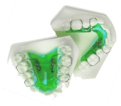
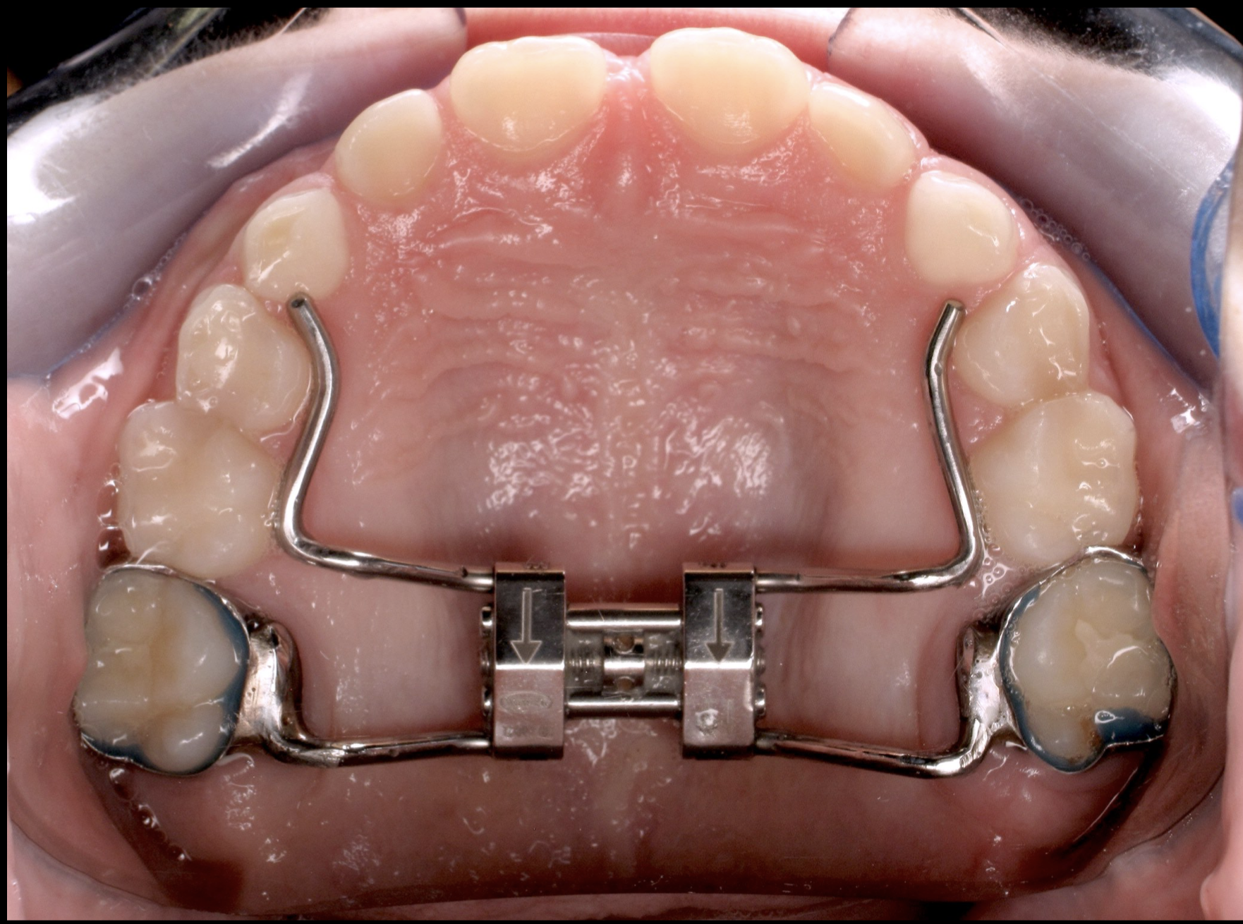
This article lists different types of expanders that are available for the process of palatal expansion in the field of orthodontics. There can be many types of transverse dimension appliances. These appliances can be used to achieve expansion in the maxillary arch; there are devices for mandibular expansion or lower expansion too. In past many years, different types of appliances have been made. These types are: tissue-borne, tooth-borne, slow maxillary expansion, rapid maxillary expansion, and bone-anchored. Types of expanders Tissue-borne expander Tissue supported expanders allow the forces to be applied directly to the tissues of palatal mucosa instead of teeth. The most common type of tissue-borne expander is known as the Haas Appliance. This appliance was popularized by Andrew Haas in 1961. This appliance involves bands placed on maxillary first premolar and first molars on each side. Haas appliance has palatal acrylic that is in contact with palatal mucosa. Inside the ac ... [...More Info...] [...Related Items...] OR: [Wikipedia] [Google] [Baidu] |
Upper And Lower Jaw Functional Expanders
Upper may refer to: * Shoe upper or ''vamp'', the part of a shoe on the top of the foot * Stimulant, drugs which induce temporary improvements in either mental or physical function or both * ''Upper'', the original film title for the 2013 found footage film ''The Upper Footage ''The Upper Footage'' (also known as ''Upper'') is a 2013 found footage film written and directed by Justin Cole. First released on January 31, 2013 to a limited run of midnight theatrical screenings at Landmark’s Sunshine Cinema in New York Cit ...'' See also {{Disambiguation ... [...More Info...] [...Related Items...] OR: [Wikipedia] [Google] [Baidu] |
Palatal Expansion
A palatal expander is a device in the field of orthodontics which is used to widen the upper jaw (maxilla) so that the bottom and upper teeth will fit together better. This is a common orthodontic procedure. Although the use of an expander is most common in children and adolescents 8-18 years of age, it can also be used in adults, although expansion is slightly more uncomfortable and takes longer. A patient who would rather not wait several months for the end result by a palatal expander may be able to opt for a surgical separation of the maxilla. Use of a palatal expander is most often followed by braces to then straighten the teeth. It is believed that expansion therapy should be started in patients either before or during their peak growth spurt. To obtain maximal skeletal changes, the therapy is typically initiated at a very early age. Expansion therapy performed after the peak growth spurt will lead to more dental changes than skeletal which leads to tipping of buccal teeth. ... [...More Info...] [...Related Items...] OR: [Wikipedia] [Google] [Baidu] |
Mandibular Expansion
In anatomy, the mandible, lower jaw or jawbone is the largest, strongest and lowest bone in the human facial skeleton. It forms the lower jaw and holds the lower tooth, teeth in place. The mandible sits beneath the maxilla. It is the only movable bone of the skull (discounting the ossicles of the middle ear). It is connected to the temporal bones by the temporomandibular joints. The bone is formed prenatal development, in the fetus from a fusion of the left and right mandibular prominences, and the point where these sides join, the mandibular symphysis, is still visible as a faint ridge in the midline. Like other symphyses in the body, this is a midline articulation where the bones are joined by fibrocartilage, but this articulation fuses together in early childhood.Illustrated Anatomy of the Head and Neck, Fehrenbach and Herring, Elsevier, 2012, p. 59 The word "mandible" derives from the Latin word ''mandibula'', "jawbone" (literally "one used for chewing"), from ''wikt:mandere ... [...More Info...] [...Related Items...] OR: [Wikipedia] [Google] [Baidu] |
Palatal Mucosa
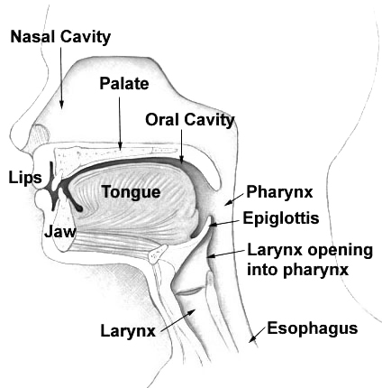
The palate () is the roof of the mouth in humans and other mammals. It separates the oral cavity from the nasal cavity. A similar structure is found in crocodilians, but in most other tetrapods, the oral and nasal cavities are not truly separated. The palate is divided into two parts, the anterior, bony hard palate and the posterior, fleshy soft palate (or velum). Structure Innervation The maxillary nerve branch of the trigeminal nerve supplies sensory innervation to the palate. Development The hard palate forms before birth. Variation If the fusion is incomplete, a cleft palate results. Function When functioning in conjunction with other parts of the mouth, the palate produces certain sounds, particularly velar, palatal, palatalized, postalveolar, alveolopalatal, and uvular consonants. History Etymology The English synonyms palate and palatum, and also the related adjective palatine (as in palatine bone), are all from the Latin ''palatum'' via Old French ''palat'' ... [...More Info...] [...Related Items...] OR: [Wikipedia] [Google] [Baidu] |
Andrew Haas
Andrew is the English form of a given name common in many countries. In the 1990s, it was among the top ten most popular names given to boys in English-speaking countries. "Andrew" is frequently shortened to "Andy" or "Drew". The word is derived from the el, Ἀνδρέας, ''Andreas'', itself related to grc, ἀνήρ/ἀνδρός ''aner/andros'', "man" (as opposed to "woman"), thus meaning "manly" and, as consequence, "brave", "strong", "courageous", and "warrior". In the King James Bible, the Greek "Ἀνδρέας" is translated as Andrew. Popularity Australia In 2000, the name Andrew was the second most popular name in Australia. In 1999, it was the 19th most common name, while in 1940, it was the 31st most common name. Andrew was the first most popular name given to boys in the Northern Territory in 2003 to 2015 and continuing. In Victoria, Andrew was the first most popular name for a boy in the 1970s. Canada Andrew was the 20th most popular name chosen for male ... [...More Info...] [...Related Items...] OR: [Wikipedia] [Google] [Baidu] |
Maxilla
The maxilla (plural: ''maxillae'' ) in vertebrates is the upper fixed (not fixed in Neopterygii) bone of the jaw formed from the fusion of two maxillary bones. In humans, the upper jaw includes the hard palate in the front of the mouth. The two maxillary bones are fused at the intermaxillary suture, forming the anterior nasal spine. This is similar to the mandible (lower jaw), which is also a fusion of two mandibular bones at the mandibular symphysis. The mandible is the movable part of the jaw. Structure In humans, the maxilla consists of: * The body of the maxilla * Four processes ** the zygomatic process ** the frontal process of maxilla ** the alveolar process ** the palatine process * three surfaces – anterior, posterior, medial * the Infraorbital foramen * the maxillary sinus * the incisive foramen Articulations Each maxilla articulates with nine bones: * two of the cranium: the frontal and ethmoid * seven of the face: the nasal, zygomatic, lacrimal, inferior n ... [...More Info...] [...Related Items...] OR: [Wikipedia] [Google] [Baidu] |
Surgically Assisted Rapid Palatal Expansion
Surgically assisted rapid palatal expansion (SARPE), also known as surgically assisted rapid maxillary expansion (SARME), is a technique in the field of orthodontics which is used to expand the maxillary arch. This technique is a combination of both Oral and Maxillofacial Surgery and Orthodontics. This procedure is primarily done in adult patients whose maxillary sutures are fused and cannot be expanded via other techniques. History Orthodontic expansion was first described by Emersen Angell about 145 years ago. Kole in 1959 was the first person to speak about the procedure of corticotomy in adults with maxillary constriction. Brown first described the surgical technique for SARPE in 1938. Steinhauser first described the technique involving the segmental left/right split of maxilla along with placement of the graft in 1972. Indications * Skeletal maturity or adult patients * Fused intermaxillary suture * Transverse maxillary hypoplasia * Bilateral posterior crossbite * Previou ... [...More Info...] [...Related Items...] OR: [Wikipedia] [Google] [Baidu] |
Pharynx
The pharynx (plural: pharynges) is the part of the throat behind the mouth and nasal cavity, and above the oesophagus and trachea (the tubes going down to the stomach and the lungs). It is found in vertebrates and invertebrates, though its structure varies across species. The pharynx carries food and air to the esophagus and larynx respectively. The flap of cartilage called the epiglottis stops food from entering the larynx. In humans, the pharynx is part of the digestive system and the conducting zone of the respiratory system. (The conducting zone—which also includes the nostrils of the nose, the larynx, trachea, bronchi, and bronchioles—filters, warms and moistens air and conducts it into the lungs). The human pharynx is conventionally divided into three sections: the nasopharynx, oropharynx, and laryngopharynx. It is also important in vocalization. In humans, two sets of pharyngeal muscles form the pharynx and determine the shape of its lumen. They are arranged as an ... [...More Info...] [...Related Items...] OR: [Wikipedia] [Google] [Baidu] |
Nasal Cavity
The nasal cavity is a large, air-filled space above and behind the nose in the middle of the face. The nasal septum divides the cavity into two cavities, also known as fossae. Each cavity is the continuation of one of the two nostrils. The nasal cavity is the uppermost part of the respiratory system and provides the nasal passage for inhaled air from the nostrils to the nasopharynx and rest of the respiratory tract. The paranasal sinuses surround and drain into the nasal cavity. Structure The term "nasal cavity" can refer to each of the two cavities of the nose, or to the two sides combined. The lateral wall of each nasal cavity mainly consists of the maxilla. However, there is a deficiency that is compensated for by the perpendicular plate of the palatine bone, the medial pterygoid plate, the labyrinth of ethmoid and the inferior concha. The paranasal sinuses are connected to the nasal cavity through small orifices called ostia. Most of these ostia communicate with the n ... [...More Info...] [...Related Items...] OR: [Wikipedia] [Google] [Baidu] |


