|
List Of Fetal Abnormalities
Fetal abnormalities are conditions that affect a fetus or embryo, are able to be diagnosed prenatally, and may be fatal or cause disease after birth. They may include aneuploidies, structural abnormalities, or neoplasms. * Acardiac twin * Achondrogenesis * Achondroplasia * Adrenal hematoma * Agenesis of the corpus callosum * Amniotic band syndrome * Anal atresia * Anencephaly * Angelman syndrome * Aqueductal stenosis * Arachnoid cyst * Arthrogryposis * Bilateral multicystic dysplastic kidneys * Camptomelic dysplasia * Cardiac rhabdomyoma * Caudal regression syndrome * Chorioangioma * Cleft palate * Club foot * Coarctation of the aorta * Conjoined twins * Cystic hygroma * Dandy–Walker malformation * Diaphragmatic hernia * Diastrophic dysplasia * Double outlet right ventricle * Duodenal atresia * Ebstein's anomaly * Ectopia cordis * Encephalocele * Endocardial cushion defect * Esophageal atresia * Exstrophy of the bladder * Fetal alcohol syndrome * First arch syndrome * Foc ... [...More Info...] [...Related Items...] OR: [Wikipedia] [Google] [Baidu] |
Fetus
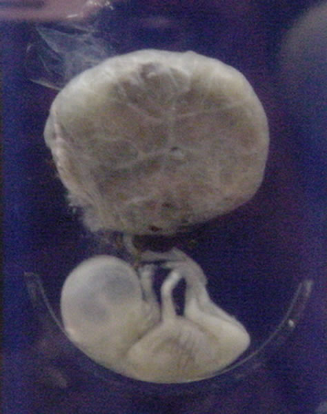
A fetus or foetus (; plural fetuses, feti, foetuses, or foeti) is the unborn offspring that develops from an animal embryo. Following embryonic development the fetal stage of development takes place. In human prenatal development, fetal development begins from the ninth week after fertilization (or eleventh week gestational age) and continues until birth. Prenatal development is a continuum, with no clear defining feature distinguishing an embryo from a fetus. However, a fetus is characterized by the presence of all the major body organs, though they will not yet be fully developed and functional and some not yet situated in their final anatomical location. Etymology The word ''fetus'' (plural ''fetuses'' or '' feti'') is related to the Latin '' fētus'' ("offspring", "bringing forth", "hatching of young") and the Greek "φυτώ" to plant. The word "fetus" was used by Ovid in Metamorphoses, book 1, line 104. The predominant British, Irish, and Commonwealth spelling is '' ... [...More Info...] [...Related Items...] OR: [Wikipedia] [Google] [Baidu] |
Cardiac Rhabdomyoma
A rhabdomyoma is a benign tumor of striated muscle. Rhabdomyomas may be either "cardiac" or "extra cardiac" (occurring outside the heart). Extracardiac forms of rhabdomyoma are sub classified into three distinct types: adult type, fetal type, and genital type. Cardiac rhabdomyomas are the most common primary tumor of the heart in infants and children. It has an association with tuberous sclerosis. In those with tuberous sclerosis, the tumor may regress and disappear completely, or remain consistent in size. A common histological feature is the presence of Spider Cells, which are cardiac myocytes with enlarged glycogen vacuoles separated by eosinophilic strands, resembling the legs of a spider. It is most commonly associated with the tongue, and heart, but can also occur in other locations, such as the vagina. Malignant skeletal muscle tumors are referred to as rhabdomyosarcoma. Only rare cases of possible malignant change have been reported in fetal rhabdomyoma. The differential ... [...More Info...] [...Related Items...] OR: [Wikipedia] [Google] [Baidu] |
Ebstein's Anomaly
Ebstein's anomaly is a congenital heart defect in which the septal and posterior leaflets of the tricuspid valve are displaced towards the apex of the right ventricle of the heart. It is classified as a critical congenital heart defect accounting for less than 1% of all congenital heart defects presenting in around per 200,000 live births. Ebstein anomaly is the congenital heart lesion most commonly associated with supraventricular tachycardia. Signs and symptoms The annulus of the valve is still in the normal position. The valve leaflets, however, are to a varying degree, attached to the walls and septum of the right ventricle. A subsequent "atrialization" of a portion of the morphologic right ventricle (which is then contiguous with the right atrium) is seen. This causes the right atrium to be large and the anatomic right ventricle to be small in size. * S3 heart sound * S4 heart sound * Triple or quadruple gallop due to widely split S1 and S2 sounds plus a loud S3 and/or S4 ... [...More Info...] [...Related Items...] OR: [Wikipedia] [Google] [Baidu] |
Duodenal Atresia
Duodenal atresia is the congenital absence or complete closure of a portion of the lumen of the duodenum. It causes increased levels of amniotic fluid during pregnancy ( polyhydramnios) and intestinal obstruction in newborn babies. Newborns present with bilious or non-bilous vomiting (depending on where in the duodenum the obstruction is) within the first 24 to 48 hours after birth, typically after their first oral feeding. Radiography shows a distended stomach and distended duodenum, which are separated by the pyloric valve, a finding described as the double-bubble sign. Treatment includes suctioning out any fluid that is trapped in the stomach, providing fluids intravenously, and surgical repair of the intestinal closure. Signs and symptoms History and physical examination During pregnancy, duodenal atresia is associated with increased amniotic fluid in the uterus, which is called polyhydramnios. This increase in amniotic fluid is caused by the inability of the fetus to ... [...More Info...] [...Related Items...] OR: [Wikipedia] [Google] [Baidu] |
Double Outlet Right Ventricle
Double outlet right ventricle (DORV) is a form of congenital heart disease where both of the great arteries connect (in whole or in part) to the right ventricle (RV). In some cases it is found that this occurs on the left side of the heart rather than the right side. Cause Pathogenesis DORV occurs in multiple forms, with variability of great artery position and size, as well as of ventricular septal defect (VSD) location. It can occur with or without transposition of the great arteries. The clinical manifestations are similarly variable, depending on how the anatomical defects affect the physiology of the heart, in terms of altering the normal flow of blood from the RV and left ventricle (LV) to the aorta and pulmonary artery. For example: :*in DORV with a subaortic VSD, blood from the LV flows through the VSD to the aorta and blood from the RV flows mainly to the pulmonary artery, yielding physiology similar to ventricular septal defect :*in DORV with a subpulmonic VSD (cal ... [...More Info...] [...Related Items...] OR: [Wikipedia] [Google] [Baidu] |
Diastrophic Dysplasia
Diastrophic dysplasia is an autosomal recessive dysplasia which affects cartilage and bone development. ("Diastrophism" is a general word referring to a twisting.) Diastrophic dysplasia is due to mutations in the ''SLC26A2'' gene. Affected individuals have short stature with very short arms and legs and joint problems that restrict mobility. Signs and symptoms This condition is also characterized by an unusual clubfoot with twisting of the metatarsals, inward- and upward-turning foot, tarsus varus and inversion adducted appearances. Furthermore, they classically present with scoliosis (progressive curvature of the spine) and unusually positioned thumbs (hitchhiker's thumbs). About half of infants with diastrophic dysplasia are born with an opening in the roof of the mouth called a cleft palate. Swelling of the external ears is also common in newborns and can lead to thickened, deformed ears. The signs and symptoms of diastrophic dysplasia are similar to those of another skeletal ... [...More Info...] [...Related Items...] OR: [Wikipedia] [Google] [Baidu] |
Diaphragmatic Hernia
Diaphragmatic hernia is a defect or hole in the diaphragm that allows the abdominal contents to move into the chest cavity. Treatment is usually surgical. Types * Congenital diaphragmatic hernia ** Morgagni's hernia ** Bochdalek hernia * Hiatal hernia * Iatrogenic diaphragmatic hernia * Traumatic diaphragmatic hernia Signs and symptoms A scaphoid abdomen (sucked inwards) may be the presenting symptom in a newborn. Diagnosis Diagnosis can be made by either CT or X-ray. Treatment Treatment for a diaphragmatic hernia usually involves surgery, with acute injuries often repaired with monofilament permanent sutures. Other animals Peritoneopericardial diaphragmatic hernia is a type of hernia more common in other mammals Mammals () are a group of vertebrate animals constituting the class Mammalia (), characterized by the presence of mammary glands which in females produce milk for feeding (nursing) their young, a neocortex (a region of the brain), fur or .... This is us ... [...More Info...] [...Related Items...] OR: [Wikipedia] [Google] [Baidu] |
Dandy–Walker Malformation
Dandy–Walker malformation (DWM), also known as Dandy–Walker syndrome (DWS), is a rare congenital brain malformation in which the part joining the two hemispheres of the cerebellum (the cerebellar vermis) does not fully form, and the fourth ventricle and space behind the cerebellum (the posterior fossa) are enlarged with cerebrospinal fluid. Most of those affected develop hydrocephalus within the first year of life, which can present as increasing head size, vomiting, excessive sleepiness, irritability, downward deviation of the eyes and seizures. Other, less common symptoms are generally associated with comorbid genetic conditions and can include congenital heart defects, eye abnormalities, intellectual disability, congenital tumours, other brain defects such as agenesis of the corpus callosum, skeletal abnormalities, an occipital encephalocele or underdeveloped genitalia or kidneys. It is sometimes discovered in adolescents or adults due to mental health problems. DWM i ... [...More Info...] [...Related Items...] OR: [Wikipedia] [Google] [Baidu] |
Cystic Hygroma
A cystic hygroma is an abnormal growth that usually appears on a baby's neck or head. It consists of one or more cysts and tends to grow larger over time. The disorder usually develops while the fetus is still in the uterus, but can also appear after birth. Also known as cystic lymphangioma and macrocystic lymphatic malformation, the growth is often a congenital lymphatic lesion of many small cavities (multiloculated) that can arise anywhere, but is classically found in the left posterior triangle of the neck and armpits. The malformation contains large cyst-like cavities containing lymph, a watery fluid that circulates throughout the lymphatic system. Microscopically, cystic hygroma consists of multiple locules filled with lymph. Deep locules are quite big, but they decrease in size towards the surface. Cystic hygromas are benign, but can be disfiguring. It is a condition which usually affects children; very rarely it can be present in adulthood. Currently, the medical field p ... [...More Info...] [...Related Items...] OR: [Wikipedia] [Google] [Baidu] |
Conjoined Twins
Conjoined twins – sometimes popularly referred to as Siamese twins – are twins joined ''in utero''. A very rare phenomenon, the occurrence is estimated to range from 1 in 49,000 births to 1 in 189,000 births, with a somewhat higher incidence in Southwest Asia and Africa. Approximately half are stillborn, and an additional one-third die within 24 hours. Most live births are female, with a ratio of 3:1. Two theories exist to explain the origins of conjoined twins. The more generally accepted theory is ''fission'', in which the fertilized egg splits partially. The other theory, no longer believed to be the basis of conjoined twinning, is ''fusion'', in which a fertilized egg completely separates, but stem cells (which search for similar cells) find similar stem cells on the other twin and fuse the twins together. Conjoined twins share a single common chorion, placenta, and amniotic sac, although these characteristics are not exclusive to conjoined twins, as there are some monozyg ... [...More Info...] [...Related Items...] OR: [Wikipedia] [Google] [Baidu] |
Coarctation Of The Aorta
Coarctation of the aorta (CoA or CoAo), also called aortic narrowing, is a congenital condition whereby the aorta is narrow, usually in the area where the ductus arteriosus (ligamentum arteriosum after regression) inserts. The word ''coarctation'' means "pressing or drawing together; narrowing". Coarctations are most common in the aortic arch. The arch may be small in babies with coarctations. Other heart defects may also occur when coarctation is present, typically occurring on the left side of the heart. When a patient has a coarctation, the left ventricle has to work harder. Since the aorta is narrowed, the left ventricle must generate a much higher pressure than normal in order to force enough blood through the aorta to deliver blood to the lower part of the body. If the narrowing is severe enough, the left ventricle may not be strong enough to push blood through the coarctation, thus resulting in a lack of blood to the lower half of the body. Physiologically its complete form ... [...More Info...] [...Related Items...] OR: [Wikipedia] [Google] [Baidu] |
Club Foot
Clubfoot is a birth defect where one or both feet are rotated inward and downward. Congenital clubfoot is the most common congenital malformation of the foot with an incidence of 1 per 1000 births. In approximately 50% of cases, clubfoot affects both feet, but it can present unilaterally causing one leg or foot to be shorter than the other. Most of the time, it is not associated with other problems. Without appropriate treatment, the foot deformity will persist and lead to pain and impaired ability to walk, which can have a dramatic impact on the quality of life. The exact cause is usually not identified. Both genetic and environmental factors are believed to be involved. There are two main types of congenital clubfoot: idiopathic (80% of cases) and secondary clubfoot (20% of cases). The idiopathic congenital clubfoot is a multifactorial condition that includes environmental, vascular, positional, and genetic factors. There appears to be hereditary component for this birth d ... [...More Info...] [...Related Items...] OR: [Wikipedia] [Google] [Baidu] |


.jpg)

