|
List Of Anatomical Isthmi
{{set index article In anatomy, isthmus refers to a constriction between organs. This is a list of anatomical isthmi: * Aortic isthmus, section of the aortic arch * Cavo-tricuspid isthmus of the right atrium of the heart, a body of fibrous tissue in the lower atrium between the inferior vena cava, and the tricuspid valve * Isthmus, the ear side of the eustachian tube * Isthmus, narrowed part between the trunk and the splenium of the corpus callosum * Isthmus, formation of the shell membrane in birds oviduct's * Isthmus lobe, lobe in the prostate * Isthmus of cingulate gyrus * Isthmus of fauces, opening at the back of the mouth into the throat * Isthmus organizer, secondary organizer region at the junction of the midbrain and metencephalon * Isthmus tubae uterinae, links the fallopian tube to the uterus * Kronig isthmus, band of resonance representing the apex of lung * Thyroid isthmus, thin band of tissue connecting some of the lobes that make up the thyroid * Uterine isth ... [...More Info...] [...Related Items...] OR: [Wikipedia] [Google] [Baidu] |
Aortic Isthmus
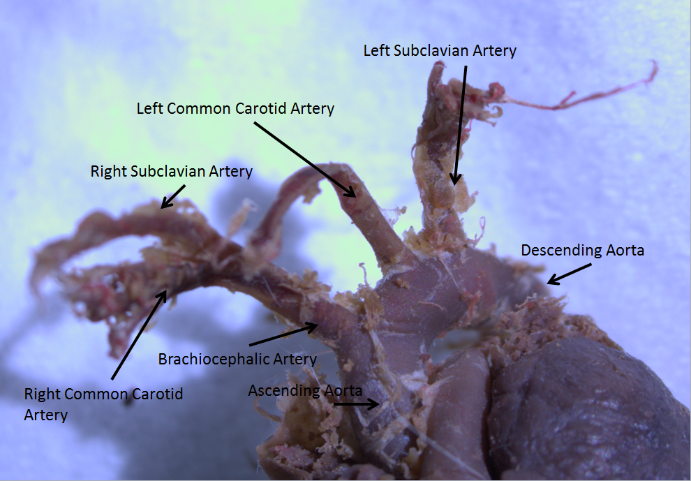
The aortic arch, arch of the aorta, or transverse aortic arch () is the part of the aorta between the ascending and descending aorta. The arch travels backward, so that it ultimately runs to the left of the trachea. Structure The aorta begins at the level of the upper border of the second/third sternocostal articulation of the right side, behind the ventricular outflow tract and pulmonary trunk. The right atrial appendage overlaps it. The first few centimeters of the ascending aorta and pulmonary trunk lies in the same pericardial sheath. and runs at first upward, arches over the pulmonary trunk, right pulmonary artery, and right main bronchus to lie behind the right second coastal cartilage. The right lung and sternum lies anterior to the aorta at this point. The aorta then passes posteriorly and to the left, anterior to the trachea, and arches over left main bronchus and left pulmonary artery, and reaches to the left side of the T4 vertebral body. Apart from T4 vertebral bo ... [...More Info...] [...Related Items...] OR: [Wikipedia] [Google] [Baidu] |
Cavo-tricuspid Isthmus
The cavo-tricuspid isthmus is a body of fibrous tissue in the lower right atrium between the inferior vena cava, and the tricuspid valve. It is a target for ablation for treating atrial flutter Atrial flutter (AFL) is a common abnormal heart rhythm that starts in the atrial chambers of the heart. When it first occurs, it is usually associated with a fast heart rate and is classified as a type of supraventricular tachycardia. Atrial f .... Cardiac anatomy {{cardiology-stub ... [...More Info...] [...Related Items...] OR: [Wikipedia] [Google] [Baidu] |
Eustachian Tube
In anatomy, the Eustachian tube, also known as the auditory tube or pharyngotympanic tube, is a tube that links the nasopharynx to the middle ear, of which it is also a part. In adult humans, the Eustachian tube is approximately long and in diameter. It is named after the sixteenth-century Italian anatomist Bartolomeo Eustachi. In humans and other tetrapods, both the middle ear and the ear canal are normally filled with air. Unlike the air of the ear canal, however, the air of the middle ear is not in direct contact with the atmosphere outside the body; thus, a pressure difference can develop between the atmospheric pressure of the ear canal and the middle ear. Normally, the Eustachian tube is collapsed, but it gapes open with swallowing and with positive pressure, allowing the middle ear's pressure to adjust to the atmospheric pressure. When taking off in an aircraft, the ambient air pressure goes from higher (on the ground) to lower (in the sky). The air in the middle ear ... [...More Info...] [...Related Items...] OR: [Wikipedia] [Google] [Baidu] |
Corpus Callosum
The corpus callosum (Latin for "tough body"), also callosal commissure, is a wide, thick nerve tract, consisting of a flat bundle of commissural fibers, beneath the cerebral cortex in the brain. The corpus callosum is only found in placental mammals. It spans part of the longitudinal fissure, connecting the left and right cerebral hemispheres, enabling communication between them. It is the largest white matter structure in the human brain, about in length and consisting of 200–300 million axonal projections. A number of separate nerve tracts, classed as subregions of the corpus callosum, connect different parts of the hemispheres. The main ones are known as the genu, the rostrum, the trunk or body, and the splenium. Structure The corpus callosum forms the floor of the longitudinal fissure that separates the two cerebral hemispheres. Part of the corpus callosum forms the roof of the lateral ventricles. The corpus callosum has four main parts – individual nerve tracts ... [...More Info...] [...Related Items...] OR: [Wikipedia] [Google] [Baidu] |
Oviduct
The oviduct in mammals, is the passageway from an ovary. In human females this is more usually known as the Fallopian tube or uterine tube. The eggs travel along the oviduct. These eggs will either be fertilized by spermatozoa to become a zygote, or will degenerate in the body. Normally, these are paired structures, but in birds and some cartilaginous fishes, one or the other side fails to develop (together with the corresponding ovary), and only one functional oviduct can be found. Except in teleosts, the oviduct is not directly in contact with the ovary. Instead, the most anterior portion ends in a funnel-shaped structure called the infundibulum, which collects eggs as they are released by the ovary into the body cavity. The only female vertebrates to lack oviducts are the jawless fishes. In these species, the single fused ovary releases eggs directly into the body cavity. The fish eventually extrudes the eggs through a small genital pore towards the rear of the body. Fish and ... [...More Info...] [...Related Items...] OR: [Wikipedia] [Google] [Baidu] |
Prostate
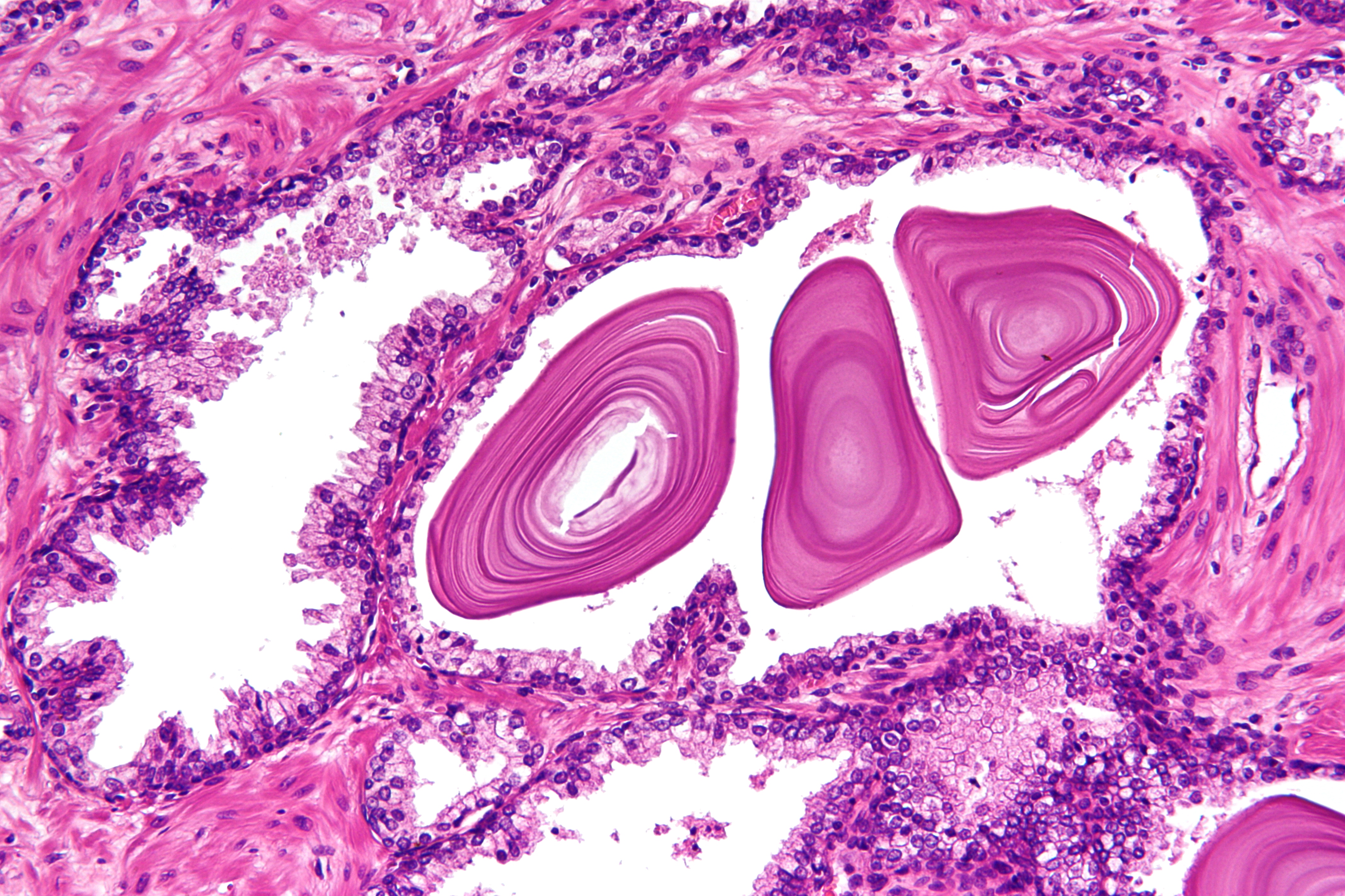
The prostate is both an Male accessory gland, accessory gland of the male reproductive system and a muscle-driven mechanical switch between urination and ejaculation. It is found only in some mammals. It differs between species anatomically, chemically, and physiologically. Anatomically, the prostate is found below the Urinary bladder, bladder, with the urethra passing through it. It is described in gross anatomy as consisting of lobes and in microanatomy by zone. It is surrounded by an elastic, fibromuscular capsule and contains glandular tissue as well as connective tissue. The prostate glands produce and contain fluid that forms part of semen, the substance emitted during ejaculation as part of the male Human sexual response cycle, sexual response. This prostatic fluid is slightly alkaline, milky or white in appearance. The alkalinity of semen helps neutralize the acidity of the vagina, vaginal tract, prolonging the lifespan of sperm. The prostatic fluid is expelled in the ... [...More Info...] [...Related Items...] OR: [Wikipedia] [Google] [Baidu] |
Isthmus Of Cingulate Gyrus
The cingulate gyrus commences below the rostrum of the corpus callosum, curves around in front of the genu, extends along the upper surface of the body, and finally turns downward behind the splenium, where it is connected by a narrow isthmus with the parahippocampal gyrus The parahippocampal gyrus (or hippocampal gyrus') is a grey matter cortical region of the brain that surrounds the hippocampus and is part of the limbic system. The region plays an important role in memory encoding and retrieval. It has been in .... Additional images File:Medial surface of cerebral cortex - gyri.png, Medial surface of cerebral cortex - gyri File:Isthmus_Cingulate_-_DK_ATLAS.png, Isthmus of the cingulate gyrus, medial surface of right hemisphere References External links * http://braininfo.rprc.washington.edu/whereisit.aspx?language=0&requestID=ID163&parentID=ID159&originalID=ID163 * https://web.archive.org/web/20091029114245/http://bstr431.biostr.washington.edu/syl/lab7/lab7.htm { ... [...More Info...] [...Related Items...] OR: [Wikipedia] [Google] [Baidu] |
Isthmus Faucium
The fauces, isthmus of fauces, or the oropharyngeal isthmus, is the opening at the back of the mouth into the throat. It is a narrow passage between the velum and the base of the tongue. The fauces is a part of the oropharynx directly behind the oral cavity as a subdivision, bounded superiorly by the soft palate, laterally by the palatoglossal and palatopharyngeal arches, and inferiorly by the tongue. The arches form the pillars of the fauces. The anterior pillar is the palatoglossal arch formed of the palatoglossus muscle. The posterior pillar is the palatopharyngeal arch formed of the palatopharyngeus muscle. Between these two arches on the lateral walls of the oropharynx is the tonsillar fossa which is the location of the palatine tonsil. The arches are also known together as the palatine arches. Each arch runs downwards, laterally and forwards, from the soft palate to the side of the tongue. The approximation of the arches due to the contraction of the palatoglossal ... [...More Info...] [...Related Items...] OR: [Wikipedia] [Google] [Baidu] |
Isthmus Of Uterine Tube
The fallopian tubes, also known as uterine tubes, oviducts or salpinges (singular salpinx), are paired tubes in the human female that stretch from the uterus to the ovaries. The fallopian tubes are part of the female reproductive system. In other mammals they are only called oviducts. Each tube is a muscular hollow organ that is on average between 10 and 14 cm in length, with an external diameter of 1 cm. It has four described parts: the intramural part, isthmus, ampulla, and infundibulum with associated fimbriae. Each tube has two openings a proximal opening nearest and opening to the uterus, and a distal opening furthest and opening to the abdomen. The fallopian tubes are held in place by the mesosalpinx, a part of the broad ligament mesentery that wraps around the tubes. Another part of the broad ligament, the mesovarium suspends the ovaries in place. An egg cell is transported from an ovary to a fallopian tube where it may be fertilized in the ampulla of the ... [...More Info...] [...Related Items...] OR: [Wikipedia] [Google] [Baidu] |
Kronig Isthmus
Kronig isthmus is a band of resonance representing the apex of lung, also it's described as the narrow strap-like portion of the resonant field that extends over the shoulder, that connect the larger areas of resonance over the pulmonary apex in front and behind. Boundaries * Medially: Scalene * Laterally: Acromion process * Anteriorly: Clavicle * Posteriorly: Trapezius The trapezius is a large paired trapezoid-shaped surface muscle that extends longitudinally from the occipital bone to the lower thoracic vertebrae of the spine and laterally to the spine of the scapula. It moves the scapula and supports th ... References Lung anatomy {{Lung-stub ... [...More Info...] [...Related Items...] OR: [Wikipedia] [Google] [Baidu] |
Thyroid Isthmus
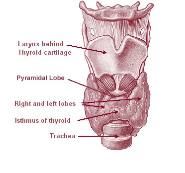
The thyroid, or thyroid gland, is an endocrine gland in vertebrates. In humans it is in the neck and consists of two connected lobes. The lower two thirds of the lobes are connected by a thin band of tissue called the thyroid isthmus. The thyroid is located at the front of the neck, below the Adam's apple. Microscopically, the functional unit of the thyroid gland is the spherical thyroid follicle, lined with follicular cells (thyrocytes), and occasional parafollicular cells that surround a lumen containing colloid. The thyroid gland secretes three hormones: the two thyroid hormones triiodothyronine (T3) and thyroxine (T4)and a peptide hormone, calcitonin. The thyroid hormones influence the metabolic rate and protein synthesis, and in children, growth and development. Calcitonin plays a role in calcium homeostasis. Secretion of the two thyroid hormones is regulated by thyroid-stimulating hormone (TSH), which is secreted from the anterior pituitary gland. TSH is regulated by th ... [...More Info...] [...Related Items...] OR: [Wikipedia] [Google] [Baidu] |


