|
List Of MeSH Codes (A08)
The following is a partial list of the "A" codes for Medical Subject Headings (MeSH), as defined by the United States National Library of Medicine (NLM). This list continues the information at List of MeSH codes (A07). Codes following these are found at List of MeSH codes (A09). For other MeSH codes, see List of MeSH codes. The source for this content is the set o2006 MeSH Treesfrom the NLM. – nervous system – central nervous system – brain * – blood–brain barrier * – brain stem * – mesencephalon * – corpora quadrigemina * – inferior colliculus * – superior colliculus * – locus coeruleus * – raphe nuclei * – substantia nigra * – tegmentum mesencephali * – cerebral aqueduct * – pedunculopontine tegmental nucleus * – periaqueductal gray * – red nucleus * – ventral tegmental area * – reticular formation * – respiratory center * – rhombencephalon * – medulla oblongata * – area postrema * – ... [...More Info...] [...Related Items...] OR: [Wikipedia] [Google] [Baidu] |
Medical Subject Headings
Medical Subject Headings (MeSH) is a comprehensive controlled vocabulary for the purpose of indexing journal articles and books in the life sciences. It serves as a thesaurus that facilitates searching. Created and updated by the United States National Library of Medicine (NLM), it is used by the MEDLINE/PubMed article database and by NLM's catalog of book holdings. MeSH is also used by ClinicalTrials.gov registry to classify which diseases are studied by trials registered in ClinicalTrials. MeSH was introduced in the 1960s, with the NLM's own index catalogue and the subject headings of the Quarterly Cumulative Index Medicus (1940 edition) as precursors. The yearly printed version of MeSH was discontinued in 2007; MeSH is now available only online. It can be browsed and downloaded free of charge through PubMed. Originally in English, MeSH has been translated into numerous other languages and allows retrieval of documents from different origins. Structure MeSH vocabulary is divi ... [...More Info...] [...Related Items...] OR: [Wikipedia] [Google] [Baidu] |
Tegmentum Mesencephali
The midbrain is anatomically delineated into the tectum (roof) and the tegmentum (floor). The midbrain tegmentum extends from the substantia nigra to the cerebral aqueduct in a horizontal section of the midbrain. It forms the floor of the midbrain that surrounds below the cerebral aqueduct as well as the floor of the fourth ventricle while the midbrain tectum forms the roof of the fourth ventricle. The tegmentum contains a collection of tracts and nuclei with movement-related, species-specific, and pain-perception functions. The general structures of midbrain tegmentum include red nucleus and the periaqueductal grey matter. Unlike the midbrain tectum (which is a sensory structure located posteriorly), the midbrain tegmentum, which locates anteriorly, is related to a number of motor functions. Within the tegmentum, the red nucleus is in charge of motor coordination (specifically for limb movements) and the periaqueductal gray matter (PAG) contains critical circuits for modulating b ... [...More Info...] [...Related Items...] OR: [Wikipedia] [Google] [Baidu] |
Metencephalon
The metencephalon is the embryonic part of the hindbrain that differentiates into the pons and the cerebellum. It contains a portion of the fourth ventricle and the trigeminal nerve (CN V), abducens nerve (CN VI), facial nerve (CN VII), and a portion of the vestibulocochlear nerve (CN VIII). Embryology The metencephalon develops from the higher/rostral half of the embryonic rhombencephalon, and is differentiated from the myelencephalon in the embryo by approximately 5 weeks of age. By the third month, the metencephalon differentiates into its two main structures, the pons and the cerebellum. Functions The pons regulates breathing through particular nuclei that regulate the breathing center of the medulla oblongata. The cerebellum works to coordinate muscle movements, maintain posture, and integrate sensory information from the inner ear and proprioceptors in the muscles and joints. Development At the early stages of brain development, the brain vesicles that are formed ar ... [...More Info...] [...Related Items...] OR: [Wikipedia] [Google] [Baidu] |
Solitary Nucleus
In the human brainstem, the solitary nucleus, also called nucleus of the solitary tract, nucleus solitarius, and nucleus tractus solitarii, (SN or NTS) is a series of purely sensory nuclei (clusters of nerve cell bodies) forming a vertical column of grey matter embedded in the medulla oblongata. Through the center of the SN runs the solitary tract, a white bundle of nerve fibers, including fibers from the facial, glossopharyngeal and vagus nerves, that innervate the SN. The SN projects to, among other regions, the reticular formation, parasympathetic preganglionic neurons, hypothalamus and thalamus, forming circuits that contribute to autonomic regulation. Cells along the length of the SN are arranged roughly in accordance with function; for instance, cells involved in taste are located in the rostral part, while those receiving information from cardio-respiratory and gastrointestinal processes are found in the caudal part. Inputs * Taste information from the facial nerve ... [...More Info...] [...Related Items...] OR: [Wikipedia] [Google] [Baidu] |
Olivary Nucleus
In anatomy, the olivary bodies or simply olives (Latin ''oliva'' and ''olivae'', singular and plural, respectively) are a pair of prominent oval structures in the medulla oblongata, the lower portion of the brainstem. They contain the olivary nuclei. Structure The olivary body is located on the anterior surface of the medulla lateral to the pyramid, from which it is separated by the antero-lateral sulcus and the fibers of the hypoglossal nerve. Behind (dorsally), it is separated from the postero-lateral sulcus by the ventral spinocerebellar fasciculus. In the depression between the upper end of the olive and the pons lies the vestibulocochlear nerve. In humans, it measures about 1.25 cm. in length, and between its upper end and the pons there is a slight depression to which the roots of the facial nerve are attached. The external arcuate fibers wind across the lower part of the pyramid and olive and enter the inferior peduncle. Olivary nuclei The olive consists of two par ... [...More Info...] [...Related Items...] OR: [Wikipedia] [Google] [Baidu] |
Area Postrema
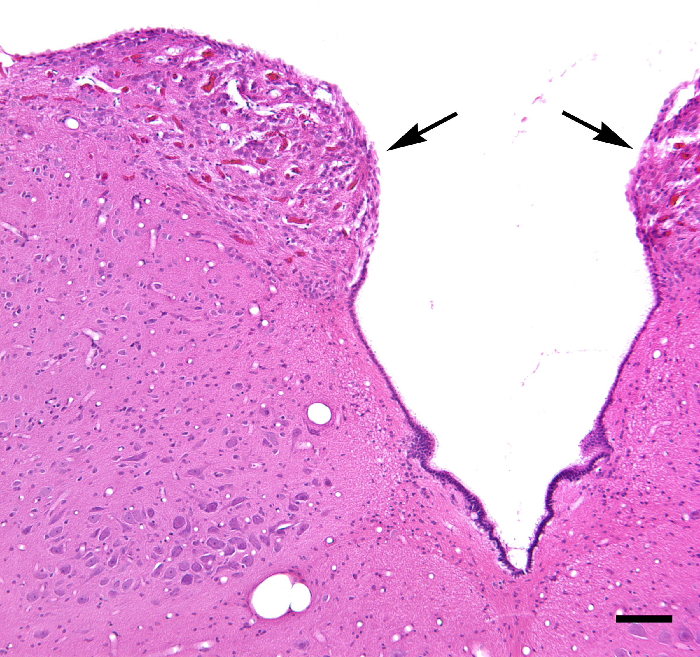
The area postrema, a paired structure in the medulla oblongata of the brainstem, is a circumventricular organ having permeable capillaries and sensory neurons that enable its dual role to detect circulating chemical messengers in the blood and transduce them into neural signals and networks. Its position adjacent to the bilateral nuclei of the solitary tract and role as a sensory transducer allow it to integrate blood-to-brain autonomic functions. Such roles of the area postrema include its detection of circulating hormones involved in vomiting, thirst, hunger, and blood pressure control. Structure The area postrema is a paired protuberance found at the inferoposterior limit of the fourth ventricle. Specialized ependymal cells are found within the area postrema. These cells differ slightly from the majority of ependymal cells (ependymocytes), forming a unicellular epithelial lining of the ventricles and central canal. The area postrema is separated from the vagal trigone by ... [...More Info...] [...Related Items...] OR: [Wikipedia] [Google] [Baidu] |
Medulla Oblongata
The medulla oblongata or simply medulla is a long stem-like structure which makes up the lower part of the brainstem. It is anterior and partially inferior to the cerebellum. It is a cone-shaped neuronal mass responsible for autonomic (involuntary) functions, ranging from vomiting to sneezing. The medulla contains the cardiac, respiratory, vomiting and vasomotor centers, and therefore deals with the autonomic functions of breathing, heart rate and blood pressure as well as the sleep–wake cycle. During embryonic development, the medulla oblongata develops from the myelencephalon. The myelencephalon is a secondary vesicle which forms during the maturation of the rhombencephalon, also referred to as the hindbrain. The bulb is an archaic term for the medulla oblongata. In modern clinical usage, the word bulbar (as in bulbar palsy) is retained for terms that relate to the medulla oblongata, particularly in reference to medical conditions. The word bulbar can refer to the nerves ... [...More Info...] [...Related Items...] OR: [Wikipedia] [Google] [Baidu] |
Rhombencephalon
The hindbrain or rhombencephalon or lower brain is a developmental categorization of portions of the central nervous system in vertebrates. It includes the medulla, pons, and cerebellum. Together they support vital bodily processes. Metencephalon Rhombomeres Rh3-Rh1 form the metencephalon. The metencephalon is composed of the pons and the cerebellum; it contains: * a portion of the fourth (IV) ventricle, * the trigeminal nerve (CN V), * abducens nerve (CN VI), * facial nerve (CN VII), * and a portion of the vestibulocochlear nerve (CN VIII). Myelencephalon Rhombomeres Rh8-Rh4 form the myelencephalon. The myelencephalon forms the medulla oblongata in the adult brain; it contains: * a portion of the fourth ventricle, * the glossopharyngeal nerve (CN IX), * vagus nerve (CN X), * accessory nerve (CN XI), * hypoglossal nerve (CN XII), * and a portion of the vestibulocochlear nerve (CN VIII). Evolution The hindbrain is homologous to a part of the arthropod brain known as the sub-oes ... [...More Info...] [...Related Items...] OR: [Wikipedia] [Google] [Baidu] |
Respiratory Center
The respiratory center is located in the medulla oblongata and pons, in the brainstem. The respiratory center is made up of three major respiratory groups of neurons, two in the medulla and one in the pons. In the medulla they are the dorsal respiratory group, and the ventral respiratory group. In the pons, the pontine respiratory group includes two areas known as the pneumotaxic centre and the apneustic centre. The respiratory centre is responsible for generating and maintaining the rhythm of respiration, and also of adjusting this in homeostatic response to physiological changes. The respiratory center receives input from chemoreceptors, mechanoreceptors, the cerebral cortex, and the hypothalamus in order to regulate the rate and depth of breathing. Input is stimulated by altered levels of oxygen, carbon dioxide, and blood pH, by hormonal changes relating to stress and anxiety from the hypothalamus, and also by signals from the cerebral cortex to give a conscious control of r ... [...More Info...] [...Related Items...] OR: [Wikipedia] [Google] [Baidu] |
Reticular Formation
The reticular formation is a set of interconnected nuclei that are located throughout the brainstem. It is not anatomically well defined, because it includes neurons located in different parts of the brain. The neurons of the reticular formation make up a complex set of networks in the core of the brainstem that extend from the upper part of the midbrain to the lower part of the medulla oblongata. The reticular formation includes ascending pathways to the cerebral cortex, cortex in the ascending reticular activating system (ARAS) and descending pathways to the spinal cord via the reticulospinal tracts. Neurons of the reticular formation, particularly those of the ascending reticular activating system, play a crucial role in maintaining behavioral arousal and consciousness. The overall functions of the reticular formation are modulatory and premotor, involving somatic motor control, cardiovascular control, pain modulation, sleep and consciousness, and habituation. The modulatory ... [...More Info...] [...Related Items...] OR: [Wikipedia] [Google] [Baidu] |
Ventral Tegmental Area
The ventral tegmental area (VTA) (tegmentum is Latin for ''covering''), also known as the ventral tegmental area of Tsai, or simply ventral tegmentum, is a group of neurons located close to the midline on the floor of the midbrain. The VTA is the origin of the dopaminergic cell bodies of the mesocorticolimbic dopamine system and other dopamine pathways; it is widely implicated in the drug and natural reward circuitry of the brain. The VTA plays an important role in a number of processes, including reward cognition ( motivational salience, associative learning, and positively-valenced emotions) and orgasm, among others, as well as several psychiatric disorders. Neurons in the VTA project to numerous areas of the brain, ranging from the prefrontal cortex to the caudal brainstem and several regions in between. Structure Neurobiologists have often had great difficulty distinguishing the VTA in humans and other primate brains from the substantia nigra (SN) and surrounding nucl ... [...More Info...] [...Related Items...] OR: [Wikipedia] [Google] [Baidu] |
Red Nucleus
The red nucleus or nucleus ruber is a structure in the rostral midbrain involved in motor coordination. The red nucleus is pale pink, which is believed to be due to the presence of iron in at least two different forms: hemoglobin and ferritin. The structure is located in the tegmentum of the midbrain next to the substantia nigra and comprises caudal magnocellular and rostral parvocellular components. The red nucleus and substantia nigra are subcortical centers of the extrapyramidal motor system. Function In a vertebrate without a significant corticospinal tract, gait is mainly controlled by the red nucleus. However, in primates, where the corticospinal tract is dominant, the rubrospinal tract may be regarded as vestigial in motor function. Therefore, the red nucleus is less important in primates than in many other mammals. Nevertheless, the crawling of babies is controlled by the red nucleus, as is arm swinging in typical walking. The red nucleus may play an additional role ... [...More Info...] [...Related Items...] OR: [Wikipedia] [Google] [Baidu] |


