|
List Of MeSH Codes (A05)
The following is a partial list of the "A" codes for Medical Subject Headings (MeSH), as defined by the United States National Library of Medicine (NLM). This list continues the information at List of MeSH codes (A04). Codes following these are found at List of MeSH codes (A06). For other MeSH codes, see List of MeSH codes. The source for this content is the set o2006 MeSH Treesfrom the NLM. – urogenital system – genitalia – genitalia, female * – adnexa uteri * – broad ligament * – fallopian tubes * – ovary * – corpus luteum * – luteal cells * – ovarian follicle * – follicular fluid * – granulosa cells * – theca cells * – round ligament * – uterus * – cervix uteri * – endometrium * – decidua * – deciduoma * – myometrium * – vagina * – hymen * – vulva * – Bartholin's glands * – clitoris – genitalia, male * – bulbourethral glands * – ejaculatory ducts * – epididymis * ... [...More Info...] [...Related Items...] OR: [Wikipedia] [Google] [Baidu] |
Medical Subject Headings
Medical Subject Headings (MeSH) is a comprehensive controlled vocabulary for the purpose of indexing journal articles and books in the life sciences. It serves as a thesaurus that facilitates searching. Created and updated by the United States National Library of Medicine (NLM), it is used by the MEDLINE/PubMed article database and by NLM's catalog of book holdings. MeSH is also used by ClinicalTrials.gov registry to classify which diseases are studied by trials registered in ClinicalTrials. MeSH was introduced in the 1960s, with the NLM's own index catalogue and the subject headings of the Quarterly Cumulative Index Medicus (1940 edition) as precursors. The yearly printed version of MeSH was discontinued in 2007; MeSH is now available only online. It can be browsed and downloaded free of charge through PubMed. Originally in English, MeSH has been translated into numerous other languages and allows retrieval of documents from different origins. Structure MeSH vocabulary is divi ... [...More Info...] [...Related Items...] OR: [Wikipedia] [Google] [Baidu] |
Theca Cells
In biology, a theca (plural thecae) is a sheath or a covering. Botany In botany, the theca is related to plant's flower anatomy. The theca of an angiosperm consists of a pair of microsporangia that are adjacent to each other and share a common area of dehiscence called the stomium. Larry Hufford, "The origin and early evolution of angiosperm stamens" i''The Anther: form, function, and phylogeny'' William G. D'Arcy and Richard C. Keating (editors), Cambridge University Press, 1996, 351pp, p.60, (from Google Books) Any part of a microsporophyll that bears microsporangia is called an anther. Most anthers are formed on the apex of a filament. An anther and its filament together form a typical (or filantherous) stamen, part of the male floral organ. The typical anther is bilocular, i.e. it consists of two thecae. Each theca contains two microsporangia, also known as pollen sacs. The microsporangia produce the microspores, which for seed plants are known as pollen grains. If th ... [...More Info...] [...Related Items...] OR: [Wikipedia] [Google] [Baidu] |
Bulbourethral Glands
The bulbourethral glands or Cowper's glands (named for English anatomist William Cowper) are two small exocrine glands in the reproductive system of many male mammals (of all domesticated animals, they are absent only in dogs). They are homologous to Bartholin's glands in females. The bulbouretheral glands are responsible for producing a pre-ejaculate fluid called Cowper's fluid (known colloquially as ''pre-ejaculate'' or ''pre-cum''), which is secreted during sexual arousal, neutralizing the acidity of the urethra in preparation for the passage of sperm cells. Location Bulbourethral glands are located posterior and lateral to the membranous portion of the urethra at the base of the penis, between the two layers of the fascia of the urogenital diaphragm, in the deep perineal pouch. They are enclosed by transverse fibers of the sphincter urethrae membranaceae muscle. Structure The bulbourethral glands are compound tubulo-alveolar glands, each approximately the size of a pea i ... [...More Info...] [...Related Items...] OR: [Wikipedia] [Google] [Baidu] |
Human Male Genitalia
The male reproductive system consists of a number of sex organs that play a role in the process of human reproduction. These organs are located on the outside of the body and within the pelvis. The main male sex organs are the penis and the testicles which produce semen and sperm, which, as part of sexual intercourse, fertilize an ovum in the female's body; the fertilized ovum (zygote) develops into a fetus, which is later born as an infant. The corresponding system in females is the female reproductive system. External genital organs Penis The penis is the male intromittent organ. It has a long shaft and an enlarged bulbous-shaped tip called the glans penis, which supports and is protected by the foreskin. When the male becomes sexually aroused, the penis becomes erect and ready for sexual activity. Erection occurs because sinuses within the erectile tissue of the penis become filled with blood. The arteries of the penis are dilated while the veins are compressed so that ... [...More Info...] [...Related Items...] OR: [Wikipedia] [Google] [Baidu] |
Clitoris
The clitoris ( or ) is a female sex organ present in mammals, ostriches and a limited number of other animals. In humans, the visible portion – the glans – is at the front junction of the labia minora (inner lips), above the opening of the urethra. Unlike the penis, the male homologue (equivalent) to the clitoris, it usually does not contain the distal portion (or opening) of the urethra and is therefore not used for urination. In most species, the clitoris lacks any reproductive function. While few animals urinate through the clitoris or use it reproductively, the spotted hyena, which has an especially large clitoris, urinates, mates, and gives birth via the organ. Some other mammals, such as lemurs and spider monkeys, also have a large clitoris. The clitoris is the human female's most sensitive erogenous zone and generally the primary anatomical source of human female sexual pleasure. In humans and other mammals, it develops from an outgrowth in the embry ... [...More Info...] [...Related Items...] OR: [Wikipedia] [Google] [Baidu] |
Bartholin's Glands
The Bartholin's glands (named after Caspar Bartholin the Younger; also called Bartholin glands or greater vestibular glands) are two pea sized compound alveolar glandsManual of Obstetrics. (3rd ed.). Elsevier. pp. 1-16. . located slightly posterior and to the left and right of the opening of the vagina. They secrete mucus to lubricate the vagina. They are homologous to bulbourethral glands in males. However, while Bartholin's glands are located in the superficial perineal pouch in females, bulbourethral glands are located in the deep perineal pouch in males. Their duct length is 1.5 to 2.0 cm and they open into navicular fossa. The ducts are paired and they open on the surface of the vulva. History Bartholin's glands were first described in the 17th century by the Danish anatomist Caspar Bartholin the Younger (1655–1738). Some sources mistakenly ascribe their discovery to his grandfather, theologian and anatomist Caspar Bartholin the Elder (1585–1629). Function Ba ... [...More Info...] [...Related Items...] OR: [Wikipedia] [Google] [Baidu] |
Vulva
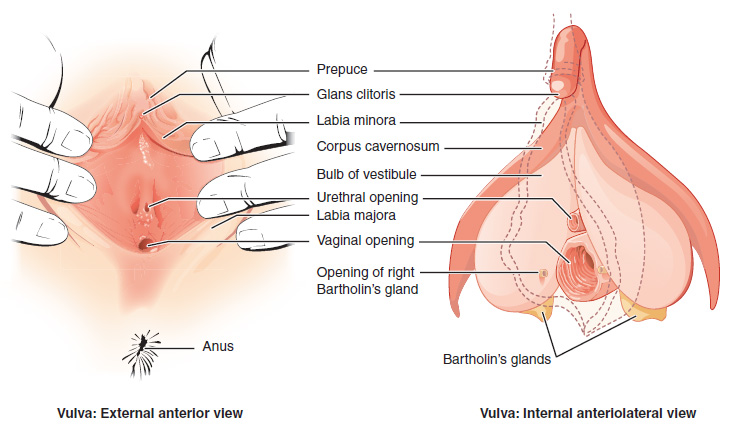
The vulva (plural: vulvas or vulvae; derived from Latin for wrapper or covering) consists of the external sex organ, female sex organs. The vulva includes the mons pubis (or mons veneris), labia majora, labia minora, clitoris, bulb of vestibule, vestibular bulbs, vulval vestibule, urinary meatus, the Vagina#Vaginal opening and hymen, vaginal opening, hymen, and Bartholin's gland, Bartholin's and Skene's gland, Skene's vestibular glands. The urinary meatus is also included as it opens into the vulval vestibule. Other features of the vulva include the pudendal cleft, sebaceous glands, the urogenital triangle (anterior part of the perineum), and pubic hair. The vulva includes the entrance to the vagina, which leads to the uterus, and provides a double layer of protection for this by the folds of the outer and inner labia. Pelvic floor muscles support the structures of the vulva. Other muscles of the urogenital triangle also give support. Blood supply to the vulva comes from the t ... [...More Info...] [...Related Items...] OR: [Wikipedia] [Google] [Baidu] |
Hymen
The hymen is a thin piece of mucosal tissue that surrounds or partially covers the external vaginal opening. It forms part of the vulva, or external genitalia, and is similar in structure to the vagina. In children, a common appearance of the hymen is crescent-shaped, although many shapes are possible. During puberty, estrogen causes the hymen to change in appearance and become very elastic. Normal variations of the post-pubertal hymen range from thin and stretchy to thick and somewhat rigid. Very rarely, it may be completely absent. The hymen can rip or tear during first penetrative intercourse, which usually results in pain and, sometimes, mild temporary bleeding or spotting. Sources differ on how common tearing or bleeding after first intercourse are. The state of the hymen is not a reliable indicator of virginity, though "virginity testing" remains a common practice in some cultures, sometimes accompanied by surgical restoration of hymen to give the appearance of virginity. ... [...More Info...] [...Related Items...] OR: [Wikipedia] [Google] [Baidu] |
Vagina
In mammals, the vagina is the elastic, muscular part of the female genital tract. In humans, it extends from the vestibule to the cervix. The outer vaginal opening is normally partly covered by a thin layer of mucosal tissue called the hymen. At the deep end, the cervix (neck of the uterus) bulges into the vagina. The vagina allows for sexual intercourse and birth. It also channels menstrual flow, which occurs in humans and closely related primates as part of the menstrual cycle. Although research on the vagina is especially lacking for different animals, its location, structure and size are documented as varying among species. Female mammals usually have two external openings in the vulva; these are the urethral opening for the urinary tract and the vaginal opening for the genital tract. This is different from male mammals, who usually have a single urethral opening for both urination and reproduction. The vaginal opening is much larger than the nearby urethral opening, an ... [...More Info...] [...Related Items...] OR: [Wikipedia] [Google] [Baidu] |
Myometrium
The myometrium is the middle layer of the uterine wall, consisting mainly of uterine smooth muscle cells (also called uterine myocytes) but also of supporting stromal and vascular tissue. Its main function is to induce uterine contractions. Structure The myometrium is located between the endometrium (the inner layer of the uterine wall) and the serosa or perimetrium (the outer uterine layer). The inner one-third of the myometrium (termed the ''junctional'' or ''sub-endometrial'' layer) appears to be derived from the Müllerian duct, while the outer, more predominant layer of the myometrium appears to originate from non-Müllerian tissue and is the major contractile tissue during parturition and abortion. The junctional layer appears to function like a circular muscle layer, capable of peristaltic and anti-peristaltic activity, equivalent to the muscular layer of the intestines. Muscular structure The molecular structure of the smooth muscle of myometrium is very similar to tha ... [...More Info...] [...Related Items...] OR: [Wikipedia] [Google] [Baidu] |
Decidua
The decidua is the modified mucosal lining of the uterus (that is, modified endometrium) that forms every month, in preparation for pregnancy. It is shed off each month when there is no fertilised egg to support. The decidua is under the influence of progesterone. Endometrial cells become highly characteristic. The decidua forms the maternal part of the placenta and remains for the duration of the pregnancy. After birth the decidua is shed together with the placenta. Structure The part of the decidua that interacts with the trophoblast is the ''decidua basalis'' (also called ''decidua placentalis''), while the ''decidua capsularis'' grows over the embryo on the luminal side, enclosing it into the endometrium. The remainder of the decidua is termed the ''decidua parietalis'' or ''decidua vera'', and it will fuse with the decidua capsularis by the fourth month of gestation. Three morphologically distinct layers of the decidua basalis can then be described: * Compact outer laye ... [...More Info...] [...Related Items...] OR: [Wikipedia] [Google] [Baidu] |
Endometrium
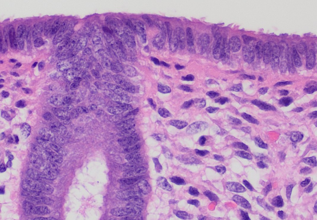
The endometrium is the inner epithelial layer, along with its mucous membrane, of the mammalian uterus. It has a basal layer and a functional layer: the basal layer contains stem cells which regenerate the functional layer. The functional layer thickens and then is shed during menstruation in humans and some other mammals, including apes, Old World monkeys, some species of bat, the elephant shrew and the Cairo spiny mouse. In most other mammals, the endometrium is reabsorbed in the estrous cycle. During pregnancy, the glands and blood vessels in the endometrium further increase in size and number. Vascular spaces fuse and become interconnected, forming the placenta, which supplies oxygen and nutrition to the embryo and fetus.Blue Histology - Female Reproductive System . School ... [...More Info...] [...Related Items...] OR: [Wikipedia] [Google] [Baidu] |


.png)


