|
List Of MeSH Codes (A02)
The following is a partial list of the "A" codes for Medical Subject Headings (MeSH), as defined by the United States National Library of Medicine (NLM). This list continues the information at List of MeSH codes (A01). Codes following these are found at List of MeSH codes (A03). For other MeSH codes, see List of MeSH codes. The source for this content is the set o2006 MeSH Treesfrom the NLM. – musculoskeletal system – aponeurosis – cartilage – elastic cartilage * – ear cartilages * – laryngeal cartilages ** – arytenoid cartilage ** – cricoid cartilage ** – epiglottis ** – thyroid cartilage – fibrocartilage * – intervertebral disc ** – annulus fibrosus ** – nucleus fibrosus * – meniscus ** – tibial menisci * – palmar plate * – plantar plate * – triangular fibrocartilage – hyaline cartilage * – articular cartilage * – costal cartilage * – laryngeal cartilages ** – arytenoid cartilage ** – ... [...More Info...] [...Related Items...] OR: [Wikipedia] [Google] [Baidu] |
Medical Subject Headings
Medical Subject Headings (MeSH) is a comprehensive controlled vocabulary for the purpose of indexing journal articles and books in the life sciences. It serves as a thesaurus that facilitates searching. Created and updated by the United States National Library of Medicine (NLM), it is used by the MEDLINE/PubMed article database and by NLM's catalog of book holdings. MeSH is also used by ClinicalTrials.gov registry to classify which diseases are studied by trials registered in ClinicalTrials. MeSH was introduced in the 1960s, with the NLM's own index catalogue and the subject headings of the Quarterly Cumulative Index Medicus (1940 edition) as precursors. The yearly printed version of MeSH was discontinued in 2007; MeSH is now available only online. It can be browsed and downloaded free of charge through PubMed. Originally in English, MeSH has been translated into numerous other languages and allows retrieval of documents from different origins. Structure MeSH vocabulary is divi ... [...More Info...] [...Related Items...] OR: [Wikipedia] [Google] [Baidu] |
Anulus Fibrosus Disci Intervertebralis
An intervertebral disc (or intervertebral fibrocartilage) lies between adjacent vertebrae in the vertebral column. Each disc forms a fibrocartilaginous joint (a symphysis), to allow slight movement of the vertebrae, to act as a ligament to hold the vertebrae together, and to function as a shock absorber for the spine. Structure Intervertebral discs consist of an outer fibrous ring, the anulus fibrosus disci intervertebralis, which surrounds an inner gel-like center, the nucleus pulposus. The ''anulus fibrosus'' consists of several layers (laminae) of fibrocartilage made up of both type I and type II collagen. Type I is concentrated toward the edge of the ring, where it provides greater strength. The stiff laminae can withstand compressive forces. The fibrous intervertebral disc contains the ''nucleus pulposus'' and this helps to distribute pressure evenly across the disc. This prevents the development of stress concentrations which could cause damage to the underlying vertebrae ... [...More Info...] [...Related Items...] OR: [Wikipedia] [Google] [Baidu] |
Tenon Capsule
Tenon's capsule (), also known as the Tenon capsule, fascial sheath of the eyeball () or the fascia bulbi, is a thin membrane which envelops the eyeball from the optic nerve to the corneal limbus, separating it from the orbital fat and forming a socket in which it moves. The inner surface of Tenon's capsule is smooth and is separated from the outer surface of the sclera by the periscleral lymph space. This lymph space is continuous with the subdural and subarachnoid cavities and is traversed by delicate bands of connective tissue which extend between the capsule and the sclera. The capsule is perforated behind by the ciliary vessels and nerves and fuses with the sheath of the optic nerve and with the sclera around the entrance of the optic nerve. In front it adheres to the conjunctiva, and both structures are attached to the ciliary region of the eyeball. The structure was named after Jacques-René Tenon (1724–1816), a French surgeon and pathologist. Structure Relations Te ... [...More Info...] [...Related Items...] OR: [Wikipedia] [Google] [Baidu] |
Fascia Lata
The fascia lata is the deep fascia of the thigh. It encloses the thigh muscles and forms the outer limit of the fascial compartments of thigh, which are internally separated by the medial intermuscular septum and the lateral intermuscular septum. The fascia lata is thickened at its lateral side where it forms the iliotibial tract, a structure that runs to the tibia and serves as a site of muscle attachment. Structure The fascia lata is an investment for the whole of the thigh, but varies in thickness in different parts. It is thicker in the upper and lateral part of the thigh, where it receives a fibrous expansion from the gluteus maximus, and where the tensor fasciae latae is inserted between its layers; it is very thin behind and at the upper and medial part, where it covers the adductor muscles, and again becomes stronger around the knee, receiving fibrous expansions from the tendon of the biceps femoris laterally, from the sartorius medially, and from the quadriceps femoris ... [...More Info...] [...Related Items...] OR: [Wikipedia] [Google] [Baidu] |
Fascia
A fascia (; plural fasciae or fascias; adjective fascial; from Latin: "band") is a band or sheet of connective tissue, primarily collagen, beneath the skin that attaches to, stabilizes, encloses, and separates muscles and other internal organs. Fascia is classified by layer, as superficial fascia, deep fascia, and ''visceral'' or ''parietal'' fascia, or by its function and anatomical location. Like ligaments, aponeuroses, and tendons, fascia is made up of fibrous connective tissue containing closely packed bundles of collagen fibers oriented in a wavy pattern parallel to the direction of pull. Fascia is consequently flexible and able to resist great unidirectional tension forces until the wavy pattern of fibers has been straightened out by the pulling force. These collagen fibers are produced by fibroblasts located within the fascia. Fasciae are similar to ligaments and tendons as they have collagen as their major component. They differ in their location and function: ligament ... [...More Info...] [...Related Items...] OR: [Wikipedia] [Google] [Baidu] |
Nasal Cartilages
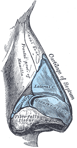
The nasal cartilages are structures within the nose that provide form and support to the nasal cavity. The nasal cartilages are made up of a flexible material called hyaline cartilage (packed collagen) in the distal portion of the nose. There are five individual cartilages that make up the nasal cavity: septal nasal cartilage, lateral nasal cartilage, major alar cartilage (greater alar cartilage, or cartilage of the aperture), minor alar cartilage (lesser alar cartilage, sesamoid, or accessory cartilage), and vomeronasal cartilage (Jacobson's cartilage). The nasal cartilages associate with other cartilage structures of the nose or with bones of the facial skeleton. These associations create vent-like structures within the nose so that air can flow from the nasal cavity to the lungs or vice versa. Therefore, the nasal cartilages are structures that aid the body in respiratory functions to intake oxygen or expire carbon dioxide. Abnormalities or defects in the nasal cartilages affect ... [...More Info...] [...Related Items...] OR: [Wikipedia] [Google] [Baidu] |
Costal Cartilage
The costal cartilages are bars of hyaline cartilage that serve to prolong the ribs forward and contribute to the elasticity of the walls of the thorax. Costal cartilage is only found at the anterior ends of the ribs, providing medial extension. Differences from Ribs 1-12 The first seven pairs are connected with the sternum; the next three are each articulated with the lower border of the cartilage of the preceding rib; the last two have pointed extremities, which end in the wall of the abdomen. Like the ribs, the costal cartilages vary in their length, breadth, and direction. They increase in length from the first to the seventh, then gradually decrease to the twelfth. Their breadth, as well as that of the intervals between them, diminishes from the first to the last. They are broad at their attachments to the ribs, and taper toward their sternal extremities, excepting the first two, which are of the same breadth throughout, and the sixth, seventh, and eighth, which are enlarge ... [...More Info...] [...Related Items...] OR: [Wikipedia] [Google] [Baidu] |
Articular Cartilage
Hyaline cartilage is the glass-like (hyaline) and translucent cartilage found on many joint surfaces. It is also most commonly found in the ribs, nose, larynx, and trachea. Hyaline cartilage is pearl-gray in color, with a firm consistency and has a considerable amount of collagen. It contains no nerves or blood vessels, and its structure is relatively simple. Structure Hyaline cartilage is covered externally by a fibrous membrane known as the perichondrium or, when it's along articulating surfaces, the synovial membrane. This membrane contains vessels that provide the cartilage with nutrition through diffusion. Hyaline cartilage matrix is primarily made of type II collagen and chondroitin sulphate, both of which are also found in elastic cartilage. Hyaline cartilage exists on the sternal ends of the ribs, in the larynx, trachea, and bronchi, and on the articulating surfaces of bones. It gives the structures a definite but pliable form. The presence of collagen fibres makes ... [...More Info...] [...Related Items...] OR: [Wikipedia] [Google] [Baidu] |
Hyaline Cartilage
Hyaline cartilage is the glass-like (hyaline) and translucent cartilage found on many joint surfaces. It is also most commonly found in the ribs, nose, larynx, and trachea. Hyaline cartilage is pearl-gray in color, with a firm consistency and has a considerable amount of collagen. It contains no nerves or blood vessels, and its structure is relatively simple. Structure Hyaline cartilage is covered externally by a fibrous membrane known as the perichondrium or, when it's along articulating surfaces, the synovial membrane. This membrane contains vessels that provide the cartilage with nutrition through diffusion. Hyaline cartilage matrix is primarily made of type II collagen and chondroitin sulphate, both of which are also found in elastic cartilage. Hyaline cartilage exists on the sternal ends of the ribs, in the larynx, trachea, and bronchi, and on the articulating surfaces of bones. It gives the structures a definite but pliable form. The presence of collagen fibres makes suc ... [...More Info...] [...Related Items...] OR: [Wikipedia] [Google] [Baidu] |
Triangular Fibrocartilage
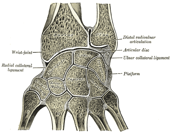
The triangular fibrocartilage complex (TFCC) is formed by the triangular fibrocartilage discus (TFC), the radioulnar ligaments (RULs) and the ulnocarpal ligaments (UCLs). Structure Triangular fibrocartilage disc The triangular fibrocartilage disc (TFC) is an articular discus that lies on the pole of the distal ulna. It has a triangular shape and a biconcave body; the periphery is thicker than its center. The central portion of the TFC is thin and consists of chondroid fibrocartilage; this type of tissue is often seen in structures that can bear compressive loads. This central area is often so thin that it is translucent and in some cases it is even absent. The peripheral portion of the TFC is well vascularized, while the central portion has no blood supply. This discus is attached by thick tissue to the base of the ulnar styloid and by thinner tissue to the edge of the radius just proximal to the radiocarpal articular surface. Radioulnar ligaments The radioulnar ligaments ... [...More Info...] [...Related Items...] OR: [Wikipedia] [Google] [Baidu] |
Plantar Plate
In the human foot, the plantar or volar plates (also called plantar or volar ligaments) are fibrocartilaginous structures found in the metatarsophalangeal (MTP) and interphalangeal (IP) joints. The anatomy and composition of the plantar plates are similar to the palmar plates in the metacarpophalangeal (MCP) and interphalangeal joints in the hand; the proximal origin is thin but the distal insertion is stout. Due to the weight-bearing nature of the human foot, the plantar plates are exposed to extension forces not present in the human hand. The plantar plate supports the weight of the body and restricts dorsiflexion, whilst the main collateral ligament and the accessory collateral ligament (together referred as the collateral ligament complex, CLC) prevent motions in the transverse and sagittal planes. The major difference between the plantar plates of the MTP and IP joints is that they blend with the transverse metatarsal ligament in the MTP joints (not present in the toes). ... [...More Info...] [...Related Items...] OR: [Wikipedia] [Google] [Baidu] |
Palmar Plate
In the human hand, palmar or volar plates (also referred to as palmar or volar ligaments) are found in the metacarpophalangeal (MCP) and interphalangeal (IP) joints, where they reinforce the joint capsules, enhance joint stability, and limit hyperextension. The plates of the MCP and IP joints are structurally and functionally similar, except that in the MCP joints they are interconnected by a deep transverse ligament. In the MCP joints, they also indirectly provide stability to the longitudinal palmar arches of the hand. (MCP joints) (IP joints) The volar plate of the thumb MCP joint has a transverse longitudinal rectangular shape, shorter than those in the fingers. Structure This fibrocartilaginous structure is attached to the volar base of the phalanx distal to the joint. From there, it forms a palmar continuation of the articular surface of the phalanx bone and its inner surface thus adds to the articular surface during extension. In its proximal end, the volar plat ... [...More Info...] [...Related Items...] OR: [Wikipedia] [Google] [Baidu] |


