|
Lisfranc Ligament
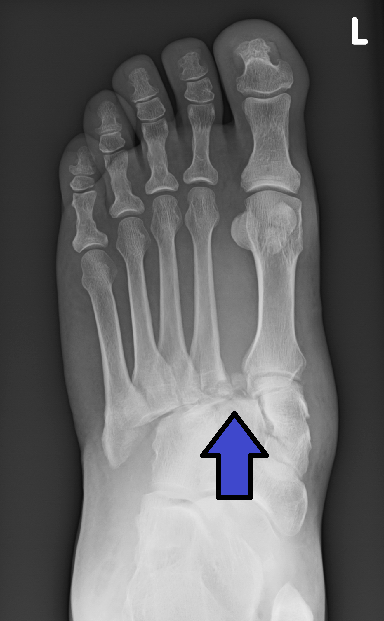
The Lisfranc ligament is one of several ligaments which connects the medial cuneiform bone to the second metatarsal. Sometimes, the term ''Lisfranc ligament'' refers specifically to the ligament that connects the superior, lateral surface of the medial cuneiform to the superior, medial surface of the base of the second metatarsal. Structure The Lisfranc ligament connects the medial cuneiform bone to the second metatarsal. It is a complex of 3 ligaments: the dorsal Lisfranc ligament, the interosseous Lisfranc ligament, and the plantar Lisfranc ligament. Variation In 20% of people, there are two bands of each component of the ligament, usually of the dorsal Lisfranc ligament or the plantar Lisfranc ligament. Function The Lisfranc ligament maintains proper alignment between the metatarsal bones and the tarsal bones. It acts as a shock absorber during the weight bearing phase of the bipedal gait cycle. It also compensates for the lack of an intermetatarsal ligament between th ... [...More Info...] [...Related Items...] OR: [Wikipedia] [Google] [Baidu] |
Medical Ultrasonography
Medical ultrasound includes diagnostic techniques (mainly medical imaging, imaging techniques) using ultrasound, as well as therapeutic ultrasound, therapeutic applications of ultrasound. In diagnosis, it is used to create an image of internal body structures such as tendons, muscles, joints, blood vessels, and internal organs, to measure some characteristics (e.g. distances and velocities) or to generate an informative audible sound. Its aim is usually to find a source of disease or to exclude pathology. The usage of ultrasound to produce visual images for medicine is called medical ultrasonography or simply sonography. The practice of examining pregnant women using ultrasound is called obstetric ultrasonography, and was an early development of clinical ultrasonography. Ultrasound is composed of sound waves with frequency, frequencies which are significantly higher than the range of human hearing (>20,000 Hz). Ultrasonic images, also known as sonograms, are created by se ... [...More Info...] [...Related Items...] OR: [Wikipedia] [Google] [Baidu] |
Cuneiform Bones
There are three cuneiform ("wedge-shaped") bones in the human foot: * the first or medial cuneiform * the second or intermediate cuneiform, also known as the middle cuneiform * the third or lateral cuneiform They are located between the navicular bone and the first, second and third metatarsal bones and are medial to the cuboid bone. Structure There are three cuneiform bones: # The medial cuneiform (also known as first cuneiform) is the largest of the cuneiforms. It is situated at the medial side of the foot, anterior to the navicular bone and posterior to the base of the first metatarsal. Lateral to it is the intermediate cuneiform. It articulates with four bones: the navicular, second cuneiform, and first and second metatarsals. The tibialis anterior and fibularis longus muscle inserts at the medial cuneiform bone. # The intermediate cuneiform (second cuneiform or middle cuneiform) is shaped like a wedge, the thin end pointing downwards. The intermediate cuneiform is situate ... [...More Info...] [...Related Items...] OR: [Wikipedia] [Google] [Baidu] |
Metatarsal
The metatarsal bones, or metatarsus, are a group of five long bones in the foot, located between the tarsal bones of the hind- and mid-foot and the phalanges of the toes. Lacking individual names, the metatarsal bones are numbered from the medial side (the side of the great toe): the first, second, third, fourth, and fifth metatarsal (often depicted with Roman numerals). The metatarsals are analogous to the metacarpal bones of the hand. The lengths of the metatarsal bones in humans are, in descending order, second, third, fourth, fifth, and first. Structure The five metatarsals are dorsal convex long bones consisting of a shaft or body, a base (proximally), and a head (distally).Platzer 2004, p. 220 The body is prismoid in form, tapers gradually from the tarsal to the phalangeal extremity, and is curved longitudinally, so as to be concave below, slightly convex above. The base or posterior extremity is wedge-shaped, articulating proximally with the tarsal bones, and by it ... [...More Info...] [...Related Items...] OR: [Wikipedia] [Google] [Baidu] |
Metatarsal Bones
The metatarsal bones, or metatarsus, are a group of five long bones in the foot, located between the tarsal bones of the hind- and mid-foot and the phalanges of the toes. Lacking individual names, the metatarsal bones are numbered from the medial side (the side of the great toe): the first, second, third, fourth, and fifth metatarsal (often depicted with Roman numerals). The metatarsals are analogous to the metacarpal bones of the hand. The lengths of the metatarsal bones in humans are, in descending order, second, third, fourth, fifth, and first. Structure The five metatarsals are dorsal convex long bones consisting of a shaft or body, a base (proximally), and a head (distally).Platzer 2004, p. 220 The body is prismoid in form, tapers gradually from the tarsal to the phalangeal extremity, and is curved longitudinally, so as to be concave below, slightly convex above. The base or posterior extremity is wedge-shaped, articulating proximally with the tarsal bones, and by its ... [...More Info...] [...Related Items...] OR: [Wikipedia] [Google] [Baidu] |
Tarsus (skeleton)
In the human body, the tarsus is a cluster of seven articulating bones in each foot situated between the lower end of the tibia and the fibula of the lower leg and the metatarsus. It is made up of the midfoot (Cuboid bone, cuboid, medial, intermediate, and lateral cuneiform bone, cuneiform, and navicular) and hindfoot (Talus bone, talus and calcaneus). The tarsus articulates with the bones of the metatarsus, which in turn articulate with the proximal phalanges of the toes. The joint between the tibia and fibula above and the tarsus below is referred to as the ankle, ankle joint proper. In humans the largest bone in the tarsus is the calcaneus, which is the weight-bearing bone within the heel of the foot. Human anatomy Bones The talus bone or ankle bone is connected superiorly to the two bones of the lower leg, the tibia and fibula, to form the ankle, ankle joint or talocrural joint; inferiorly, at the subtalar joint, to the calcaneus or heel bone. Together, the talus and ... [...More Info...] [...Related Items...] OR: [Wikipedia] [Google] [Baidu] |
Shock Absorber
A shock absorber or damper is a mechanical or hydraulic device designed to absorb and damp shock impulses. It does this by converting the kinetic energy of the shock into another form of energy (typically heat) which is then dissipated. Most shock absorbers are a form of dashpot (a damper which resists motion via viscous friction). Description Pneumatic and hydraulic shock absorbers are used in conjunction with cushions and springs. An automobile shock absorber contains spring-loaded check valves and orifices to control the flow of oil through an internal piston (see below). One design consideration, when designing or choosing a shock absorber, is where that energy will go. In most shock absorbers, energy is converted to heat inside the viscous fluid. In hydraulic cylinders, the hydraulic fluid heats up, while in air cylinders, the hot air is usually exhausted to the atmosphere. In other types of shock absorbers, such as electromagnetic types, the dissipated energy can be ... [...More Info...] [...Related Items...] OR: [Wikipedia] [Google] [Baidu] |
Bipedal Gait Cycle
A (bipedal) gait cycle is the time period or sequence of events or movements during locomotion in which one foot contacts the ground to when that same foot again contacts the ground, and involves propulsion of the centre of gravity In physics, the center of mass of a distribution of mass in space (sometimes referred to as the balance point) is the unique point where the weighted relative position of the distributed mass sums to zero. This is the point to which a force ma ... in the direction of motion. A gait cycle usually involves co-operative movements of both the left and right legs and feet. A single gait cycle is also known as a stride. Each gait cycle or stride has two major phases:CASTERMANS, T., DUVINAGE, M., CHERON, G. & DUTOIT, T. 2013. Towards Effective Non-Invasive Brain-Computer Interfaces Dedicated to Gait Rehabilitation Systems. Brain Sciences, 4, 1-48.BAKER, R. 2013. Measuring Walking : A Handbook of Clinical Gait Analysis, London, Mac Keith Press.PERRY, J. 199 ... [...More Info...] [...Related Items...] OR: [Wikipedia] [Google] [Baidu] |
Intermetatarsal Joints
The intermetatarsal joints are the articulations between the base of metatarsal bones. The base of the first metatarsal is not connected with that of the second by any ligaments; in this respect the great toe resembles the thumb. The bases of the other four metatarsals are connected by the dorsal, plantar, and interosseous ligaments. * The '' dorsal ligaments'' pass transversely between the dorsal surfaces of the bases of the adjacent metatarsal bones. * The '' plantar ligaments'' have a similar arrangement to the dorsal. * The '' interosseous ligaments'' consist of strong transverse fibers which connect the rough non-articular portions of the adjacent surfaces. Synovial membranes The synovial membranes between the second and third, and the third and fourth metatarsal bones are part of the great tarsal synovial membrane; that between the fourth and fifth is a prolongation of the synovial membrane of the cuboideometatarsal joint. Movements The movement permitted between the tar ... [...More Info...] [...Related Items...] OR: [Wikipedia] [Google] [Baidu] |
First Metatarsal Bone
The first metatarsal bone is the bone in the foot just behind the big toe. The first metatarsal bone is the shortest of the metatarsal bones and by far the thickest and strongest of them. Like the four other metatarsals, it can be divided into three parts: base, body and head. The base is the part closest to the ankle and the head is closest to the big toe. The narrowed part in the middle is referred to as the body of the bone. The bone is somewhat flattened, giving it two sides: the plantar (towards the sole of the foot) and the dorsal side (the area facing upwards while standing). The base presents, as a rule, no articular facets (joint surfaces) on its sides, but occasionally on the lateral side there is an oval facet, by which it articulates with the second metatarsal. On the lateral part of the plantar surface there is a rough oval prominence, or tuberosity, for the insertion of the tendon of the fibularis longus. The first metatarsal articulates (forms joints) with the medi ... [...More Info...] [...Related Items...] OR: [Wikipedia] [Google] [Baidu] |
Second Metatarsal Bone
The second metatarsal bone is a long bone in the foot. It is the longest of the metatarsal, metatarsal bones, being prolonged backward and held firmly into the recess formed by the three cuneiform bones. The second metatarsal forms joints with the Phalanges of the foot, second proximal phalanx (a bone in the second toe) through the metatarsophalangeal joint, the cuneiform bones, third metatarsal bone, third metatarsal and occasionally the first metatarsal bone. Structure Like the four other metatarsal bones, it can be divided into three parts: base, body and head. The base is the part closest to the ankle and the head is closest to the big toe. The narrowed part in the middle is referred to as the body of the bone. The bone is somewhat flattened, giving it two sides: the plantar (towards the Sole (foot), sole of the foot) and the dorsal side (the area facing upwards while standing). Its base is broad above, narrow and rough below. It presents four articular surfaces: one behind, ... [...More Info...] [...Related Items...] OR: [Wikipedia] [Google] [Baidu] |
Lisfranc Fracture
A Lisfranc injury, also known as Lisfranc fracture, is an injury of the foot in which one or more of the metatarsal bones are displaced from the tarsus. The injury is named after Jacques Lisfranc de St. Martin, a French surgeon and gynecologist who noticed this fracture pattern amongst cavalry men, in 1815, after the War of the Sixth Coalition. Causes The midfoot consists of five bones that form the arches of the foot (the cuboid, navicular, and three cuneiform bones) and their articulations with the bases of the five metatarsal bones. It is these articulations that are damaged in a Lisfranc injury. Such injuries typically involve the ligaments between the medial cuneiform bone and the bases of the second and third metatarsal bones, and each of these ligaments is called Lisfranc ligament. Lisfranc injuries are caused when excessive kinetic energy is applied either directly or indirectly to the midfoot and are often seen in traffic collisions or industrial accidents. Di ... [...More Info...] [...Related Items...] OR: [Wikipedia] [Google] [Baidu] |


