|
Limbic Lobe
The limbic lobe is an arc-shaped region of cortex on the medial surface of each cerebral hemisphere of the mammalian brain, consisting of parts of the frontal, parietal and temporal lobes. The term is ambiguous, with some authors including the paraterminal gyrus, the subcallosal area, the cingulate gyrus, the parahippocampal gyrus, the dentate gyrus, the hippocampus and the subiculum; while the Terminologia Anatomica includes the cingulate sulcus, the cingulate gyrus, the isthmus of cingulate gyrus, the fasciolar gyrus, the parahippocampal gyrus, the parahippocampal sulcus, the dentate gyrus, the fimbrodentate sulcus, the fimbria of hippocampus, the collateral sulcus, and the rhinal sulcus, and omits the hippocampus. History Broca named the limbic lobe in 1878, identifying it with the cingulate and parahippocampal gyri, and associating it with the sense of smell - Treviranus having earlier noted that, between species, the size of the parahippocampal gyrus varies with the s ... [...More Info...] [...Related Items...] OR: [Wikipedia] [Google] [Baidu] |
Cerebral Hemisphere
The vertebrate cerebrum (brain) is formed by two cerebral hemispheres that are separated by a groove, the longitudinal fissure. The brain can thus be described as being divided into left and right cerebral hemispheres. Each of these hemispheres has an outer layer of grey matter, the cerebral cortex, that is supported by an inner layer of white matter. In eutherian (placental) mammals, the hemispheres are linked by the corpus callosum, a very large bundle of nerve fibers. Smaller commissures, including the anterior commissure, the posterior commissure and the fornix, also join the hemispheres and these are also present in other vertebrates. These commissures transfer information between the two hemispheres to coordinate localized functions. There are three known poles of the cerebral hemispheres: the ''occipital pole'', the '' frontal pole'', and the ''temporal pole''. The central sulcus is a prominent fissure which separates the parietal lobe from the frontal lobe and the ... [...More Info...] [...Related Items...] OR: [Wikipedia] [Google] [Baidu] |
Collateral Fissure
The collateral fissure (or sulcus) is on the tentorial surface of the hemisphere and extends from near the occipital pole to within a short distance of the temporal pole. Behind, it lies below and lateral to the calcarine fissure, from which it is separated by the lingual gyrus; in front, it is situated between the parahippocampal gyrus and the anterior part of the fusiform gyrus. Additional images File:Gray738.png, Coronal section through posterior cornua of lateral ventricle The lateral ventricles are the two largest ventricles of the brain and contain cerebrospinal fluid (CSF). Each cerebral hemisphere contains a lateral ventricle, known as the left or right ventricle, respectively. Each lateral ventricle resemble .... (Collateral fissure labeled at bottom center.) File:Hippocampal Limbic Connections Functions - Sanjoy Sanyal (Cropped from 5m28s to 6m30s) Collateral sulcus.webm, Human brain dissection video (62 sec). Demonstrating location of collateral sulcus. R ... [...More Info...] [...Related Items...] OR: [Wikipedia] [Google] [Baidu] |
Amygdala
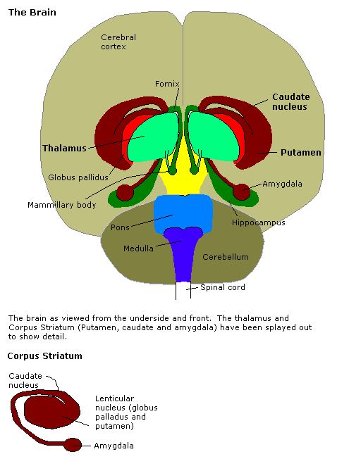
The amygdala (; plural: amygdalae or amygdalas; also '; Latin from Greek, , ', 'almond', 'tonsil') is one of two almond-shaped clusters of nuclei located deep and medially within the temporal lobes of the brain's cerebrum in complex vertebrates, including humans. Shown to perform a primary role in the processing of memory, decision making, and emotional responses (including fear, anxiety, and aggression), the amygdalae are considered part of the limbic system. The term "amygdala" was first introduced by Karl Friedrich Burdach in 1822. Structure The regions described as amygdala nuclei encompass several structures of the cerebrum with distinct connectional and functional characteristics in humans and other animals. Among these nuclei are the basolateral complex, the cortical nucleus, the medial nucleus, the central nucleus, and the intercalated cell clusters. The basolateral complex can be further subdivided into the lateral, the basal, and the accessory basal ... [...More Info...] [...Related Items...] OR: [Wikipedia] [Google] [Baidu] |
Segmental Resection
Segmental resection (or segmentectomy) is a surgical procedure to remove part of an organ or gland, as a sub-type of a resection, which might involve removing the whole body part. It may also be used to remove a tumor and normal tissue around it. In lung cancer surgery, segmental resection refers to removing a section of a lobe of the lung. The resection margin is the edge of the removed tissue; it is important that this shows free of cancerous cells on examination by a pathologist Pathology is the study of the causes and effects of disease or injury. The word ''pathology'' also refers to the study of disease in general, incorporating a wide range of biology research fields and medical practices. However, when used in th .... References * External links Segmental resectionentry in the public domain NCI Dictionary of Cancer Terms Surgical procedures and techniques Surgical removal procedures {{oncology-stub ... [...More Info...] [...Related Items...] OR: [Wikipedia] [Google] [Baidu] |
Paul Bucy
Paul Bucy (; November 13, 1904 – September 22, 1992) was an American neurosurgeon and neuropathologist who was a native of Hubbard, Iowa. He is known both for his part in describing the Klüver–Bucy syndrome, his academic life as a teacher in the neurosciences, and for his founding in 1972 and editing ''Surgical Neurology'' – An International Journal of Neurosurgery and Neuroscience" from 1972 to 1987. Academic life Bucy grew up and was educated in Iowa. He received his bachelor's degree, a master's in neuropathology and his doctorate from the University of Iowa. He interned and trained at Ford Hospital in Detroit. He was an assistant to neurosurgeon Percival Bailey (1892–1973) at the University of Chicago. In the early 1930s he traveled to Europe, and studied with Gordon Morgan Holmes (1876–1965) in London and Otfrid Foerster (1874–1941) in Breslau. In 1941, he became Professor of Neurology and Neurological Surgery at the University of Illinois in Chicago, where ... [...More Info...] [...Related Items...] OR: [Wikipedia] [Google] [Baidu] |
Heinrich Klüver
Heinrich Klüver (; May 25, 1897 – February 8, 1979) was a German-American biological psychologist and philosopher born in Holstein. After having served in the Imperial German Army during World War I, he studied at both the University of Hamburg and the University of Berlin from 1920-23. In the latter year, he arrived in the United States to attend Stanford University. He received his Ph.D. in physiological psychology from Stanford University. In 1927 he married Cessa Feyerabend and settled in the United States permanently, becoming a naturalized U.S. citizen in 1934. Klüver was a member of the 'core group' of cybernetics pioneers that participated in the Macy Conferences of the 1940s and 1950s. He collaborated most often and fruitfully with Paul Bucy and made various contributions to neuroanatomy throughout his career among others the Klüver–Bucy syndrome. His expositions of and experiments with mescaline were also groundbreaking at the time. He coined the term "co ... [...More Info...] [...Related Items...] OR: [Wikipedia] [Google] [Baidu] |
Hippocampal Formation
The hippocampal formation is a compound structure in the medial temporal lobe of the brain. It forms a c-shaped bulge on the floor of the temporal horn of the lateral ventricle. There is no consensus concerning which brain regions are encompassed by the term, with some authors defining it as the dentate gyrus, the hippocampus proper and the subiculum; and others including also the presubiculum, parasubiculum, and entorhinal cortex. The hippocampal formation is thought to play a role in memory, spatial navigation and control of attention. The neural layout and pathways within the hippocampal formation are very similar in all mammals. History and function During the nineteenth and early twentieth centuries, based largely on the observation that, between species, the size of the olfactory bulb varies with the size of the parahippocampal gyrus, the hippocampal formation was thought to be part of the olfactory system. In 1937 Papez theorized that a circuit including the hippocamp ... [...More Info...] [...Related Items...] OR: [Wikipedia] [Google] [Baidu] |
Neural Circuit
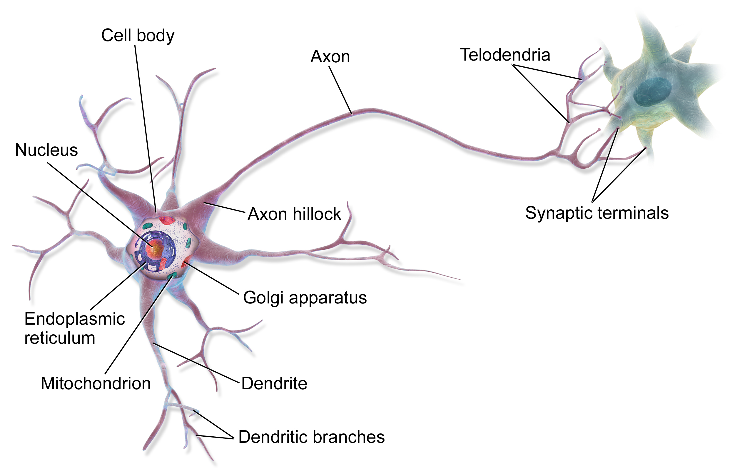
A neural circuit is a population of neurons interconnected by synapses to carry out a specific function when activated. Neural circuits interconnect to one another to form large scale brain networks. Biological neural networks have inspired the design of artificial neural networks, but artificial neural networks are usually not strict copies of their biological counterparts. Early study Early treatments of neural networks can be found in Herbert Spencer's ''Principles of Psychology'', 3rd edition (1872), Theodor Meynert's ''Psychiatry'' (1884), William James' ''Principles of Psychology'' (1890), and Sigmund Freud's Project for a Scientific Psychology (composed 1895). The first rule of neuronal learning was described by Hebb in 1949, in the Hebbian theory. Thus, Hebbian pairing of pre-synaptic and post-synaptic activity can substantially alter the dynamic characteristics of the synaptic connection and therefore either facilitate or inhibit signal transmission. In 1959, the ... [...More Info...] [...Related Items...] OR: [Wikipedia] [Google] [Baidu] |
James Papez
James Wenceslas Papez (;Livingston, Kenneth E. '. U.S. National Library of Medicine, 1981 1883–1958) was an American neuroanatomist, most famous for his 1937 description of the Papez circuit, a neural pathway in the brain thought to be involved in the cortical control of emotion. Specifically, Papez hypothesized that the hippocampus, the cingulate gyrus (Broca's callosal lobe), the hypothalamus, the anterior thalamic nuclei, and the interconnections among these structures constituted a harmonious mechanism which elaborate the functions of emotions.Papez JW. 1937. A proposed mechanism of emotion. 1937. J Neuropsychiatry Clin Neurosci. 1995 Winter;7(1):103-12. Papez never mentioned Broca's limbic lobe but others noted that his circuit was very similar to Broca's great limbic lobe.Lima, D.R.,2004. History of Medicine, Medsi, RJ. http://www.editoraguanabara.com.br/ Papez received his MD from the University of Minnesota College of Medicine and Surgery. He was a neurologist ... [...More Info...] [...Related Items...] OR: [Wikipedia] [Google] [Baidu] |
Gottfried Reinhold Treviranus
Gottfried Reinhold Treviranus (4 February 1776, Bremen – 16 February 1837, Bremen) was a German physician, naturalist, and proto-evolutionary biologist. His younger brother, Ludolph Christian Treviranus (1779–1864), was also a naturalist and botanist, and also a notable taxonomist and zoologist. History Treviranus was born in Bremen and studied medicine at the University of Göttingen, where he took his doctor's degree in 1796. During the following year, he was appointed professor of medicine and mathematics at the Bremen lyceum. In 1816, he was elected a corresponding member of the Royal Swedish Academy of Sciences. Works Treviranus was a proponent of the theory of the transmutation of species, a theory of evolution held by some biologists prior to the work of Charles Darwin. He put forward this belief in the first volume of his ''Biologie; oder die Philosophie der lebenden Natur'', published in 1802, the same year similar opinions were expressed by Jean-Baptiste Lam ... [...More Info...] [...Related Items...] OR: [Wikipedia] [Google] [Baidu] |
Paul Broca
Pierre Paul Broca (, also , , ; 28 June 1824 – 9 July 1880) was a French physician, anatomist and anthropologist. He is best known for his research on Broca's area, a region of the frontal lobe that is named after him. Broca's area is involved with language. His work revealed that the brains of patients with aphasia contained lesions in a particular part of the cortex, in the left frontal region. This was the first anatomical proof of localization of brain function. Broca's work also contributed to the development of physical anthropology, advancing the science of anthropometry. Biography Paul Broca was born on 28 June 1824 in Sainte-Foy-la-Grande, Bordeaux, France, the son of Jean Pierre "Benjamin" Broca, a medical practitioner and former surgeon in Napoleon's service, and Annette Thomas, a well-educated daughter of a Calvinist, Reformed Protestant, preacher. Huguenot Broca received basic education in the school in his hometown, earning a bachelor's degree at the age of 16. ... [...More Info...] [...Related Items...] OR: [Wikipedia] [Google] [Baidu] |
_-_inferiror_view.png)

.png)

_(14782023652).jpg)