|
Leukemia Cells
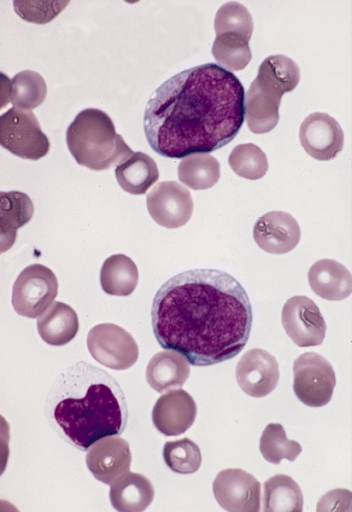
In cell biology, a precursor cell, also called a blast cell or simply blast, is a partially differentiated cell, usually referred to as a unipotent cell that has lost most of its stem cell properties. A precursor cell is also known as a progenitor cell but progenitor cells are multipotent. Precursor cells are known as the intermediate cell before they become differentiated after being a stem cell. Usually, a precursor cell is a stem cell with the capacity to differentiate into only one cell type. Sometimes, ''precursor cell'' is used as an alternative term for unipotent stem cells. In embryology, precursor cells are a group of cells that later differentiate into one organ. A blastoma is any cancer created by malignancies of precursor cells. Precursor cells, and progenitor cells, have many potential uses in medicine. , there is research being done to use these cells to build heart valves, blood vessels and other tissues, by using blood and muscle precursor, or progenitor cel ... [...More Info...] [...Related Items...] OR: [Wikipedia] [Google] [Baidu] |
Two Myeloblasts With Auer Rods
2 (two) is a number, numeral (linguistics), numeral and numerical digit, digit. It is the natural number following 1 and preceding 3. It is the smallest and only even prime number. Because it forms the basis of a Dualistic cosmology, duality, it has Religion, religious and Spirituality, spiritual significance in many Culture, cultures. Evolution Arabic digit The digit used in the modern Western world to represent the number 2 traces its roots back to the Indic Brahmic script, where "2" was written as two horizontal lines. The modern Chinese written language, Chinese and Japanese writing system, Japanese languages (and Korean Hanja) still use this method. The Gupta script rotated the two lines 45 degrees, making them diagonal. The top line was sometimes also shortened and had its bottom end curve towards the center of the bottom line. In the Devanagari, Nagari script, the top line was written more like a curve connecting to the bottom line. In the Arabic Ghubār numerals, G ... [...More Info...] [...Related Items...] OR: [Wikipedia] [Google] [Baidu] |
Megakaryoblast
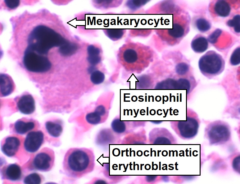
A megakaryoblast is a precursor cell to a promegakaryocyte, which in turn becomes a megakaryocyte during haematopoiesis. It is the beginning of the thrombocytic series. Development The megakaryoblast derives from a CFU-Meg colony unit of pluripotential hemopoietic stem cells. (Some sources use the term "CFU-Meg" to identify the CFU.) The CFU-Meg derives from the CFU-GEMM CFU-GEMM is a colony forming unit that generates myeloid cells. CFU-GEMM cells are the oligopotential progenitor cells for myeloid cells; they are thus also called common myeloid progenitor cells or myeloid stem cells. "GEMM" stands for granulocyte ... (common myeloid progenitor). Structure These cells tend to range from 8μm to 30μm, owing to the variation in size between different megakaryoblasts. The nucleus is three to five times the size of the cytoplasm, and is generally round or oval in shape. Several nucleoli are visible, while the chromatin varies from cell to cell, ranging from fine to heavy and dense. ... [...More Info...] [...Related Items...] OR: [Wikipedia] [Google] [Baidu] |
Neuroscience Information Framework
The Neuroscience Information Framework is a repository of global neuroscience web resources, including experimental, clinical, and translational neuroscience databases, knowledge bases, atlases, and genetic/ genomic resources and provides many authoritative links throughout the neuroscience portal of Wikipedia. Description The Neuroscience Information Framework (NIF) is an initiative of the NIH Blueprint for Neuroscience Research, which was established in 2004 by the National Institutes of Health. Development of the NIF started in 2008, when the University of California, San Diego School of Medicine obtained an NIH contract to create and maintain "a dynamic inventory of web-based neurosciences data, resources, and tools that scientists and students can access via any computer connected to the Internet". The project is headed by Maryann Martone, co-director of the National Center for Microscopy and Imaging Research (NCMIR), part of the multi-disciplinary Center for Research in Bio ... [...More Info...] [...Related Items...] OR: [Wikipedia] [Google] [Baidu] |
Plasmablast
Plasma cells, also called plasma B cells or effector B cells, are white blood cells that originate in the lymphoid organs as B lymphocytes and secrete large quantities of proteins called antibodies in response to being presented specific substances called antigens. These antibodies are transported from the plasma cells by the blood plasma and the lymphatic system to the site of the target antigen (foreign substance), where they initiate its neutralization or destruction. B cells differentiate into plasma cells that produce antibody molecules closely modeled after the receptors of the precursor B cell. Structure Plasma cells are large lymphocytes with abundant cytoplasm and a characteristic appearance on light microscopy. They have basophilic cytoplasm and an eccentric nucleus with heterochromatin in a characteristic cartwheel or clock face arrangement. Their cytoplasm also contains a pale zone that on electron microscopy contains an extensive Golgi apparatus and centriolesEM ... [...More Info...] [...Related Items...] OR: [Wikipedia] [Google] [Baidu] |
Myeloid
Myeloid tissue, in the bone marrow sense of the word '' myeloid'' ('' myelo-'' + ''-oid''), is tissue of bone marrow, of bone marrow cell lineage, or resembling bone marrow, and myelogenous tissue (''myelo-'' + '' -genous'') is any tissue of, or arising from, bone marrow; in these senses the terms are usually used synonymously, as for example with chronic myeloid/myelogenous leukemia. In hematopoiesis, myeloid or myelogenous cells are blood cells that arise from a progenitor cell for granulocytes, monocytes, erythrocytes, or platelets (the common myeloid progenitor, that is, CMP or CFU-GEMM), or in a narrower sense also often used, specifically from the lineage of the myeloblast (the myelocytes, monocytes, and their daughter types). Thus, although all blood cells, even lymphocytes, are normally born in the bone marrow in adults, myeloid cells in the narrowest sense of the term can be distinguished from lymphoid cells, that is, lymphocytes, which come from common lymphoid pro ... [...More Info...] [...Related Items...] OR: [Wikipedia] [Google] [Baidu] |
Endothelial
The endothelium is a single layer of squamous endothelial cells that line the interior surface of blood vessels and lymphatic vessels. The endothelium forms an interface between circulating blood or lymph in the lumen and the rest of the vessel wall. Endothelial cells form the barrier between vessels and tissue and control the flow of substances and fluid into and out of a tissue. Endothelial cells in direct contact with blood are called vascular endothelial cells whereas those in direct contact with lymph are known as lymphatic endothelial cells. Vascular endothelial cells line the entire circulatory system, from the heart to the smallest capillaries. These cells have unique functions that include fluid filtration, such as in the glomerulus of the kidney, blood vessel tone, hemostasis, neutrophil recruitment, and hormone trafficking. Endothelium of the interior surfaces of the heart chambers is called endocardium. An impaired function can lead to serious health issues throug ... [...More Info...] [...Related Items...] OR: [Wikipedia] [Google] [Baidu] |
Angioblast
Angioblasts (or vasoformative cells) are embryonic cells from which the endothelium of blood vessels arises. They are derived from embryonic mesoderm. Blood vessels first make their appearance in several scattered vascular areas that are developed simultaneously between the endoderm and the mesoderm of the yolk-sac, i. e., outside the body of the embryo. Here a new type of cell, the angioblast, is differentiated from the mesoderm. These cells as they divide form small, dense syncytial masses, which soon join with similar masses by means of fine processes to form plexuses. They form capillaries through vasculogenesis and angiogenesis. Angioblasts are one of the two products formed from hemangioblasts (the other being multipotential hemopoietic stem cells). See also * Blood islands See also *List of human cell types derived from the germ layers This is a list of cells in humans derived from the three embryonic germ layers – ectoderm, mesoderm, and endoderm. Cells derived ... [...More Info...] [...Related Items...] OR: [Wikipedia] [Google] [Baidu] |
Normoblast
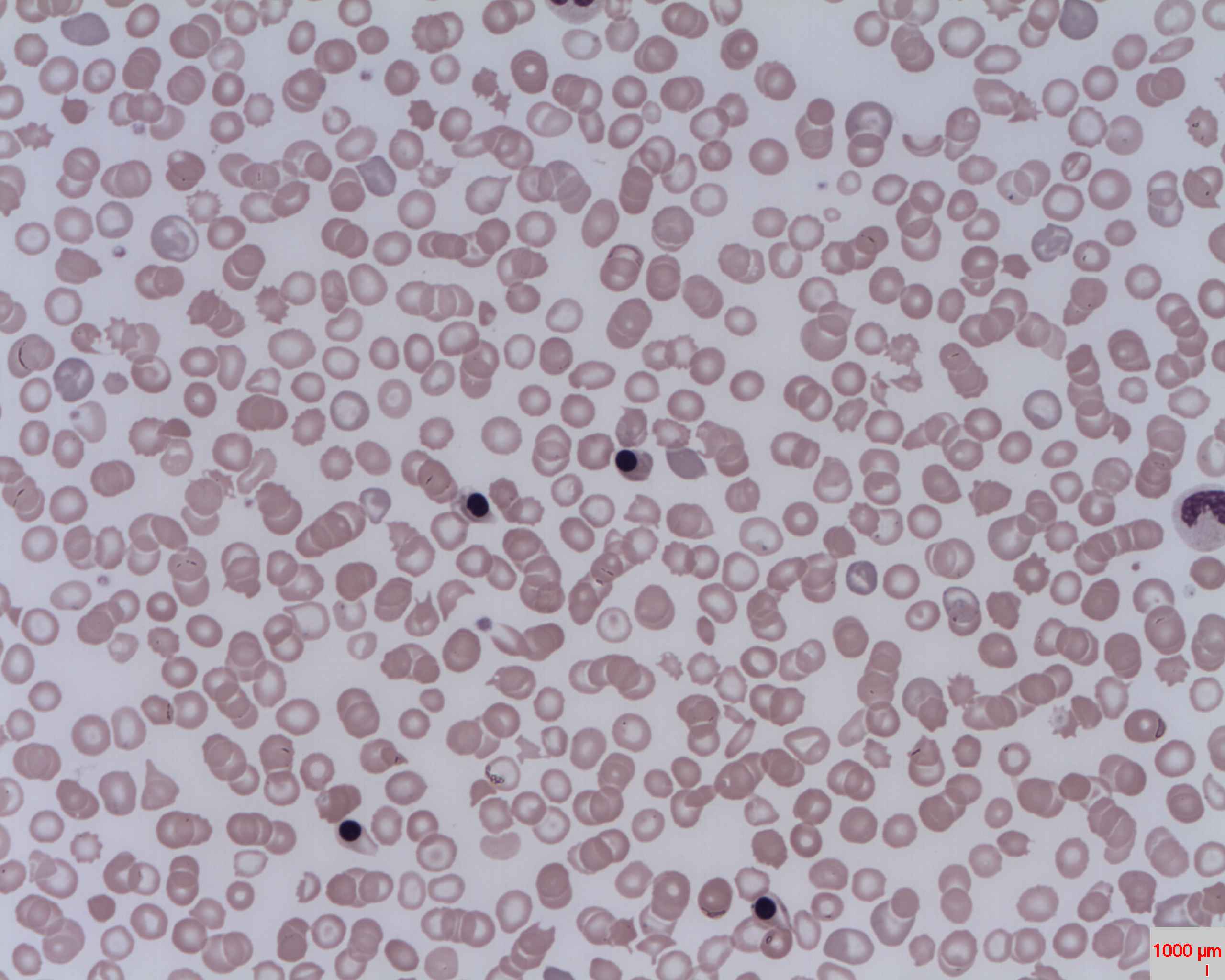
A nucleated red blood cell (NRBC), also known by several other names, is a red blood cell that contains a cell nucleus. Almost all vertebrate organisms have hemoglobin-containing cells in their blood, and with the exception of mammals, all of these red blood cells are nucleated. In mammals, NRBCs occur in normal development as precursors to mature red blood cells in erythropoiesis, the process by which the body produces red blood cells. NRBCs are normally found in the bone marrow of humans of all ages and in the blood of fetuses and newborn infants. After infancy, RBCs normally contain a nucleus only during the very early stages of the cell's life, and the nucleus is ejected as a normal part of cellular differentiation before the cell is released into the bloodstream. Thus, if NRBCs are identified on an adult's complete blood count or peripheral blood smear, it suggests that there is a very high demand for the bone marrow to produce RBCs, and immature RBCs are being released in ... [...More Info...] [...Related Items...] OR: [Wikipedia] [Google] [Baidu] |
Bone Marrow
Bone marrow is a semi-solid tissue found within the spongy (also known as cancellous) portions of bones. In birds and mammals, bone marrow is the primary site of new blood cell production (or haematopoiesis). It is composed of hematopoietic cells, marrow adipose tissue, and supportive stromal cells. In adult humans, bone marrow is primarily located in the ribs, vertebrae, sternum, and bones of the pelvis. Bone marrow comprises approximately 5% of total body mass in healthy adult humans, such that a man weighing 73 kg (161 lbs) will have around 3.7 kg (8 lbs) of bone marrow. Human marrow produces approximately 500 billion blood cells per day, which join the systemic circulation via permeable vasculature sinusoids within the medullary cavity. All types of hematopoietic cells, including both myeloid and lymphoid lineages, are created in bone marrow; however, lymphoid cells must migrate to other lymphoid organs (e.g. thymus) in order to complete maturation. ... [...More Info...] [...Related Items...] OR: [Wikipedia] [Google] [Baidu] |
Lymphoblast
__NOTOC__ A lymphoblast is a modified naive lymphocyte with altered cell morphology. It occurs when the lymphocyte is activated by an antigen (from antigen-presenting cells) and increased in volume by nucleus and cytoplasm growth as well as new mRNA and protein synthesis. The lymphoblast then starts dividing two to four times every 24 hours for three to five days, with a single lymphoblast making approximately 1000 clones of its original naive lymphocyte, with each clone sharing the originally unique antigen specificity. Finally the dividing cells differentiate into effector cells, known as plasma cells (for B cells), cytotoxic T cells, and helper T cells. Lymphoblasts can also refer to immature cells which typically differentiate to form mature lymphocytes. Normally lymphoblasts are found in the bone marrow, but in acute lymphoblastic leukemia (ALL), lymphoblasts proliferate uncontrollably and are found in large numbers in the peripheral blood. The size is between 10 and 20 μm. ... [...More Info...] [...Related Items...] OR: [Wikipedia] [Google] [Baidu] |
Melanoblast
A melanoblast is a precursor cell of a melanocyte. These cells migrate from the trunk neural crest cells (in terms of axial level from neck to posterior end) dorsolaterally between the ectoderm and dorsal surface of the somites. See also *Biological pigment *List of human cell types derived from the germ layers This is a list of cells in humans derived from the three embryonic germ layers – ectoderm, mesoderm, and endoderm. Cells derived from ectoderm Surface ectoderm Skin * Trichocyte * Keratinocyte Anterior pituitary * Gonadotrope * Corticotro ... References Pigments Biomolecules Pigmentation {{developmental-biology-stub ... [...More Info...] [...Related Items...] OR: [Wikipedia] [Google] [Baidu] |
Promegakaryocyte
A promegakaryocyte is a precursor cell for a megakaryocyte. It arises from a megakaryoblast, into a promegakaryocyte and then into a megakaryocyte, which will eventually break off and become a platelet. The developmental stages of the megakaryocyte are: CFU-Me (pluripotential hemopoietic stem cell or hemocytoblast) → megakaryoblast → promegakaryocyte → megakaryocyte. When the megakaryoblast matures into the promegakaryocyte, it undergoes endoreduplication and forms a promegakaryocyte which has multiple nuclei, azurophilic granule An azurophilic granule is a cellular object readily stainable with a Romanowsky stain. In white blood cells and hyperchromatin, staining imparts a burgundy or merlot coloration. Neutrophils in particular are known for containing azurophils load ...s, and a basophilic cytoplasm. The promegakaryocyte has rotary motion, but no forward migration. Promegakaryocytes and other precursor cells to megakaryocytes arise from pluripotential hematopoietic ... [...More Info...] [...Related Items...] OR: [Wikipedia] [Google] [Baidu] |


