|
Left Gastroepiploic Artery
The left gastroepiploic artery (or left gastro-omental artery), the largest branch of the splenic artery, runs from left to right about a finger's breadth or more from the greater curvature of the stomach, between the layers of the greater omentum, and anastomoses with the right gastroepiploic (a branch of the right gastro-duodenal artery originating from the hepatic branch of the coeliac trunk). In its course it distributes: * "Gastric branches": several ascending branches to both surfaces of the stomach; * "Omental branches": descend to supply the greater omentum and anastomose with branches of the middle colic. Additional images File:Gray533.png, Branches of the celiac artery The celiac () artery (also spelled ''coeliac''), also known as the celiac trunk or truncus coeliacus, is the first major branch of the abdominal aorta. It is about 1.25 cm in length. Branching from the aorta at thoracic vertebra 12 (T12) in .... References External links * - "Stomach, Sp ... [...More Info...] [...Related Items...] OR: [Wikipedia] [Google] [Baidu] |
Celiac Artery
The celiac () artery (also spelled ''coeliac''), also known as the celiac trunk or truncus coeliacus, is the first major branch of the abdominal aorta. It is about 1.25 cm in length. Branching from the aorta at thoracic vertebra 12 (T12) in humans, it is one of three anterior/ midline branches of the abdominal aorta (the others are the superior and inferior mesenteric arteries). Structure The celiac artery is the first major branch of the descending abdominal aorta, branching at a 90° angle. This occurs just below the crus of the diaphragm. This is around the first lumbar vertebra. There are three main divisions of the celiac artery, and each in turn has its own named branches: The celiac artery may also give rise to the inferior phrenic arteries. Function The celiac artery supplies oxygenated blood to the liver, stomach, abdominal esophagus, spleen, and the superior half of both the duodenum and the pancreas. These structures correspond to the embryonic foregut. (Si ... [...More Info...] [...Related Items...] OR: [Wikipedia] [Google] [Baidu] |
Splenic Artery
In human anatomy, the splenic artery or lienal artery is the blood vessel that supplies oxygenated blood to the spleen. It branches from the celiac artery, and follows a course superior to the pancreas. It is known for its tortuous path to the spleen. Structure The splenic artery gives off branches to the stomach and pancreas before reaching the spleen. Note that the branches of the splenic artery do not reach all the way to the lower part of the greater curvature of the stomach. Instead, that region is supplied by the right gastroepiploic artery, a branch of the gastroduodenal artery. The two gastroepiploic arteries anastomose with each other at that point. Relations The splenic artery passes between the layers of the lienorenal ligament. Along its course, it is accompanied by a similarly named vein, the splenic vein, which drains into the hepatic portal vein. Clinical significance Splenic artery aneurysms are rare, but still the third most common abdominal aneurysm, afte ... [...More Info...] [...Related Items...] OR: [Wikipedia] [Google] [Baidu] |
Left Gastroepiploic Vein .
The splenic vein and superior mesenteric vein join to form the hepatic portal vein.
The left gastroepiploic vein (left gastro-omental vein) receives branches from the antero-superior and postero-inferior surfaces of the stomach and from the greater omentum; it runs from right to left along the greater curvature of the stomach and ends in the commencement of the splenic vein The spleen is an organ (biology), organ found in almost all vertebrates. Similar in structure to a large lymph node, it acts primarily as a blood filter. The word spleen comes . References Veins of the torso Stomach {{circulatory-stub ...[...More Info...] [...Related Items...] OR: [Wikipedia] [Google] [Baidu] |
Greater Curvature Of The Stomach
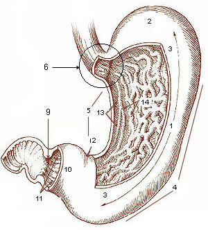
The curvatures of the stomach refer to the greater and lesser curvatures. The greater curvature of the stomach is four or five times as long as the lesser curvature. Greater curvature The greater curvature of the stomach forms the lower left or lateral border of the stomach. Surface Starting from the cardiac orifice at the incisura cardiaca, it forms an arch backward, upward, and to the left; the highest point of the convexity is on a level with the sixth left costal cartilage. From this level it may be followed downward and forward, with a slight convexity to the left as low as the cartilage of the ninth rib; it then turns to the right, to the end of the pylorus. Directly opposite the incisura angularis of the lesser curvature the greater curvature presents a dilatation, which is the left extremity of the pyloric part; this dilatation is limited on the right by a slight groove, the sulcus intermedius, which is about 2.5 cm, from the duodenopyloric constriction. The ... [...More Info...] [...Related Items...] OR: [Wikipedia] [Google] [Baidu] |
Splenic Artery
In human anatomy, the splenic artery or lienal artery is the blood vessel that supplies oxygenated blood to the spleen. It branches from the celiac artery, and follows a course superior to the pancreas. It is known for its tortuous path to the spleen. Structure The splenic artery gives off branches to the stomach and pancreas before reaching the spleen. Note that the branches of the splenic artery do not reach all the way to the lower part of the greater curvature of the stomach. Instead, that region is supplied by the right gastroepiploic artery, a branch of the gastroduodenal artery. The two gastroepiploic arteries anastomose with each other at that point. Relations The splenic artery passes between the layers of the lienorenal ligament. Along its course, it is accompanied by a similarly named vein, the splenic vein, which drains into the hepatic portal vein. Clinical significance Splenic artery aneurysms are rare, but still the third most common abdominal aneurysm, afte ... [...More Info...] [...Related Items...] OR: [Wikipedia] [Google] [Baidu] |
Stomach
The stomach is a muscular, hollow organ in the gastrointestinal tract of humans and many other animals, including several invertebrates. The stomach has a dilated structure and functions as a vital organ in the digestive system. The stomach is involved in the gastric phase of digestion, following chewing. It performs a chemical breakdown by means of enzymes and hydrochloric acid. In humans and many other animals, the stomach is located between the oesophagus and the small intestine. The stomach secretes digestive enzymes and gastric acid to aid in food digestion. The pyloric sphincter controls the passage of partially digested food ( chyme) from the stomach into the duodenum, where peristalsis takes over to move this through the rest of intestines. Structure In the human digestive system, the stomach lies between the oesophagus and the duodenum (the first part of the small intestine). It is in the left upper quadrant of the abdominal cavity. The top of the stomach lies ag ... [...More Info...] [...Related Items...] OR: [Wikipedia] [Google] [Baidu] |
Greater Omentum
The greater omentum (also the great omentum, omentum majus, gastrocolic omentum, epiploon, or, especially in animals, caul) is a large apron-like fold of visceral peritoneum that hangs down from the stomach. It extends from the greater curvature of the stomach, passing in front of the small intestines and doubles back to ascend to the transverse colon before reaching to the posterior abdominal wall. The greater omentum is larger than the lesser omentum, which hangs down from the liver to the lesser curvature. The common anatomical term "epiploic" derives from "epiploon", from the Greek ''epipleein'', meaning to float or sail on, since the greater omentum appears to float on the surface of the intestines. It is the first structure observed when the abdominal cavity is opened anteriorly (from the front). Structure The greater omentum is the larger of the two peritoneal folds. It consists of a double sheet of peritoneum, folded on itself so that it has four layers. The two layers o ... [...More Info...] [...Related Items...] OR: [Wikipedia] [Google] [Baidu] |
Right Gastroepiploic
The right gastroepiploic artery (or right gastro-omental artery) is one of the two terminal branches of the gastroduodenal artery In anatomy, the gastroduodenal artery is a small blood vessel in the abdomen. It supplies blood directly to the pylorus (distal part of the stomach) and proximal part of the duodenum. It also indirectly supplies the pancreatic head (via the anteri .... It runs from right to left along the greater curvature of the stomach, between the layers of the greater omentum, anastomosing with the left gastroepiploic artery, a branch of the splenic artery. Except at the pylorus where it is in contact with the stomach, it lies about a finger's breadth from the greater curvature. Branches This vessel gives off numerous branches: * "gastric branches": ascend to supply both surfaces of the stomach. * "omental branches": descend to supply the greater omentum and anastomose with branches of the middle colic. Use in coronary artery surgery The right gastroepiploic ar ... [...More Info...] [...Related Items...] OR: [Wikipedia] [Google] [Baidu] |
Middle Colic
The middle colic artery is an artery of the abdomen; a branch of the superior mesenteric artery distributed to parts of the ascending and transverse colon. It usually divides into two terminal branches - a left one and a right one - which go on to form anastomoses with the left colic artery, and right colic artery (respectively), thus participating in the formation of the marginal artery of the colon. Parts of the artery may be removed in different types of hemicolectomy. Structure The middle colic artery supplies the superior/distal part of the ascending colon and right/proximal two-thirds of the transverse colon. Origin The middle colic artery is a branch of the superior mesenteric artery, branching off from its right aspect. Its origin is situated just inferior the neck of the pancreas. It may share a common origin with the right colic artery. Course The middle colic artery passes anterosuperiorly between the layers of the transverse mesocolon just right of the midlin ... [...More Info...] [...Related Items...] OR: [Wikipedia] [Google] [Baidu] |
Stomach Blood Supply
The stomach is a muscular, hollow organ in the gastrointestinal tract of humans and many other animals, including several invertebrates. The stomach has a dilated structure and functions as a vital organ in the digestive system. The stomach is involved in the gastric phase of digestion, following chewing. It performs a chemical breakdown by means of enzymes and hydrochloric acid. In humans and many other animals, the stomach is located between the oesophagus and the small intestine. The stomach secretes digestive enzymes and gastric acid to aid in food digestion. The pyloric sphincter controls the passage of partially digested food (chyme) from the stomach into the duodenum, where peristalsis takes over to move this through the rest of intestines. Structure In the human digestive system, the stomach lies between the oesophagus and the duodenum (the first part of the small intestine). It is in the left upper quadrant of the abdominal cavity. The top of the stomach lies against ... [...More Info...] [...Related Items...] OR: [Wikipedia] [Google] [Baidu] |
Arteries Of The Abdomen
An artery (plural arteries) () is a blood vessel in humans and most animals that takes blood away from the heart to one or more parts of the body (tissues, lungs, brain etc.). Most arteries carry oxygenated blood; the two exceptions are the pulmonary and the umbilical arteries, which carry deoxygenated blood to the organs that oxygenate it (lungs and placenta, respectively). The effective arterial blood volume is that extracellular fluid which fills the arterial system. The arteries are part of the circulatory system, that is responsible for the delivery of oxygen and nutrients to all cells, as well as the removal of carbon dioxide and waste products, the maintenance of optimum blood pH, and the circulation of proteins and cells of the immune system. Arteries contrast with veins, which carry blood back towards the heart. Structure The anatomy of arteries can be separated into gross anatomy, at the macroscopic level, and microanatomy, which must be studied with a microscop ... [...More Info...] [...Related Items...] OR: [Wikipedia] [Google] [Baidu] |


