|
Latissimus
The latissimus dorsi () is a large, flat muscle on the back that stretches to the sides, behind the arm, and is partly covered by the trapezius on the back near the midline. The word latissimus dorsi (plural: ''latissimi dorsorum'') comes from Latin and means "broadest uscleof the back", from "latissimus" ( la, broadest)' and "dorsum" ( la, back). The pair of muscles are commonly known as "lats", especially among bodybuilders. The latissimus dorsi is the largest muscle in the upper body. The latissimus dorsi is responsible for extension, adduction, transverse extension also known as horizontal abduction (or horizontal extension), flexion from an extended position, and (medial) internal rotation of the shoulder joint. It also has a synergistic role in extension and lateral flexion of the lumbar spine. Due to bypassing the scapulothoracic joints and attaching directly to the spine, the actions the latissimi dorsi have on moving the arms can also influence the movement of th ... [...More Info...] [...Related Items...] OR: [Wikipedia] [Google] [Baidu] |
Iliac Crest
The crest of the ilium (or iliac crest) is the superior border of the wing of ilium and the superiolateral margin of the greater pelvis. Structure The iliac crest stretches posteriorly from the anterior superior iliac spine (ASIS) to the posterior superior iliac spine (PSIS). Behind the ASIS, it divides into an outer and inner lip separated by the intermediate zone. The outer lip bulges laterally into the iliac tubercle. Platzer (2004), p 186 Palpable in its entire length, the crest is convex superiorly but is sinuously curved, being concave inward in front, concave outward behind. Palastanga (2006), p 243 It is thinner at the center than at the extremities. Development The iliac crest is derived from endochondral bone. Function To the external lip are attached the ''Tensor fasciae latae'', '' Obliquus externus abdominis'', and '' Latissimus dorsi'', and along its whole length the '' fascia lata''; to the intermediate line, the '' Obliquus internus abdominis''. To the inter ... [...More Info...] [...Related Items...] OR: [Wikipedia] [Google] [Baidu] |
Thoracodorsal Nerve
The thoracodorsal nerve is a nerve present in humans and other animals, also known as the middle subscapular nerve or the long subscapular nerve. It supplies the latissimus dorsi muscle. Structure The thoracodorsal nerve arises from the brachial plexus. It derives its fibers from the sixth, seventh, and eighth cervical nerves. It is derived from their ventral rami, in spite of the fact that the latissimus dorsi is found in the back. The thoracodorsal nerve is a branch of the posterior cord of the brachial plexus, and is made up of fibres from the posterior divisions of all three trunks of the brachial plexus. It follows the course of the subscapular artery, along the posterior wall of the axilla to the latissimus dorsi muscle, in which it may be traced as far as the lower border of the muscle. Function The thoracodorsal nerve innervates the latissimus dorsi muscle on its deep surface. Clinical Significance The latissimus dorsi is occasionally used for transplantation, an ... [...More Info...] [...Related Items...] OR: [Wikipedia] [Google] [Baidu] |
Inferior Angle Of The Scapula
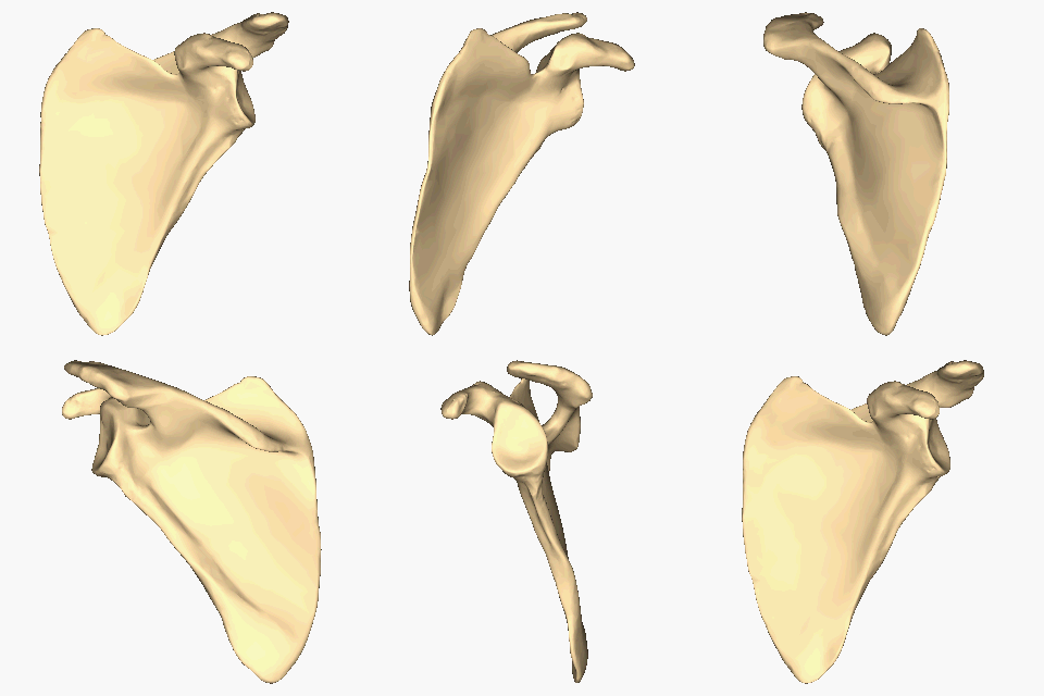
The scapula (plural scapulae or scapulas), also known as the shoulder blade, is the bone that connects the humerus (upper arm bone) with the clavicle (collar bone). Like their connected bones, the scapulae are paired, with each scapula on either side of the body being roughly a mirror image of the other. The name derives from the Classical Latin word for trowel or small shovel, which it was thought to resemble. In compound terms, the prefix omo- is used for the shoulder blade in medical terminology. This prefix is derived from ὦμος (ōmos), the Ancient Greek word for shoulder, and is cognate with the Latin , which in Latin signifies either the shoulder or the upper arm bone. The scapula forms the back of the shoulder girdle. In humans, it is a flat bone, roughly triangular in shape, placed on a posterolateral aspect of the thoracic cage. Structure The scapula is a thick, flat bone lying on the thoracic wall that provides an attachment for three groups of muscles: intrin ... [...More Info...] [...Related Items...] OR: [Wikipedia] [Google] [Baidu] |
Scapula
The scapula (plural scapulae or scapulas), also known as the shoulder blade, is the bone that connects the humerus (upper arm bone) with the clavicle (collar bone). Like their connected bones, the scapulae are paired, with each scapula on either side of the body being roughly a mirror image of the other. The name derives from the Classical Latin word for trowel or small shovel, which it was thought to resemble. In compound terms, the prefix omo- is used for the shoulder blade in medical terminology. This prefix is derived from ὦμος (ōmos), the Ancient Greek word for shoulder, and is cognate with the Latin , which in Latin signifies either the shoulder or the upper arm bone. The scapula forms the back of the shoulder girdle. In humans, it is a flat bone, roughly triangular in shape, placed on a posterolateral aspect of the thoracic cage. Structure The scapula is a thick, flat bone lying on the thoracic wall that provides an attachment for three groups of muscles: intrin ... [...More Info...] [...Related Items...] OR: [Wikipedia] [Google] [Baidu] |
Thoracodorsal Artery
The thoracodorsal artery is a branch of the subscapular artery. It travels inferiorly with the thoracodorsal nerve and supplies the latissimus dorsi The latissimus dorsi () is a large, flat muscle on the back that stretches to the sides, behind the arm, and is partly covered by the trapezius on the back near the midline. The word latissimus dorsi (plural: ''latissimi dorsorum'') comes from L .... External links * - "The axillary artery and its major branches shown in relation to major landmarks." * - "Major Branches of the Axillary Artery" * Arteries of the upper limb {{circulatory-stub ... [...More Info...] [...Related Items...] OR: [Wikipedia] [Google] [Baidu] |
Pull Up (exercise)
A pull-up is an upper-body strength exercise. The pull-up is a closed-chain movement where the body is suspended by the hands, gripping a bar or other implement at a distance typically wider than shoulder-width, and pulled up. As this happens, the elbows flex and the shoulders adduct and extend to bring the elbows to the torso. Pull-ups build up several muscles of the upper body, including the latissimus dorsi, trapezius, and biceps brachii. A pull-up may be performed with overhand (pronated), underhand (supinated)—sometimes referred to as a chin-up—neutral, or rotating hand position. Pull-ups are used by some organizations as a component of fitness tests, and as a conditioning activity for some sports. Movement Beginning by hanging from the bar, the body is pulled up vertically. From the top position, the participant lowers their body until the arms and shoulders are fully extended. The end range of motion at the top end may be chin over bar or higher, such as chest to b ... [...More Info...] [...Related Items...] OR: [Wikipedia] [Google] [Baidu] |
Subscapular Artery
The subscapular artery, the largest branch of the axillary artery, arises from the third part of the axillary artery at the lower border of the subscapularis muscle, which it follows to the inferior angle of the scapula, where it anastomoses with the lateral thoracic and intercostal arteries, and with the descending branch of the dorsal scapular artery (a.k.a. deep branch of the transverse cervical artery if it arises from the cervical trunk), and ends in the neighboring muscles. About 4 cm from its origin it gives off two branches, first the scapular circumflex artery and then the thoracodorsal artery. From the thoracodorsal artery it supplies latissimus dorsi, while the scapular circumflex artery participates in the scapular anastamosis. It terminates in an anastomosis with the dorsal scapular artery The transverse cervical artery (transverse artery of neck or transversa colli artery) is an artery in the neck and a branch of the thyrocervical trunk, running at a h ... [...More Info...] [...Related Items...] OR: [Wikipedia] [Google] [Baidu] |
Trapezius Muscle
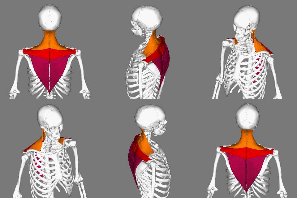
The trapezius is a large paired trapezoid-shaped surface muscle that extends longitudinally from the occipital bone to the lower thoracic vertebrae of the spine and laterally to the spine of the scapula. It moves the scapula and supports the arm. The trapezius has three functional parts: an upper (descending) part which supports the weight of the arm; a middle region (transverse), which retracts the scapula; and a lower (ascending) part which medially rotates and depresses the scapula. Name and history The trapezius muscle resembles a trapezium, also known as a trapezoid, or diamond-shaped quadrilateral. The word "spinotrapezius" refers to the human trapezius, although it is not commonly used in modern texts. In other mammals, it refers to a portion of the analogous muscle. Similarly, the term "tri-axle back plate" was historically used to describe the trapezius muscle. Structure The ''superior'' or ''upper'' (or descending) fibers of the trapezius originate from the sp ... [...More Info...] [...Related Items...] OR: [Wikipedia] [Google] [Baidu] |
Trapezius
The trapezius is a large paired trapezoid-shaped surface muscle that extends longitudinally from the occipital bone to the lower thoracic vertebrae of the spine and laterally to the spine of the scapula. It moves the scapula and supports the arm. The trapezius has three functional parts: an upper (descending) part which supports the weight of the arm; a middle region (transverse), which retracts the scapula; and a lower (ascending) part which medially rotates and depresses the scapula. Name and history The trapezius muscle resembles a trapezium, also known as a trapezoid, or diamond-shaped quadrilateral. The word "spinotrapezius" refers to the human trapezius, although it is not commonly used in modern texts. In other mammals, it refers to a portion of the analogous muscle. Similarly, the term "tri-axle back plate" was historically used to describe the trapezius muscle. Structure The ''superior'' or ''upper'' (or descending) fibers of the trapezius originate from the sp ... [...More Info...] [...Related Items...] OR: [Wikipedia] [Google] [Baidu] |
Deltoid Muscle
The deltoid muscle is the muscle forming the rounded contour of the human shoulder. It is also known as the 'common shoulder muscle', particularly in other animals such as the domestic cat. Anatomically, the deltoid muscle appears to be made up of three distinct sets of muscle fibers, namely the # anterior or clavicular part (pars clavicularis) # posterior or scapular part (pars scapularis) # intermediate or acromial part (pars acromialis) However, electromyography suggests that it consists of at least seven groups that can be independently coordinated by the nervous system. It was previously called the deltoideus (plural ''deltoidei'') and the name is still used by some anatomists. It is called so because it is in the shape of the Greek capital letter delta (Δ). Deltoid is also further shortened in slang as "delt". A study of 30 shoulders revealed an average mass of in humans, ranging from to . Structure Previous studies showed that the insertions of the tendons of the delto ... [...More Info...] [...Related Items...] OR: [Wikipedia] [Google] [Baidu] |
Humerus
The humerus (; ) is a long bone in the arm that runs from the shoulder to the elbow. It connects the scapula and the two bones of the lower arm, the radius and ulna, and consists of three sections. The humeral upper extremity consists of a rounded head, a narrow neck, and two short processes (tubercles, sometimes called tuberosities). The body is cylindrical in its upper portion, and more prismatic below. The lower extremity consists of 2 epicondyles, 2 processes (trochlea & capitulum), and 3 fossae (radial fossa, coronoid fossa, and olecranon fossa). As well as its true anatomical neck, the constriction below the greater and lesser tubercles of the humerus is referred to as its surgical neck due to its tendency to fracture, thus often becoming the focus of surgeons. Etymology The word "humerus" is derived from la, humerus, umerus meaning upper arm, shoulder, and is linguistically related to Gothic ''ams'' shoulder and Greek ''ōmos''. Structure Upper extremity The upper or pr ... [...More Info...] [...Related Items...] OR: [Wikipedia] [Google] [Baidu] |
Adduction
Motion, the process of movement, is described using specific anatomical terms. Motion includes movement of organs, joints, limbs, and specific sections of the body. The terminology used describes this motion according to its direction relative to the anatomical position of the body parts involved. Anatomists and others use a unified set of terms to describe most of the movements, although other, more specialized terms are necessary for describing unique movements such as those of the hands, feet, and eyes. In general, motion is classified according to the anatomical plane it occurs in. ''Flexion'' and ''extension'' are examples of ''angular'' motions, in which two axes of a joint are brought closer together or moved further apart. ''Rotational'' motion may occur at other joints, for example the shoulder, and are described as ''internal'' or ''external''. Other terms, such as ''elevation'' and ''depression'', describe movement above or below the horizontal plane. Many anatomic ... [...More Info...] [...Related Items...] OR: [Wikipedia] [Google] [Baidu] |


