|
Lateral Sacral Artery
The lateral sacral arteries is an artery in the pelvis that arises from the posterior division of the internal iliac artery. It later splits into two smaller branches, a superior and an inferior. Structure The lateral sacral artery is the second branch of the posterior division of the internal iliac artery. It is a parietal branch. Superior The superior, of large size, passes medialward, and, after anastomosing with branches from the middle sacral, enters the first or second anterior sacral foramen, supplies branches to the contents of the sacral canal, and, escaping by the corresponding posterior sacral foramen, is distributed to the skin and muscles on the dorsum of the sacrum, anastomosing with the superior gluteal. Inferior The inferior runs obliquely across the front of the piriformis and the sacral nerves to the medial side of the anterior sacral foramina, descends on the front of the sacrum, and anastomoses over the coccyx with the middle sacral and opposite latera ... [...More Info...] [...Related Items...] OR: [Wikipedia] [Google] [Baidu] |
Internal Iliac Artery
The internal iliac artery (formerly known as the hypogastric artery) is the main artery of the pelvis. Structure The internal iliac artery supplies the walls and viscera of the pelvis, the buttock, the reproductive organs, and the medial compartment of the thigh. The vesicular branches of the internal iliac arteries supply the bladder. It is a short, thick vessel, smaller than the external iliac artery, and about 3 to 4 cm in length. Course The internal iliac artery arises at the bifurcation of the common iliac artery, opposite the lumbosacral articulation, and, passing downward to the upper margin of the greater sciatic foramen, divides into two large trunks, an anterior and a posterior. It is posterior to the ureter, anterior to the internal iliac vein, anterior to the lumbosacral trunk, and anterior to the piriformis muscle. Near its origin, it is medial to the external iliac vein, which lies between it and the psoas major muscle. It is above the obturator nerve. Bra ... [...More Info...] [...Related Items...] OR: [Wikipedia] [Google] [Baidu] |
Posterior Sacral Foramen
The sacrum (plural: ''sacra'' or ''sacrums''), in human anatomy, is a large, triangular bone at the base of the spine that forms by the fusing of the sacral vertebrae (S1S5) between ages 18 and 30. The sacrum situates at the upper, back part of the pelvic cavity, between the two wings of the pelvis. It forms joints with four other bones. The two projections at the sides of the sacrum are called the alae (wings), and articulate with the ilium at the L-shaped sacroiliac joints. The upper part of the sacrum connects with the last lumbar vertebra (L5), and its lower part with the coccyx (tailbone) via the sacral and coccygeal cornua. The sacrum has three different surfaces which are shaped to accommodate surrounding pelvic structures. Overall it is concave (curved upon itself). The base of the sacrum, the broadest and uppermost part, is tilted forward as the sacral promontory internally. The central part is curved outward toward the posterior, allowing greater room for the pelvi ... [...More Info...] [...Related Items...] OR: [Wikipedia] [Google] [Baidu] |
Cauda Equina
The cauda equina () is a bundle of spinal nerves and spinal nerve rootlets, consisting of the second through fifth lumbar nerve pairs, the first through fifth sacral nerve pairs, and the coccygeal nerve, all of which arise from the lumbar enlargement and the conus medullaris of the spinal cord. The cauda equina occupies the lumbar cistern, a subarachnoid space inferior to the conus medullaris. The nerves that compose the cauda equina innervate the pelvic organs and lower limbs to include motor innervation of the hips, knees, ankles, feet, internal anal sphincter and external anal sphincter. In addition, the cauda equina extends to sensory innervation of the perineum and, partially, parasympathetic innervation of the bladder. Structure In adulthood, the cauda equina is made of lumbosacral spinal nerve roots. Development In humans, the spinal cord stops growing in infancy and the end of the spinal cord is about the level of the third lumbar vertebra, or L3, at birth. Because the ... [...More Info...] [...Related Items...] OR: [Wikipedia] [Google] [Baidu] |
Blood
Blood is a body fluid in the circulatory system of humans and other vertebrates that delivers necessary substances such as nutrients and oxygen to the cells, and transports metabolic waste products away from those same cells. Blood in the circulatory system is also known as ''peripheral blood'', and the blood cells it carries, ''peripheral blood cells''. Blood is composed of blood cells suspended in blood plasma. Plasma, which constitutes 55% of blood fluid, is mostly water (92% by volume), and contains proteins, glucose, mineral ions, hormones, carbon dioxide (plasma being the main medium for excretory product transportation), and blood cells themselves. Albumin is the main protein in plasma, and it functions to regulate the colloidal osmotic pressure of blood. The blood cells are mainly red blood cells (also called RBCs or erythrocytes), white blood cells (also called WBCs or leukocytes) and platelets (also called thrombocytes). The most abundant cells in vertebrate blo ... [...More Info...] [...Related Items...] OR: [Wikipedia] [Google] [Baidu] |
Posterior Sacral Foramina
The sacrum (plural: ''sacra'' or ''sacrums''), in human anatomy, is a large, triangular bone at the base of the spine that forms by the fusing of the sacral vertebrae (S1S5) between ages 18 and 30. The sacrum situates at the upper, back part of the pelvic cavity, between the two wings of the pelvis. It forms joints with four other bones. The two projections at the sides of the sacrum are called the alae (wings), and articulate with the ilium at the L-shaped sacroiliac joints. The upper part of the sacrum connects with the last lumbar vertebra (L5), and its lower part with the coccyx (tailbone) via the sacral and coccygeal cornua. The sacrum has three different surfaces which are shaped to accommodate surrounding pelvic structures. Overall it is concave (curved upon itself). The base of the sacrum, the broadest and uppermost part, is tilted forward as the sacral promontory internally. The central part is curved outward toward the posterior, allowing greater room for the pelvi ... [...More Info...] [...Related Items...] OR: [Wikipedia] [Google] [Baidu] |
Coccyx
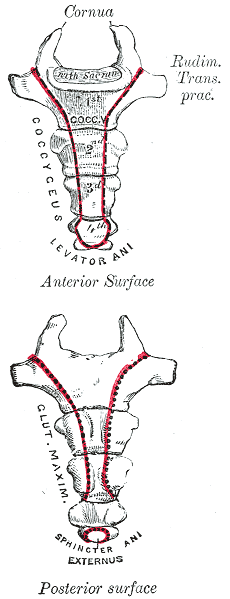
The coccyx ( : coccyges or coccyxes), commonly referred to as the tailbone, is the final segment of the vertebral column in all apes, and analogous structures in certain other mammals such as horses. In tailless primates (e.g. humans and other great apes) since ''Nacholapithecus'' (a Miocene hominoid),Nakatsukasa 2004, ''Acquisition of bipedalism'' (SeFig. 5entitled ''First coccygeal/caudal vertebra in short-tailed or tailless primates.''.) the coccyx is the remnant of a vestigial tail. In animals with bony tails, it is known as ''tailhead'' or ''dock'', in bird anatomy as ''tailfan''. It comprises three to five separate or fused coccygeal vertebrae below the sacrum, attached to the sacrum by a fibrocartilaginous joint, the sacrococcygeal symphysis, which permits limited movement between the sacrum and the coccyx. Structure The coccyx is formed of three, four or five rudimentary vertebrae. It articulates superiorly with the sacrum. In each of the first three segments may ... [...More Info...] [...Related Items...] OR: [Wikipedia] [Google] [Baidu] |
Sacral Nerves
A spinal nerve is a mixed nerve, which carries motor, sensory, and autonomic signals between the spinal cord and the body. In the human body there are 31 pairs of spinal nerves, one on each side of the vertebral column. These are grouped into the corresponding cervical, thoracic, lumbar, sacral and coccygeal regions of the spine. There are eight pairs of cervical nerves, twelve pairs of thoracic nerves, five pairs of lumbar nerves, five pairs of sacral nerves, and one pair of coccygeal nerves. The spinal nerves are part of the peripheral nervous system. Structure Each spinal nerve is a mixed nerve, formed from the combination of nerve fibers from its dorsal and ventral roots. The dorsal root is the afferent sensory root and carries sensory information to the brain. The ventral root is the efferent motor root and carries motor information from the brain. The spinal nerve emerges from the spinal column through an opening (intervertebral foramen) between adjacent vertebrae. Th ... [...More Info...] [...Related Items...] OR: [Wikipedia] [Google] [Baidu] |
Piriformis
The piriformis muscle () is a flat, pyramidally-shaped muscle in the buttock, gluteal region of the lower limbs. It is one of the six muscles in the lateral rotator group. The piriformis muscle has its origin upon the front surface of the sacrum, and inserts onto the greater trochanter of the femur. Depending upon the given position of the leg, it acts either as external (lateral) rotator of the thigh or as abductor of the thigh. It is innervated by the piriformis nerve. Structure The piriformis is a flat muscle, and is pyramidal in shape. Origin The piriformis muscle originates from the Pelvic surface of sacrum, anterior (front) surface of the sacrum by three fleshy digitations attached to the Sacrum, second, third, and fourth sacral vertebra. It also arises from the superior margin of the greater sciatic notch, the gluteal surface of the Ilium (bone), ilium (near the posterior inferior iliac spine), the sacroiliac joint capsule, and (sometimes) the sacrotuberous ligament (m ... [...More Info...] [...Related Items...] OR: [Wikipedia] [Google] [Baidu] |
Superior Gluteal Artery
The superior gluteal artery is the largest and final branch of the internal iliac artery. It is the continuation of the posterior division of that vessel. It is a short artery which runs backward between the lumbosacral trunk and the first sacral nerve. It divides into a superficial and a deep branch after passing out of the pelvis above the upper border of the piriformis muscle. Within the pelvis, it gives off branches to the iliacus, piriformis, and obturator internus muscles. Just previous to exiting the pelvic cavity, it also gives off a nutrient artery which enters the ilium. Structure The superior gluteal artery is the largest and final branch of the internal iliac artery. It branches from the posterior division of the internal iliac artery. It exits the pelvis through the greater sciatic foramen. It divides into a superficial and a deep branch. Superficial branch The superficial branch passes over the piriformis muscle. It enters the deep surface of the gluteus maximus mus ... [...More Info...] [...Related Items...] OR: [Wikipedia] [Google] [Baidu] |
Dorsum (anatomy)
Standard anatomical terms of location are used to unambiguously describe the anatomy of animals, including humans. The terms, typically derived from Latin or Greek roots, describe something in its standard anatomical position. This position provides a definition of what is at the front ("anterior"), behind ("posterior") and so on. As part of defining and describing terms, the body is described through the use of anatomical planes and anatomical axes. The meaning of terms that are used can change depending on whether an organism is bipedal or quadrupedal. Additionally, for some animals such as invertebrates, some terms may not have any meaning at all; for example, an animal that is radially symmetrical will have no anterior surface, but can still have a description that a part is close to the middle ("proximal") or further from the middle ("distal"). International organisations have determined vocabularies that are often used as standard vocabularies for subdisciplines of anatom ... [...More Info...] [...Related Items...] OR: [Wikipedia] [Google] [Baidu] |
Sacral Canal
The sacrum (plural: ''sacra'' or ''sacrums''), in human anatomy, is a large, triangular bone at the base of the spine that forms by the fusing of the sacral vertebrae (S1S5) between ages 18 and 30. The sacrum situates at the upper, back part of the pelvic cavity, between the two wings of the pelvis. It forms joints with four other bones. The two projections at the sides of the sacrum are called the alae (wings), and articulate with the ilium at the L-shaped sacroiliac joints. The upper part of the sacrum connects with the last lumbar vertebra (L5), and its lower part with the coccyx (tailbone) via the sacral and coccygeal cornua. The sacrum has three different surfaces which are shaped to accommodate surrounding pelvic structures. Overall it is concave (curved upon itself). The base of the sacrum, the broadest and uppermost part, is tilted forward as the sacral promontory internally. The central part is curved outward toward the posterior, allowing greater room for the pelvi ... [...More Info...] [...Related Items...] OR: [Wikipedia] [Google] [Baidu] |
Lateral Sacral Veins
The lateral sacral veins accompany the lateral sacral arteries on the anterior surface of the sacrum The sacrum (plural: ''sacra'' or ''sacrums''), in human anatomy, is a large, triangular bone at the base of the spine that forms by the fusing of the sacral vertebrae (S1S5) between ages 18 and 30. The sacrum situates at the upper, back part ... and end in the hypogastric vein. Additional images File:Gray612.png, Iliopelvic glands (lateral view). References Veins of the torso {{circulatory-stub ... [...More Info...] [...Related Items...] OR: [Wikipedia] [Google] [Baidu] |


