|
Laser Doppler Imaging
Laser Doppler imaging (LDI) is an imaging method that uses a laser beam to scan live tissue. When the laser light reaches the tissue, the moving blood cells generate doppler components in the reflected ( backscattered) light. The light that comes back is detected using a photodiode that converts it into an electrical signal. Then the signal is processed to calculate a signal that is proportional to the tissue perfusion in the scanned area. When the process is completed, the signal is processed to generate an image that shows the perfusion on a screen. The laser doppler effect was first used to measure microcirculation by Stern M.D. in 1975. And it is used widely in medicine, some representative research work about it are these: Use in Ophthalmology The eye offers a unique opportunity for the non-invasive exploration of cardiovascular diseases. LDI by digital holography can measure blood flow in the retina and choroid. In particular, the choroid is a highly vascularized tissue s ... [...More Info...] [...Related Items...] OR: [Wikipedia] [Google] [Baidu] |
Microangiography
Microangiography ( ) is a type of angiography that consists of the radiography of small blood or lymphatic vessels of an organ. While most other types of angiography cannot produce images of vessels smaller than 200 µm in diameter, microangiography does just that. A microangiographic image is the result of injection of a contrast medium into either the blood or the lymphatic system and, then, enlargement of the resulting radiograph. Thus, an image is obtained in which there is contrast between vessel and surrounding tissue. It is often used in order to detect microvascular lesions in organs. But, it has been suggested that microangiography can also be used to detect tumors through visualization of tumor-induced small blood vessels. This is because tumor growths require vascularization before they can develop more rapidly. A few of the commonly used types are fluorescent, silicone rubber, and synchrotron radiation microangiography. Common types Fluorescent microangiography ... [...More Info...] [...Related Items...] OR: [Wikipedia] [Google] [Baidu] |
Systole
Systole ( ) is the part of the cardiac cycle during which some chambers of the heart contract after refilling with blood. The term originates, via New Latin, from Ancient Greek (''sustolē''), from (''sustéllein'' 'to contract'; from ''sun'' 'together' + ''stéllein'' 'to send'), and is similar to the use of the English term ''to squeeze''. The mammalian heart has four chambers: the left atrium above the left ventricle (lighter pink, see graphic), which two are connected through the mitral (or bicuspid) valve; and the right atrium above the right ventricle (lighter blue), connected through the tricuspid valve. The atria are the receiving blood chambers for the circulation of blood and the ventricles are the discharging chambers. In late ventricular diastole, the atrial chambers contract and send blood to the larger, lower ventricle chambers. This flow fills the ventricles with blood, and the resulting pressure closes the valves to the atria. The ventricles now perform i ... [...More Info...] [...Related Items...] OR: [Wikipedia] [Google] [Baidu] |
Hot-wire Anemometry
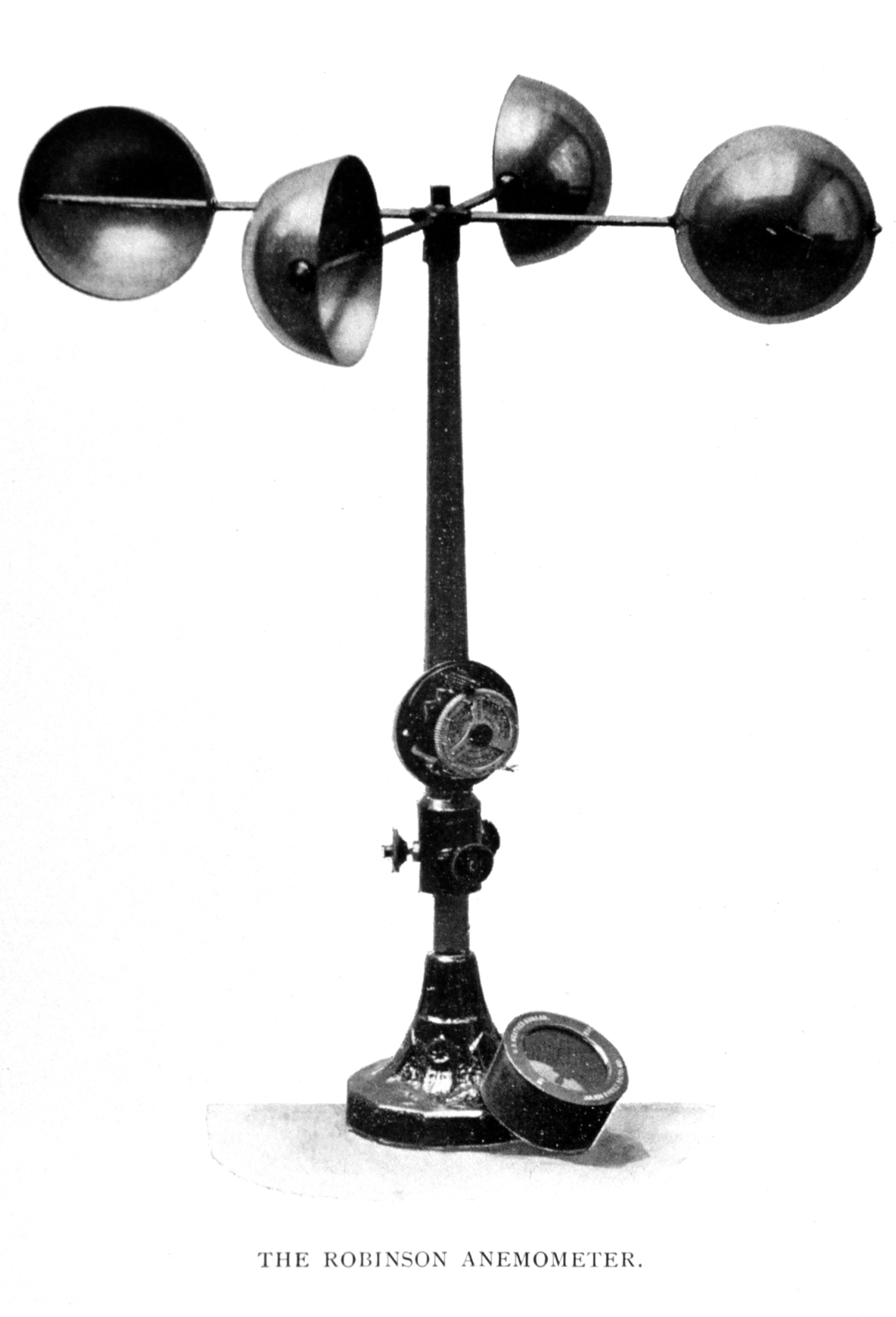
In meteorology, an anemometer () is a device that measures wind speed and direction. It is a common instrument used in weather stations. The earliest known description of an anemometer was by Italian architect and author Leon Battista Alberti (1404–1472) in 1450. History The anemometer has changed little since its development in the 15th century. Alberti is said to have invented it around 1450. In the ensuing centuries numerous others, including Robert Hooke (1635–1703), developed their own versions, with some mistakenly credited as its inventor. In 1846, John Thomas Romney Robinson (1792–1882) improved the design by using four hemispherical cups and mechanical wheels. In 1926, Canadian meteorologist John Patterson (1872–1956) developed a three-cup anemometer, which was improved by Brevoort and Joiner in 1935. In 1991, Derek Weston added the ability to measure wind direction. In 1994, Andreas Pflitsch developed the sonic anemometer. Velocity anemometers Cup anemo ... [...More Info...] [...Related Items...] OR: [Wikipedia] [Google] [Baidu] |


