|
List Of MeSH Codes (C17)
The following is a partial list of the "C" codes for Medical Subject Headings (MeSH), as defined by the United States National Library of Medicine (NLM). This list continues the information at List of MeSH codes (C16). Codes following these are found at List of MeSH codes (C18). For other MeSH codes, see List of MeSH codes. The source for this content is the set o2006 MeSH Treesfrom the NLM. ŌĆō skin and connective tissue diseases ŌĆō connective tissue diseases ŌĆō alpha 1-antitrypsin deficiency ŌĆō cartilage diseases * ŌĆō chondromalacia patellae * ŌĆō osteochondritis * ŌĆō polychondritis, relapsing * ŌĆō Tietze syndrome ŌĆō cellulitis ŌĆō collagen diseases * ŌĆō EhlersŌĆōDanlos syndrome * ŌĆō keloid * ŌĆō acne keloid * ŌĆō necrobiotic disorders * ŌĆō granuloma annulare * ŌĆō necrobiosis lipoidica * ŌĆō osteogenesis imperfecta ŌĆō cutis laxa ŌĆō dermatomyositis ŌĆō Dupuytren's contracture ŌĆō homocystinuria ŌĆō lupus erythematosus, cutaneous * Ō ... [...More Info...] [...Related Items...] OR: [Wikipedia] [Google] [Baidu] |
Medical Subject Headings
Medical Subject Headings (MeSH) is a comprehensive controlled vocabulary for the purpose of indexing journal articles and books in the life sciences. It serves as a thesaurus that facilitates searching. Created and updated by the United States National Library of Medicine (NLM), it is used by the MEDLINE/PubMed article database and by NLM's catalog of book holdings. MeSH is also used by ClinicalTrials.gov registry to classify which diseases are studied by trials registered in ClinicalTrials. MeSH was introduced in the 1960s, with the NLM's own index catalogue and the subject headings of the Quarterly Cumulative Index Medicus (1940 edition) as precursors. The yearly printed version of MeSH was discontinued in 2007; MeSH is now available only online. It can be browsed and downloaded free of charge through PubMed. Originally in English, MeSH has been translated into numerous other languages and allows retrieval of documents from different origins. Structure MeSH vocabulary is divi ... [...More Info...] [...Related Items...] OR: [Wikipedia] [Google] [Baidu] |
Acne Keloid
Keloid, also known as keloid disorder and keloidal scar, is the formation of a type of scar which, depending on its maturity, is composed mainly of either type III (early) or type I (late) collagen. It is a result of an overgrowth of granulation tissue (collagen type 3) at the site of a healed skin injury which is then slowly replaced by collagen type 1. Keloids are firm, rubbery lesions or shiny, fibrous nodules, and can vary from pink to the color of the person's skin or red to dark brown in color. A keloid scar is benign and not contagious, but sometimes accompanied by severe itchiness, pain, and changes in texture. In severe cases, it can affect movement of skin. In the United States keloid scars are seen 15 times more frequently in people of sub-Saharan African descent than in people of European descent. There is a higher tendency to develop a keloid among those with a family history of keloids and people between the ages of 10 and 30 years. Keloids should not be confused ... [...More Info...] [...Related Items...] OR: [Wikipedia] [Google] [Baidu] |
Lupus Nephritis
Lupus nephritis is an inflammation of the kidneys caused by systemic lupus erythematosus (SLE), an autoimmune disease. It is a type of glomerulonephritis in which the glomeruli become inflamed. Since it is a result of SLE, this type of glomerulonephritis is said to be ''secondary'', and has a different pattern and outcome from conditions with a ''primary'' cause originating in the kidney. The diagnosis of lupus nephritis depends on blood tests, urinalysis, X-rays, ultrasound scans of the kidneys, and a kidney biopsy. On urinalysis, a nephritic picture is found and red blood cell casts, red blood cells and proteinuria is found. Classification The World Health Organization has divided lupus nephritis into five stages based on the biopsy. This classification was defined in 1982 and revised in 1995. Class IV disease ( Diffuse proliferative nephritis) is both the most severe, and the most common subtype. Class VI (advanced sclerosing lupus nephritis) is a final class which is included ... [...More Info...] [...Related Items...] OR: [Wikipedia] [Google] [Baidu] |
Lupus Erythematosus, Systemic
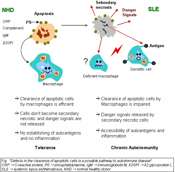
Lupus, technically known as systemic lupus erythematosus (SLE), is an autoimmune disease in which the body's immune system mistakenly attacks healthy tissue in many parts of the body. Symptoms vary among people and may be mild to severe. Common symptoms include painful and swollen joints, fever, chest pain, hair loss, mouth ulcers, swollen lymph nodes, feeling tired, and a red rash which is most commonly on the face. Often there are periods of illness, called flares, and periods of remission during which there are few symptoms. The cause of SLE is not clear. It is thought to involve a mixture of genetics combined with environmental factors. Among identical twins, if one is affected there is a 24% chance the other one will also develop the disease. Female sex hormones, sunlight, smoking, vitamin D deficiency, and certain infections are also believed to increase a person's risk. The mechanism involves an immune response by autoantibodies against a person's own tissues. These ... [...More Info...] [...Related Items...] OR: [Wikipedia] [Google] [Baidu] |
Discoid Lupus Erythematosus
Discoid lupus erythematosus is the most common type of chronic cutaneous lupus (CCLE), an autoimmune skin condition on the lupus erythematosus spectrum of illnesses. It presents with red, painful, inflamed and coin-shaped patches of skin with a scaly and crusty appearance, most often on the scalp, cheeks, and ears. Hair loss may occur if the lesions are on the scalp.James, William; Berger, Timothy; Elston, Dirk (2005). ''Andrews' Diseases of the Skin: Clinical Dermatology''. (10th ed.) Saunders. Chapter 8. . The lesions can then develop severe scarring, and the centre areas may appear lighter in color with a rim darker than the normal skin. These lesions can last for years without treatment. Patients with systemic lupus erythematous develop discoid lupus lesions with some frequency. However, patients who present initially with discoid lupus infrequently develop systemic lupus. Discoid lupus can be divided into localized, generalized, and childhood discoid lupus. The lesions are d ... [...More Info...] [...Related Items...] OR: [Wikipedia] [Google] [Baidu] |
Lupus Erythematosus, Cutaneous
Lupus, technically known as systemic lupus erythematosus (SLE), is an autoimmune disease in which the body's immune system mistakenly attacks healthy tissue in many parts of the body. Symptoms vary among people and may be mild to severe. Common symptoms include painful and swollen joints, fever, chest pain, hair loss, mouth ulcers, swollen lymph nodes, feeling tired, and a red rash which is most commonly on the face. Often there are periods of illness, called flares, and periods of remission during which there are few symptoms. The cause of SLE is not clear. It is thought to involve a mixture of genetics combined with environmental factors. Among identical twins, if one is affected there is a 24% chance the other one will also develop the disease. Female sex hormones, sunlight, smoking, vitamin D deficiency, and certain infections are also believed to increase a person's risk. The mechanism involves an immune response by autoantibodies against a person's own tissues ... [...More Info...] [...Related Items...] OR: [Wikipedia] [Google] [Baidu] |
Homocystinuria
Homocystinuria or HCU is an inherited disorder of the metabolism of the amino acid methionine due to a deficiency of cystathionine beta synthase or methionine synthase. It is an inherited autosomal recessive trait, which means a child needs to inherit a copy of the defective gene from both parents to be affected. Symptoms of homocystinuria can also be caused by a deficiency of vitamins B6, B12, or folate. Signs and symptoms This defect leads to a multi-systemic disorder of the connective tissue, muscles, central nervous system (CNS), and cardiovascular system. Homocystinuria represents a group of hereditary metabolic disorders characterized by an accumulation of the amino acid homocysteine in the serum and an increased excretion of homocysteine in the urine. Infants appear to be normal and early symptoms, if any are present, are vague. Signs and symptoms of homocystinuria that may be seen include the following: Cause It is usually caused by the deficiency of the enzyme cystathi ... [...More Info...] [...Related Items...] OR: [Wikipedia] [Google] [Baidu] |
Dupuytren's Contracture
Dupuytren's contracture (also called Dupuytren's disease, Morbus Dupuytren, Viking disease, palmar fibromatosis and Celtic hand) is a condition in which one or more fingers become progressively bent in a flexed position. It is named after Guillaume Dupuytren, who first described the underlying mechanism of action, followed by the first successful operation in 1831 and publication of the results in ''The Lancet'' in 1834. It usually begins as small, hard nodules just under the skin of the palm, then worsens over time until the fingers can no longer be fully straightened. While typically not painful, some aching or itching may be present. The ring finger followed by the little and middle fingers are most commonly affected. It can affect one or both hands. The condition can interfere with activities such as preparing food, writing, putting the hand in a tight pocket, putting on gloves, or shaking hands. The cause is unknown but might have a genetic component. Risk factors include ... [...More Info...] [...Related Items...] OR: [Wikipedia] [Google] [Baidu] |
Dermatomyositis
Dermatomyositis (DM) is a long-term inflammatory disorder which affects skin and the muscles. Its symptoms are generally a skin rash and worsening muscle weakness over time. These may occur suddenly or develop over months. Other symptoms may include weight loss, fever, lung inflammation, or light sensitivity. Complications may include calcium deposits in muscles or skin. The cause is unknown. Theories include that it is an autoimmune disease or a result of a viral infection. Dermatomyositis may develop as a paraneoplastic syndrome associated with several forms of malignancy. It is a type of inflammatory myopathy. Diagnosis is typically based on some combination of symptoms, blood tests, electromyography, and muscle biopsies. While no cure for the condition is known, treatments generally improve symptoms. Treatments may include medication, physical therapy, exercise, heat therapy, orthotics and assistive devices, and rest. Medications in the corticosteroids family are typic ... [...More Info...] [...Related Items...] OR: [Wikipedia] [Google] [Baidu] |
Cutis Laxa
Cutis laxa or pachydermatocele is a group of rare connective tissue disorders in which the skin becomes inelastic and hangs loosely in folds. Signs and symptoms It is characterised by skin that is loose, hanging, wrinkled, and lacking in elasticity. The loose skin can be either generalised or localised. Biopsies have shown reduction and degeneration of dermal elastic fibres in the affected areas of skin. The loose skin is often most noticeable on the face, resulting in a prematurely aged appearance. The affected areas of skin may be thickened and dark. In addition, the joints may be loose ( hypermobile) because of lax ligaments and tendons. When cutis laxa is severe, it can also affect the internal organs. The lungs, heart (supravalvular pulmonary stenosis), intestines, or arteries may be affected with a variety of severe impairments. In some cases, hernias and outpouching of the bladder can be observed. Patients can also present with whites of the eyes that are blue. Causes In ... [...More Info...] [...Related Items...] OR: [Wikipedia] [Google] [Baidu] |
Osteogenesis Imperfecta
Osteogenesis imperfecta (; OI), colloquially known as brittle bone disease, is a group of genetic disorders that all result in bones that break easily. The range of symptomsŌĆöon the skeleton as well as on the body's other organsŌĆömay be mild to severe. Symptoms found in various types of OI include whites of the eye (sclerae) that are blue instead, short stature, loose joints, hearing loss, breathing problems and problems with the teeth (dentinogenesis imperfecta). Potentially life-threatening complications, all of which become more common in more severe OI, include: tearing ( dissection) of the major arteries, such as the aorta; pulmonary valve insufficiency secondary to distortion of the ribcage; and basilar invagination. The underlying mechanism is usually a problem with connective tissue due to a lack of, or poorly formed, type I collagen. In more than 90% of cases, OI occurs due to mutations in the ''COL1A1'' or ''COL1A2'' genes. These mutations may be inherited ... [...More Info...] [...Related Items...] OR: [Wikipedia] [Google] [Baidu] |


