|
Limbal Relaxing Incisions
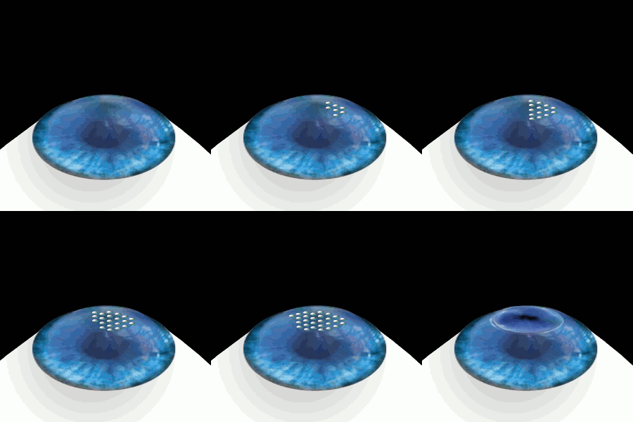
Limbal relaxing incisions (LRI) are a refractive surgical procedure to correct minor astigmatism in the eye. Incisions are made at the opposite edges of the cornea, following the curve of the iris, causing a slight flattening in that direction. Because the incisions are outside of the field of view, they do not cause glare and other visual effects that result from other corneal surgeries like radial keratotomy. LRI have become the most common technique to correct astigmatism as part of cataract surgery. They are simpler and less expensive than laser surgery such as LASIK or photorefractive keratectomy Photorefractive keratectomy (PRK) and laser-assisted sub-epithelial keratectomy (or laser epithelial keratomileusis) (LASEK) are laser eye surgery procedures intended to correct a person's vision, reducing dependency on glasses or contact lenses. .... Good results do not require the location and length of the incisions to be highly precise. And the incisions can easily be extended ... [...More Info...] [...Related Items...] OR: [Wikipedia] [Google] [Baidu] |
Refractive Surgery
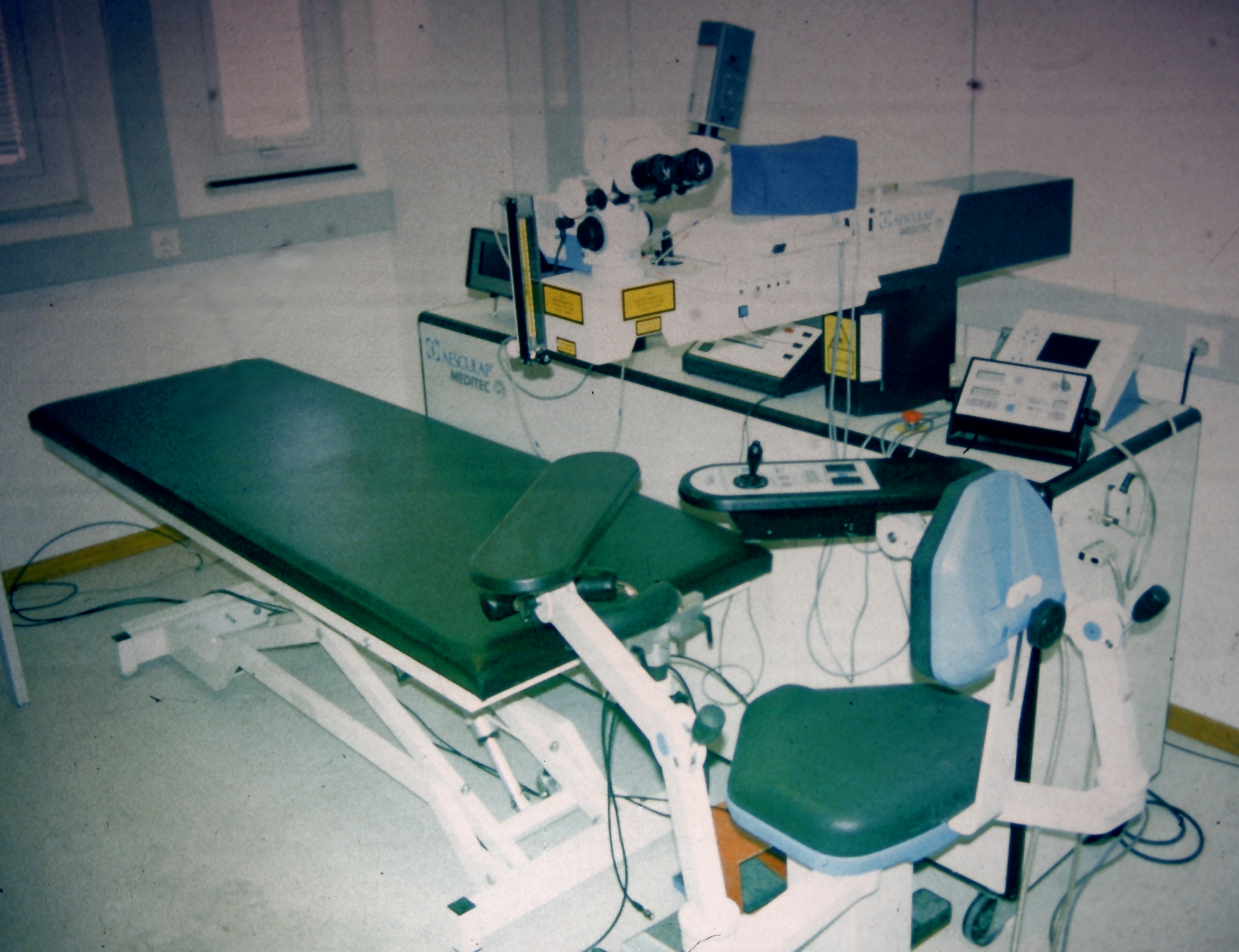
Refractive eye surgery is optional eye surgery used to improve the refractive state of the eye and decrease or eliminate dependency on glasses or contact lenses. This can include various methods of surgical remodeling of the cornea (keratomileusis), lens implantation or lens replacement. The most common methods today use excimer lasers to reshape the curvature of the cornea. Refractive eye surgeries are used to treat common vision disorders such as myopia, hyperopia, presbyopia and astigmatism. History The first theoretical work on the potential of refractive surgery was published in 1885 by Hjalmar August Schiøtz, an ophthalmologist from Norway. In 1930, the Japanese ophthalmologist Tsutomu Sato made the first attempts at performing this kind of surgery, hoping to correct the vision of military pilots. His approach was to make radial cuts in the cornea, correcting effects by up to 6 diopters. The procedure unfortunately produced a high rate of corneal degeneration, howeve ... [...More Info...] [...Related Items...] OR: [Wikipedia] [Google] [Baidu] |
Astigmatism
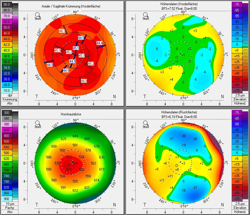
Astigmatism is a type of refractive error due to rotational asymmetry in the eye's refractive power. This results in distorted or blurred vision at any distance. Other symptoms can include eyestrain, headaches, and trouble driving at night. Astigmatism often occurs at birth and can change or develop later in life. If it occurs in early life and is left untreated, it may result in amblyopia. The cause of astigmatism is unclear; however, it is believed to be partly related to genetic factors. The underlying mechanism involves an irregular curvature of the cornea and protective reaction changes in the lens of the eye, called lens astigmatism, that has the same mechanism as spasm of accomodation. Diagnosis is by an eye examination called autorefractor keratometry (objective, allows to see lens and cornea components of astigmatism) and subjective refraction, but subjective methods are almost always inaccurate, if lens astigmatism is not fully removed first with a week of e ... [...More Info...] [...Related Items...] OR: [Wikipedia] [Google] [Baidu] |
Cornea
The cornea is the transparent front part of the eye that covers the iris, pupil, and anterior chamber. Along with the anterior chamber and lens, the cornea refracts light, accounting for approximately two-thirds of the eye's total optical power. In humans, the refractive power of the cornea is approximately 43 dioptres. The cornea can be reshaped by surgical procedures such as LASIK. While the cornea contributes most of the eye's focusing power, its focus is fixed. Accommodation (the refocusing of light to better view near objects) is accomplished by changing the geometry of the lens. Medical terms related to the cornea often start with the prefix "'' kerat-''" from the Greek word κέρας, ''horn''. Structure The cornea has unmyelinated nerve endings sensitive to touch, temperature and chemicals; a touch of the cornea causes an involuntary reflex to close the eyelid. Because transparency is of prime importance, the healthy cornea does not have or need blood vessels with ... [...More Info...] [...Related Items...] OR: [Wikipedia] [Google] [Baidu] |
Iris (anatomy)
In humans and most mammals and birds, the iris (plural: ''irides'' or ''irises'') is a thin, annular structure in the eye, responsible for controlling the diameter and size of the pupil, and thus the amount of light reaching the retina. Eye color is defined by the iris. In optical terms, the pupil is the eye's aperture, while the iris is the diaphragm. Structure The iris consists of two layers: the front pigmented fibrovascular layer known as a stroma and, beneath the stroma, pigmented epithelial cells. The stroma is connected to a sphincter muscle (sphincter pupillae), which contracts the pupil in a circular motion, and a set of dilator muscles ( dilator pupillae), which pull the iris radially to enlarge the pupil, pulling it in folds. The sphincter pupillae is the opposing muscle of the dilator pupillae. The pupil's diameter, and thus the inner border of the iris, changes size when constricting or dilating. The outer border of the iris does not change size. The constricti ... [...More Info...] [...Related Items...] OR: [Wikipedia] [Google] [Baidu] |
Cataract
A cataract is a cloudy area in the lens of the eye that leads to a decrease in vision. Cataracts often develop slowly and can affect one or both eyes. Symptoms may include faded colors, blurry or double vision, halos around light, trouble with bright lights, and trouble seeing at night. This may result in trouble driving, reading, or recognizing faces. Poor vision caused by cataracts may also result in an increased risk of falling and depression. Cataracts cause 51% of all cases of blindness and 33% of visual impairment worldwide. Cataracts are most commonly due to aging but may also occur due to trauma or radiation exposure, be present from birth, or occur following eye surgery for other problems. Risk factors include diabetes, longstanding use of corticosteroid medication, smoking tobacco, prolonged exposure to sunlight, and alcohol. The underlying mechanism involves accumulation of clumps of protein or yellow-brown pigment in the lens that reduces transmission of li ... [...More Info...] [...Related Items...] OR: [Wikipedia] [Google] [Baidu] |
LASIK
LASIK or Lasik (''laser-assisted in situ keratomileusis''), commonly referred to as laser eye surgery or laser vision correction, is a type of refractive surgery for the correction of myopia, hyperopia, and an actual cure for astigmatism, since it is in the cornea. LASIK surgery is performed by an ophthalmologist who uses a laser or microkeratome to reshape the eye's cornea in order to improve visual acuity. For most people, LASIK provides a long-lasting alternative to eyeglasses or contact lenses. LASIK is very similar to another surgical corrective procedure, photorefractive keratectomy (PRK), and LASEK. All represent advances over radial keratotomy in the surgical treatment of refractive errors of vision. For patients with moderate to high myopia or thin corneas which cannot be treated with LASIK and PRK, the phakic intraocular lens is an alternative. As of 2018, roughly 9.5 million Americans have had LASIK and, globally, between 1991 and 2016, more than 40 million proced ... [...More Info...] [...Related Items...] OR: [Wikipedia] [Google] [Baidu] |
Photorefractive Keratectomy
Photorefractive keratectomy (PRK) and laser-assisted sub-epithelial keratectomy (or laser epithelial keratomileusis) (LASEK) are laser eye surgery procedures intended to correct a person's vision, reducing dependency on glasses or contact lenses. LASEK and PRK permanently change the shape of the anterior central cornea using an excimer laser to ablate (remove by vaporization) a small amount of tissue from the corneal stroma at the front of the eye, just under the corneal epithelium. The outer layer of the cornea is removed prior to the ablation. A computer system tracks the patient's eye position 60 to 4,000 times per second, depending on the specifications of the laser that is used. The computer system redirects laser pulses for precise laser placement. Most modern lasers will automatically center on the patient's visual axis and will pause if the eye moves out of range and then resume ablating at that point after the patient's eye is re-centered. The outer layer of the cornea, ... [...More Info...] [...Related Items...] OR: [Wikipedia] [Google] [Baidu] |

.png)


_PHIL_4284_lores.jpg)

