|
Lignosus Hainanensis
''Lignosus'' is a genus of polypore fungi in the family Polyporaceae. The genus was circumscribed in 1920 by mycologists Curtis Gates Lloyd and Camille Torrend, with '' L. sacer'' as the type species. Description The fruit bodies of ''Lignosus'' fungi are annual. They have a cap that is coloured white to brown, with a central supporting stipe. The texture of the cap surface is smooth to very finely tomentose. Pores on the cap underside range in size from small to large. The stipe originates from a sclerotium in the ground. The hyphal system is trimitic. Generative hyphae have clamp connections and are hyaline. There are binding and skeletal hyphae in the context, sclerotium and the stipe. The hymenium lacks cystidia. Spores are smooth, ellipsoid, hyaline, and inamyloid. ''Lignosus'' is similar in morphology to ''Microporus'', but the fungi in this latter genus grow on wood and do not arise from a sclerotium. ''Microporus'' spores are cylindrical to allantoid (s ... [...More Info...] [...Related Items...] OR: [Wikipedia] [Google] [Baidu] |
Fungi
A fungus ( : fungi or funguses) is any member of the group of eukaryotic organisms that includes microorganisms such as yeasts and molds, as well as the more familiar mushrooms. These organisms are classified as a kingdom, separately from the other eukaryotic kingdoms, which by one traditional classification include Plantae, Animalia, Protozoa, and Chromista. A characteristic that places fungi in a different kingdom from plants, bacteria, and some protists is chitin in their cell walls. Fungi, like animals, are heterotrophs; they acquire their food by absorbing dissolved molecules, typically by secreting digestive enzymes into their environment. Fungi do not photosynthesize. Growth is their means of mobility, except for spores (a few of which are flagellated), which may travel through the air or water. Fungi are the principal decomposers in ecological systems. These and other differences place fungi in a single group of related organisms, named the ''Eumycota'' (''t ... [...More Info...] [...Related Items...] OR: [Wikipedia] [Google] [Baidu] |
Stipe (mycology)
In mycology, a stipe () is the stem or stalk-like feature supporting the cap of a mushroom. Like all tissues of the mushroom other than the hymenium, the stipe is composed of sterile hyphal tissue. In many instances, however, the fertile hymenium extends down the stipe some distance. Fungi that have stipes are said to be stipitate. The evolutionary benefit of a stipe is generally considered to be in mediating spore dispersal. An elevated mushroom will more easily release its spores into wind currents or onto passing animals. Nevertheless, many mushrooms do not have stipes, including cup fungi, puffballs, earthstars, some polypores, jelly fungi, ergots, and smuts. It is often the case that features of the stipe are required to make a positive identification of a mushroom. Such distinguishing characters include: # the texture of the stipe (fibrous, brittle, chalky, leathery, firm, etc.) # whether it has remains of a partial veil (such as an annulus or cortina) or universal ve ... [...More Info...] [...Related Items...] OR: [Wikipedia] [Google] [Baidu] |
Microporus
''Microporus'' is a genus of fungi in the family Polyporaceae. The genus has a widespread distribution and, according to a 2008 estimate, contains 11 species. The genus name combines the Ancient Greek words ("small") and ("pore"). Species , Index Fungorum accepts 12 species in ''Microporus'': *''Microporus affinis, M. affinis'' (Blume & T.Nees) Kuntze (1898) *''Microporus affinis-microloma, M. affinis-microloma'' (Lloyd) T.Hatt. & Sotome (2013) *''Microporus atroalbus, M. atroalbus'' (Henn.) Kuntze (1898) *''Microporus atrovillosus, M. atrovillosus'' Ryvarden (1975) *''Microporus concinnus, M. concinnus'' P.Beauv. (1804) *''Microporus incomptus, M. incomptus'' (Afzel. ex Fr.) Kuntze (1898) *''Microporus internuntius, M. internuntius'' (Corner) T. Hatt. (2005) *''Microporus longisporus, M. longisporus'' T.Hatt. (2000) *''Microporus luteoceraceus, M. luteoceraceus'' D.A.Reid (1986) – Peninsular Malaysia *''Microporus nipponicus, M. nipponicus'' (Yasuda) Imazeki (1943) *''Micropo ... [...More Info...] [...Related Items...] OR: [Wikipedia] [Google] [Baidu] |
Morphology (biology)
Morphology is a branch of biology dealing with the study of the form and structure of organisms and their specific structural features. This includes aspects of the outward appearance (shape, structure, colour, pattern, size), i.e. external morphology (or eidonomy), as well as the form and structure of the internal parts like bones and organs, i.e. internal morphology (or anatomy). This is in contrast to physiology, which deals primarily with function. Morphology is a branch of life science dealing with the study of gross structure of an organism or taxon and its component parts. History The etymology of the word "morphology" is from the Ancient Greek (), meaning "form", and (), meaning "word, study, research". While the concept of form in biology, opposed to function, dates back to Aristotle (see Aristotle's biology), the field of morphology was developed by Johann Wolfgang von Goethe (1790) and independently by the German anatomist and physiologist Karl Friedrich Burdach ... [...More Info...] [...Related Items...] OR: [Wikipedia] [Google] [Baidu] |
Inamyloid
In mycology a tissue or feature is said to be amyloid if it has a positive amyloid reaction when subjected to a crude chemical test using iodine as an ingredient of either Melzer's reagent or Lugol's solution, producing a blue to blue-black staining. The term "amyloid" is derived from the Latin ''amyloideus'' ("starch-like"). It refers to the fact that starch gives a similar reaction, also called an amyloid reaction. The test can be on microscopic features, such as spore walls or hyphal walls, or the apical apparatus or entire ascus wall of an ascus, or be a macroscopic reaction on tissue where a drop of the reagent is applied. Negative reactions, called inamyloid or nonamyloid, are for structures that remain pale yellow-brown or clear. A reaction producing a deep reddish to reddish-brown staining is either termed a dextrinoid reaction (pseudoamyloid is a synonym) or a hemiamyloid reaction. Melzer's reagent reactions Hemiamyloidity Hemiamyloidity in mycology refers to a special ... [...More Info...] [...Related Items...] OR: [Wikipedia] [Google] [Baidu] |
Ellipsoid
An ellipsoid is a surface that may be obtained from a sphere by deforming it by means of directional scalings, or more generally, of an affine transformation. An ellipsoid is a quadric surface; that is, a surface that may be defined as the zero set of a polynomial of degree two in three variables. Among quadric surfaces, an ellipsoid is characterized by either of the two following properties. Every planar cross section is either an ellipse, or is empty, or is reduced to a single point (this explains the name, meaning "ellipse-like"). It is bounded, which means that it may be enclosed in a sufficiently large sphere. An ellipsoid has three pairwise perpendicular axes of symmetry which intersect at a center of symmetry, called the center of the ellipsoid. The line segments that are delimited on the axes of symmetry by the ellipsoid are called the ''principal axes'', or simply axes of the ellipsoid. If the three axes have different lengths, the figure is a triaxial ellipsoid (r ... [...More Info...] [...Related Items...] OR: [Wikipedia] [Google] [Baidu] |
Basidiospore
A basidiospore is a reproductive spore produced by Basidiomycete fungi, a grouping that includes mushrooms, shelf fungi, rusts, and smuts. Basidiospores typically each contain one haploid nucleus that is the product of meiosis, and they are produced by specialized fungal cells called basidia. Typically, four basidiospores develop on appendages from each basidium, of which two are of one strain and the other two of its opposite strain. In gills under a cap of one common species, there exist millions of basidia. Some gilled mushrooms in the order Agaricales have the ability to release billions of spores. The puffball fungus ''Calvatia gigantea'' has been calculated to produce about five trillion basidiospores. Most basidiospores are forcibly discharged, and are thus considered ballistospores. These spores serve as the main air dispersal units for the fungi. The spores are released during periods of high humidity and generally have a night-time or pre-dawn peak concentration in the ... [...More Info...] [...Related Items...] OR: [Wikipedia] [Google] [Baidu] |
Cystidia
A cystidium (plural cystidia) is a relatively large cell found on the sporocarp of a basidiomycete (for example, on the surface of a mushroom gill), often between clusters of basidia. Since cystidia have highly varied and distinct shapes that are often unique to a particular species or genus, they are a useful micromorphological characteristic in the identification of basidiomycetes. In general, the adaptive significance of cystidia is not well understood. Classification of cystidia By position Cystidia may occur on the edge of a lamella (or analogous hymenophoral structure) (cheilocystidia), on the face of a lamella (pleurocystidia), on the surface of the cap (dermatocystidia or pileocystidia), on the margin of the cap (circumcystidia) or on the stipe (caulocystidia). Especially the pleurocystidia and cheilocystidia are important for identification within many genera. Sometimes the cheilocystidia give the gill edge a distinct colour which is visible to the naked eye or wit ... [...More Info...] [...Related Items...] OR: [Wikipedia] [Google] [Baidu] |
Hymenium
The hymenium is the tissue layer on the hymenophore of a fungal fruiting body where the cells develop into basidia or asci, which produce spores. In some species all of the cells of the hymenium develop into basidia or asci, while in others some cells develop into sterile cells called cystidia (basidiomycetes) or paraphyses (ascomycetes). Cystidia are often important for microscopic identification. The subhymenium consists of the supportive hyphae from which the cells of the hymenium grow, beneath which is the hymenophoral trama, the hyphae that make up the mass of the hymenophore. The position of the hymenium is traditionally the first characteristic used in the classification and identification of mushrooms. Below are some examples of the diverse types which exist among the macroscopic Basidiomycota and Ascomycota. * In agarics, the hymenium is on the vertical faces of the gills. * In boletes and polypores, it is in a spongy mass of downward-pointing tubes. * In puffballs, ... [...More Info...] [...Related Items...] OR: [Wikipedia] [Google] [Baidu] |
Trama (mycology)
In mycology, the term trama is used in two ways. In the broad sense, it is the inner, fleshy portion of a mushroom's basidiocarp, or fruit body. It is distinct from the outer layer of tissue, known as the pileipellis or cuticle, and from the spore-bearing tissue layer known as the hymenium. In essence, the trama is the tissue that is commonly referred to as the "flesh" of mushrooms and similar fungi.Largent D, Johnson D, Watling R. 1977. ''How to Identify Mushrooms to Genus III: Microscopic Features''. Arcata, CA: Mad River Press. . pp. 60–70. The second use is more specific, and refers to the "hymenophoral trama" that supports the hymenium. It is similarly interior, connective tissue, but it is more specifically the central layer of hyphae running from the underside of the mushroom cap to the lamella or gill, upon which the hymenium rests. Various types have been classified by their structure, including trametoid, cantharelloid, boletoid, and agaricoid, with agaricoid the ... [...More Info...] [...Related Items...] OR: [Wikipedia] [Google] [Baidu] |
Hyaline
A hyaline substance is one with a glassy appearance. The word is derived from el, ὑάλινος, translit=hyálinos, lit=transparent, and el, ὕαλος, translit=hýalos, lit=crystal, glass, label=none. Histopathology Hyaline cartilage is named after its glassy appearance on fresh gross pathology. On light microscopy of H&E stained slides, the extracellular matrix of hyaline cartilage looks homogeneously pink, and the term "hyaline" is used to describe similarly homogeneously pink material besides the cartilage. Hyaline material is usually acellular and proteinaceous. For example, arterial hyaline is seen in aging, high blood pressure, diabetes mellitus and in association with some drugs (e.g. calcineurin inhibitors). It is bright pink with PAS staining. Ichthyology and entomology In ichthyology and entomology, ''hyaline'' denotes a colorless, transparent substance, such as unpigmented fins of fishes or clear insect wings. Resh, Vincent H. and R. T. Cardé, Eds. Encyclo ... [...More Info...] [...Related Items...] OR: [Wikipedia] [Google] [Baidu] |
Clamp Connection
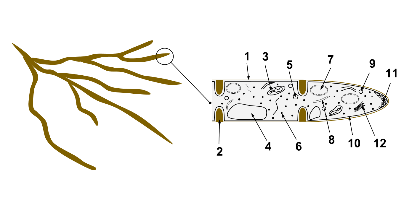
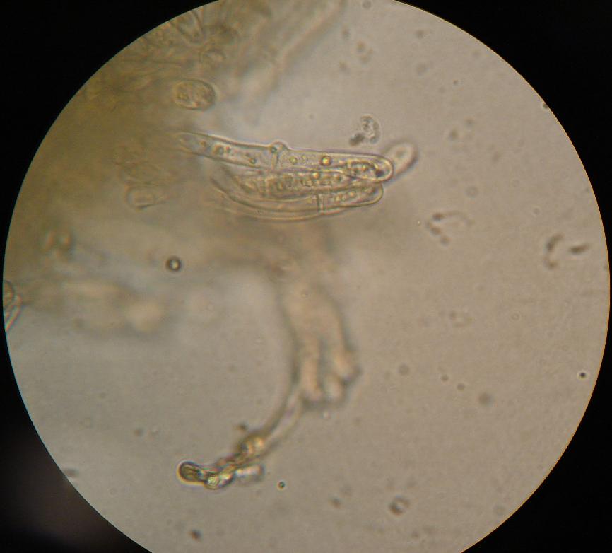
A clamp connection is a hook-like structure formed by growing hyphal cells of certain fungi. It is a characteristic feature of Basidiomycetes fungi. It is created to ensure that each cell, or segment of hypha separated by septa (cross walls), receives a set of differing nuclei, which are obtained through mating of hyphae of differing sexual types. It is used to maintain genetic variation within the hypha much like the mechanisms found in crozier (hook) during sexual reproduction. Formation Clamp connections are formed by the terminal hypha during elongation. Before the clamp connection is formed this terminal segment contains two nuclei. Once the terminal segment is long enough it begins to form the clamp connection. At the same time, each nucleus undergoes mitotic division to produce two daughter nuclei. As the clamp continues to develop it uptakes one of the daughter (green circle) nuclei and separates it from its sister nucleus. While this is occurring the remaining nuclei ... [...More Info...] [...Related Items...] OR: [Wikipedia] [Google] [Baidu] |


