|
Koller's Sickle
In avian gastrulation, Koller's sickle is a local thickening of cells at the posterior edge of the upper layer of the area pellucida called the epiblast. Koller's sickle is crucial for avian development, due to its critical role in inducing the differentiation of various avian body parts. Koller's sickle induces primitive streak and Hensen's node, which are major components of avian gastrulation. Avian gastrulation is a process by which developing cells in an avian embryo move relative to one another in order to form the three germ layers (endoderm, mesoderm, and ectoderm). In-depth definition The thickening of the epiblast in Koller's sickle acts as a margin separating sheets of cells from posterior side of avian blastoderms from hypoblasts and area opaca endoderm. The blastoderm is a single layer of cells, and the hypoblast and area opaca endoderm cells lie directly below the blastoderm. Koller's sickle arises from the midpoint, between the hypoblast cells and the area opaca en ... [...More Info...] [...Related Items...] OR: [Wikipedia] [Google] [Baidu] |
Amphibian
Amphibians are tetrapod, four-limbed and ectothermic vertebrates of the Class (biology), class Amphibia. All living amphibians belong to the group Lissamphibia. They inhabit a wide variety of habitats, with most species living within terrestrial animal, terrestrial, fossorial, arboreal or freshwater aquatic ecosystems. Thus amphibians typically start out as larvae living in water, but some species have developed behavioural adaptations to bypass this. The young generally undergo metamorphosis from larva with gills to an adult air-breathing form with lungs. Amphibians use their skin as a secondary respiratory surface and some small terrestrial salamanders and frogs lack lungs and rely entirely on their skin. They are superficially similar to reptiles like lizards but, along with mammals and birds, reptiles are amniotes and do not require water bodies in which to breed. With their complex reproductive needs and permeable skins, amphibians are often ecological indicators; in re ... [...More Info...] [...Related Items...] OR: [Wikipedia] [Google] [Baidu] |
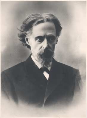
August Rauber
August Rauber (March 9, 1841 – February 16, 1917) was a German anatomist and embryologist born in Obermoschel in the Rhineland-Palatinate. Rauber was born the fourth of five children to Stephan Rauber and Rosalie née Oberlé. He studied medicine in Munich, obtaining his doctorate in 1865. At Munich his instructors included Theodor Bischoff (1807–1882), Nicolaus Rüdinger (1832–1896) and Julius Kollmann (1834–1918).Drw.saw-leipzig.de (biography) Career In 1869 he obtained his habilitation, and in 1872 worked as a dissector at the . Shortly afterwards, he relocated to the |
GSC (gene)
Homeobox protein goosecoid (GSC) is a homeobox protein that is encoded in humans by the ''GSC'' gene. Like other homeobox proteins, goosecoid functions as a transcription factor involved in morphogenesis. In ''Xenopus'', ''GSC'' is thought to play a crucial role in the phenomenon of the Spemann-Mangold organizer. Through lineage tracing and timelapse microscopy, the effects of GSC on neighboring cell fates could be observed. In an experiment that injected cells with GSC and observed the effects of uninjected cells, GSC recruited neighboring uninjected cells in the dorsal blastopore lip of the Xenopus gastrula to form a twinned dorsal axis, suggesting that the goosecoid protein plays a role in the regulation and migration of cells during gastrulation. It is not clear how GSC conducts this organizational function. Errors in the formation of goosecoid protein in mice and humans have a range of consequences on the developing embryo typically in regions of neural crest cell derivatives ... [...More Info...] [...Related Items...] OR: [Wikipedia] [Google] [Baidu] |
NODAL
Nodal homolog is a secretory protein that in humans is encoded by the ''NODAL'' gene which is located on chromosome 10q22.1. It belongs to the transforming growth factor beta superfamily (TGF-β superfamily). Like many other members of this superfamily it is involved in cell differentiation in early embryogenesis, playing a key role in signal transfer from the primitive node, in the anterior primitive streak, to lateral plate mesoderm (LPM). Nodal signaling is important very early in development for mesoderm and endoderm formation and subsequent organization of left-right axial structures. In addition, Nodal seems to have important functions in neural patterning, stem cell maintenance and many other developmental processes, including left/right handedness. Signaling Nodal can bind type I and type II serine/threonine kinase receptors, with Cripto-1 acting as its co-receptor. Signaling through SMAD 2/3 and subsequent translocation of SMAD 4 to the nucleus promotes the expressio ... [...More Info...] [...Related Items...] OR: [Wikipedia] [Google] [Baidu] |
Prechordal Plate
In the development Development or developing may refer to: Arts *Development hell, when a project is stuck in development *Filmmaking, development phase, including finance and budgeting *Development (music), the process thematic material is reshaped * Photograph ... of vertebrate animals, the prechordal plate is a "uniquely thickened portion" of the endoderm that is in contact with ectoderm immediately rostral to the cephalic tip of the notochord. It is the most likely origin of the rostral cranial mesoderm.Seifert, R; et al. ''J Anat'' 1993 183:75-89 STAGE 6 The prechordal plate is a thickening of the endoderm at the cranial end of the primitive streak seen in Embryo Beneke by Hill J.P., Florian J (1963) STAGE 7 The prechordal plate is described as a median mass of cells, located at the anterior end of the notochord, which appears in early embryos as an integral part of the roof of the foregut. e.g. Embryos Bi 24 and Manchester 1285. and Gilbert P.W., (1957) STAG ... [...More Info...] [...Related Items...] OR: [Wikipedia] [Google] [Baidu] |
Notochord
In anatomy, the notochord is a flexible rod which is similar in structure to the stiffer cartilage. If a species has a notochord at any stage of its life cycle (along with 4 other features), it is, by definition, a chordate. The notochord consists of inner, vacuolated cells covered by fibrous and elastic sheaths, lies along the anteroposterior axis (''front to back''), is usually closer to the dorsal than the ventral surface of the embryo, and is composed of cells derived from the mesoderm. The most commonly cited functions of the notochord are: as a midline tissue that provides directional signals to surrounding tissue during development, as a skeletal (structural) element, and as a vertebral precursor. In lancelets the notochord persists throughout life as the main structural support of the body. In tunicates the notochord is present only in the larval stage, being completely absent in the adult animal. In these invertebrate chordates, the notochord is not vacuolated. In all ... [...More Info...] [...Related Items...] OR: [Wikipedia] [Google] [Baidu] |
Primitive Groove
The primitive streak is a structure that forms in the early embryo in amniotes. In amphibians the equivalent structure is the blastopore. During early embryonic development, the embryonic disc becomes oval shaped, and then pear-shaped with the broad end towards the anterior, and the narrower region projected to the posterior. The primitive streak forms a longitudinal midline structure in the narrower posterior (caudal) region of the developing embryo on its dorsal side. At first formation the primitive streak extends for half the length of the embryo. In the human embryo this appears by stage 6, about 17 days. The primitive streak establishes bilateral symmetry, determines the site of gastrulation, and initiates germ layer formation. To form the primitive streak mesenchymal stem cells are arranged along the prospective midline, establishing the second embryonic axis, and the site where cells will ingress and migrate during the process of gastrulation and germ layer format ... [...More Info...] [...Related Items...] OR: [Wikipedia] [Google] [Baidu] |
FGFs
Fibroblast growth factors (FGF) are a family of cell signalling proteins produced by macrophages; they are involved in a wide variety of processes, most notably as crucial elements for normal development in animal cells. Any irregularities in their function lead to a range of developmental defects. These growth factors typically act as systemic or locally circulating molecules of extracellular origin that activate cell surface receptors. A defining property of FGFs is that they bind to heparin and to heparan sulfate. Thus, some are sequestered in the extracellular matrix of tissues that contains heparan sulfate proteoglycans and are released locally upon injury or tissue remodeling. Families In humans, 23 members of the FGF family have been identified, all of which are ''structurally'' related signaling molecules: * Members FGF1 through FGF10 all bind fibroblast growth factor receptors (FGFRs). FGF1 is also known as ''acidic fibroblast growth factor'', and FGF2 is also known a ... [...More Info...] [...Related Items...] OR: [Wikipedia] [Google] [Baidu] |
Wnt Signaling Pathway
The Wnt signaling pathways are a group of signal transduction pathways which begin with proteins that pass signals into a cell through cell surface receptors. The name Wnt is a portmanteau created from the names Wingless and Int-1. Wnt signaling pathways use either nearby cell-cell communication (paracrine) or same-cell communication (autocrine). They are highly evolutionarily conserved in animals, which means they are similar across animal species from fruit flies to humans. Three Wnt signaling pathways have been characterized: the canonical Wnt pathway, the noncanonical planar cell polarity pathway, and the noncanonical Wnt/calcium pathway. All three pathways are activated by the binding of a Wnt-protein ligand to a Frizzled family receptor, which passes the biological signal to the Dishevelled protein inside the cell. The canonical Wnt pathway leads to regulation of gene transcription, and is thought to be negatively regulated in part by the SPATS1 gene. The noncanonical plana ... [...More Info...] [...Related Items...] OR: [Wikipedia] [Google] [Baidu] |
Chordin
Chordin (from Greek χορδή, string, catgut) is a protein with a prominent role in dorsal–ventral patterning during early embryonic development. In humans it is encoded for by the ''CHRD'' gene. History Chordin was originally identified in the African clawed frog (''Xenopus laevis'') in the laboratory of Edward M. De Robertis as a key developmental protein that dorsalizes early vertebrate embryonic tissues. It was first hypothesized that chordin plays a role in the dorsal homeobox genes in Spemann's organizer. The chordin gene was discovered through its activation following use of gsc (goosecoid) and Xnot mRNA injections. The discoverers of chordin concluded that it is expressed in embryo regions where gsc and Xnot were also expressed, which included the prechordal plate, the notochord, and the chordoneural hinge. The expression of the gene in these regions led to the name chordin. Initial functions of chordin were thought to include recruitment of neighboring cells to ... [...More Info...] [...Related Items...] OR: [Wikipedia] [Google] [Baidu] |

.png)


