|
Joint Task Force For Global Network Operations
A joint or articulation (or articular surface) is the connection made between bones, ossicles, or other hard structures in the body which link an animal's skeletal system into a functional whole.Saladin, Ken. Anatomy & Physiology. 7th ed. McGraw-Hill Connect. Webp.274/ref> They are constructed to allow for different degrees and types of movement. Some joints, such as the knee, elbow, and shoulder, are self-lubricating, almost frictionless, and are able to withstand compression and maintain heavy loads while still executing smooth and precise movements. Other joints such as sutures between the bones of the skull permit very little movement (only during birth) in order to protect the brain and the sense organs. The connection between a tooth and the jawbone is also called a joint, and is described as a fibrous joint known as a gomphosis. Joints are classified both structurally and functionally. Classification The number of joints depends on if sesamoids are included, age o ... [...More Info...] [...Related Items...] OR: [Wikipedia] [Google] [Baidu] |
Synovial Joint
A synovial joint, also known as diarthrosis, joins bones or cartilage with a fibrous joint capsule that is continuous with the periosteum of the joined bones, constitutes the outer boundary of a synovial cavity, and surrounds the bones' articulating surfaces. This joint unites long bones and permits free bone movement and greater mobility. The synovial cavity/joint is filled with synovial fluid. The joint capsule is made up of an outer layer of fibrous membrane, which keeps the bones together structurally, and an inner layer, the synovial membrane, which seals in the synovial fluid. They are the most common and most movable type of joint in the body of a mammal. As with most other joints, synovial joints achieve movement at the point of contact of the articulating bones. Structure Synovial joints contain the following structures: * Synovial cavity: all diarthroses have the characteristic space between the bones that is filled with synovial fluid * Joint capsule: the fibrous cap ... [...More Info...] [...Related Items...] OR: [Wikipedia] [Google] [Baidu] |
Fibrous Joint
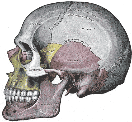
In anatomy, fibrous joints are joints connected by fibrous tissue, consisting mainly of collagen. These are fixed joints where bones are united by a layer of white fibrous tissue of varying thickness. In the skull the joints between the bones are called sutures. Such immovable joints are also referred to as synarthroses. Types Most fibrous joints are also called "fixed" or "immovable". These joints have no joint cavity and are connected via fibrous connective tissue. The skull bones are connected by fibrous joints called '' sutures''. In fetal skulls the sutures are wide to allow slight movement during birth. They later become rigid ( synarthrodial). Some of the long bones in the body such as the radius and ulna in the forearm are joined by a '' syndesmosis'' (along the interosseous membrane). Syndemoses are slightly moveable ( amphiarthrodial). The distal tibiofibular joint is another example. A '' gomphosis'' is a joint between the root of a tooth and the socket in ... [...More Info...] [...Related Items...] OR: [Wikipedia] [Google] [Baidu] |
Pivot Joint
In animal anatomy, a pivot joint (trochoid joint, rotary joint or lateral ginglymus) is a type of synovial joint whose movement axis is parallel to the long axis of the proximal bone, which typically has a convex articular surface. According to one classification system, a pivot joint like the other synovial joint —the hinge joint has one degree of freedom.Platzer, Werner (2008) ''Color Atlas of Human Anatomy'', Volume 1p.28/ref> Note that the degrees of freedom Degrees of freedom (often abbreviated df or DOF) refers to the number of independent variables or parameters of a thermodynamic system. In various scientific fields, the word "freedom" is used to describe the limits to which physical movement or ... of a joint is not the same as the same as joint's range of motion. Movements Pivot joints allow for rotation, which can be external (for example when rotating an arm outward), or internal (as in rotating an arm inward). When rotating the forearm, these movements a ... [...More Info...] [...Related Items...] OR: [Wikipedia] [Google] [Baidu] |
Hinge Joint
A hinge joint (ginglymus or ginglymoid) is a bone joint in which the articular surfaces are molded to each other in such a manner as to permit motion only in one plane. According to one classification system they are said to be uniaxial (having one degree of freedom).Platzer, Werner (2008) ''Color Atlas of Human Anatomy', Volume 1p.28/ref> The direction which the distal bone takes in this motion is seldom in the same plane as that of the axis of the proximal bone; there is usually a certain amount of deviation from the straight line during flexion. The articular surfaces of the bones are connected by strong collateral ligaments. The best examples of ginglymoid joints are the Interphalangeal joints of the hand and those of the foot and the joint between the humerus and ulna. The knee joints and ankle joints are less typical, as they allow a slight degree of rotation or of side-to-side movement in certain positions of the limb. The knee is the largest hinge joint in the h ... [...More Info...] [...Related Items...] OR: [Wikipedia] [Google] [Baidu] |
Ball And Socket Joint
The ball-and-socket joint (or spheroid joint) is a type of synovial joint in which the ball-shaped surface of one rounded bone fits into the cup-like depression of another bone. The distal bone is capable of motion around an indefinite number of axes, which have one common center. This enables the joint to move in many directions. An enarthrosis is a special kind of spheroidal joint in which the socket covers the sphere beyond its equator.Platzer, Werner (2008) ''Color Atlas of Human Anatomy'', Volume 1p.28/ref> Examples Examples of this form of articulation are found in the hip, where the round head of the femur (ball) rests in the cup-like acetabulum (socket) of the pelvis; and in the shoulder joint, where the rounded upper extremity of the humerus (ball) rests in the cup-like glenoid fossa (socket) of the shoulder blade The scapula (plural scapulae or scapulas), also known as the shoulder blade, is the bone that connects the humerus (upper arm bone) with the clavicle ... [...More Info...] [...Related Items...] OR: [Wikipedia] [Google] [Baidu] |
Plane Joint
A plane joint (arthrodial joint, gliding joint, plane articulation) is a synovial joint A synovial joint, also known as diarthrosis, joins bones or cartilage with a fibrous joint capsule that is continuous with the periosteum of the joined bones, constitutes the outer boundary of a synovial cavity, and surrounds the bones' articulat ... which, under physiological conditions, allows only gliding movement. Plane joints permit sliding movements in the plane of articular surfaces. The opposed surfaces of the bones are flat or almost flat, with movement limited by their tight joint capsules. Plane joints are numerous and are nearly always small, such as the acromioclavicular joint between the acromion of the scapula and the clavicle. Typically, they are found in the wrists, ankles, the 2nd through 7th sternocostal joints, vertebral transverse and spinous processes.Moore, et al. ''Introduction to Clinically Oriented Anatomy''. Baltimore: Lippincott Williams & Wilkins, 2006. ... [...More Info...] [...Related Items...] OR: [Wikipedia] [Google] [Baidu] |
Amphiarthrosis
Amphiarthrosis is a type of continuous, slightly movable joint. Types In amphiarthroses, the contiguous bony surfaces can be: * A symphysis: connected by broad flattened disks of fibrocartilage, of a more or less complex structure, which adhere to the ends of each bone, as in the articulations between the bodies of the vertebrae or the inferior articulation of the two hip bones (aka the pubic symphysis). * An interosseous membrane - the sheet of connective tissue Connective tissue is one of the four primary types of animal tissue, along with epithelial tissue, muscle tissue, and nervous tissue. It develops from the mesenchyme derived from the mesoderm the middle embryonic germ layer. Connective tiss ... joining neighboring bones (e.g. tibia and fibula).Principles of Anatomy & Physiology, 12th Edition, Tortora & Derrickson, Pub: Wiley & Sons References External links * Joints {{musculoskeletal-stub ... [...More Info...] [...Related Items...] OR: [Wikipedia] [Google] [Baidu] |
Synarthrosis
A synarthrosis is a type of joint which allows no movement under normal conditions. Sutures and gomphoses are both synarthroses. Joints which allow more movement are called amphiarthroses or diarthroses. Syndesmoses joints are considered to be amphiarthrotic, because they allow a small amount of movement. Types They can be categorised by how the bones are joined together: *'' Gomphosis'' is the type of joint in which a conical peg fits into a socket, for example, the socket of a tooth. Normally, there is very little movement of the teeth in the mandible or maxilla. *'' Synostosis'' is where two bones that are initially separated eventually fuse, essentially becoming one bone. In humans, as in other animals, the plates of the cranium fuse with dense fibrous connective tissue as a child approaches adulthood.Principles of Anatomy & Physiology, 12th Edition, Tortora & Derrickson, Pub: Wiley & Sons Children whose cranial plates fuse too early may suffer deformities and brain damage ... [...More Info...] [...Related Items...] OR: [Wikipedia] [Google] [Baidu] |
Anatomical Planes
An anatomical plane is a hypothetical plane used to transect the body, in order to describe the location of structures or the direction of movements. In human and animal anatomy, three principal planes are used: * The sagittal plane or lateral plane (''longitudinal, anteroposterior'') is a plane parallel to the sagittal suture. It divides the body into left and right. * The coronal plane or frontal plane (''vertical'') divides the body into dorsal and ventral (back and front, or posterior and anterior) portions. * The transverse plane or axial plane (''horizontal'') divides the body into cranial and caudal (head and tail) portions. Terminology There could be any number of sagittal planes; however, there is only one cardinal sagittal plane. The term ''cardinal'' refers to the one plane that divides the body into equal segments, with exactly one half of the body on either side of the cardinal plane. The term ''cardinal plane'' appears in some texts as the ''principal plane'' ... [...More Info...] [...Related Items...] OR: [Wikipedia] [Google] [Baidu] |
Process (anatomy)
In anatomy, a process ( la, processus) is a projection or outgrowth of tissue from a larger body. For instance, in a vertebra, a process may serve for muscle attachment and leverage (as in the case of the transverse and spinous processes), or to fit (forming a synovial joint), with another vertebra (as in the case of the articular processes).Moore, Keith L. et al. (2010) ''Clinically Oriented Anatomy'', 6th Ed, p.442 fig. 4.2 The word is used even at the microanatomic level, where cells can have processes such as cilia or pedicels. Depending on the tissue, processes may also be called by other terms, such as ''apophysis'', '' tubercle'', or ''protuberance''. Examples Examples of processes include: *The many processes of the human skull: ** The mastoid and styloid processes of the temporal bone ** The zygomatic process of the temporal bone ** The zygomatic process of the frontal bone ** The orbital, temporal, lateral, frontal, and maxillary processes of the zygomati ... [...More Info...] [...Related Items...] OR: [Wikipedia] [Google] [Baidu] |
Facet Joint
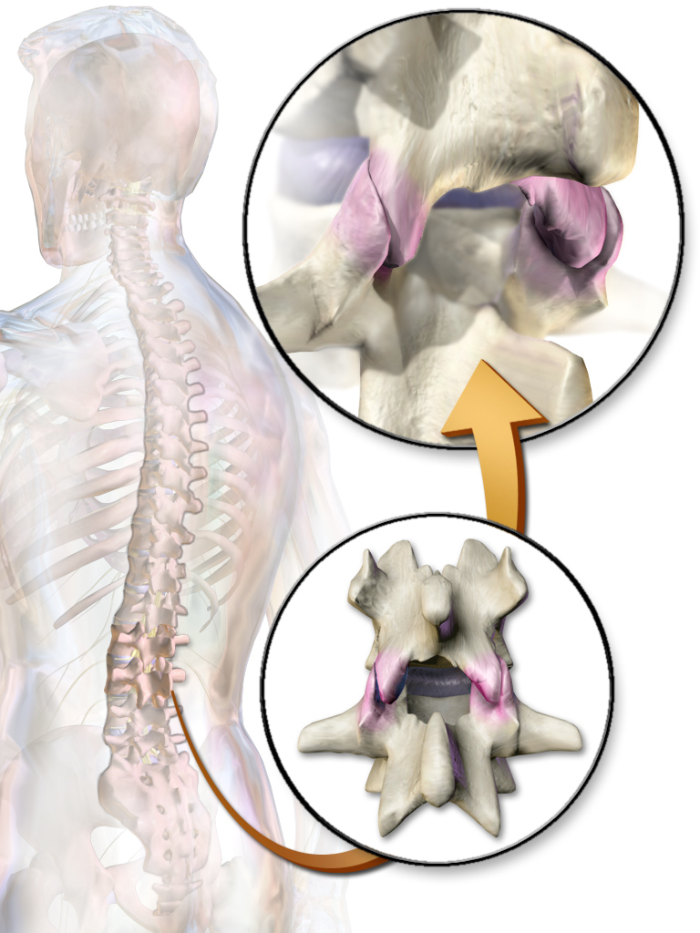
The facet joints (or zygapophysial joints, zygapophyseal, apophyseal, or Z-joints) are a set of synovial, plane joints between the articular processes of two adjacent vertebrae. There are two facet joints in each spinal motion segment and each facet joint is innervated by the recurrent meningeal nerves. Innervation Innervation to the facet joints vary between segments of the spinal, but they are generally innervated by medial branch nerves that come off the dorsal rami. It is thought that these nerves are for primary sensory input, though there is some evidence that they have some motor input local musculature. Within the cervical spine, most joints are innervated by the medial branch nerve (a branch of the dorsal rami) from the same levels. In other words, the facet joint between C4 and C5 vertebral segments is innervated by the C4 and C5 medial branch nerves. However, there are two exceptions: # The facet joint between C2 and C3 is innervated by the third occipital ner ... [...More Info...] [...Related Items...] OR: [Wikipedia] [Google] [Baidu] |
Fibrocartilage
Fibrocartilage consists of a mixture of white fibrous tissue and cartilaginous tissue in various proportions. It owes its inflexibility and toughness to the former of these constituents, and its elasticity to the latter. It is the only type of cartilage that contains type I collagen in addition to the normal type II. Structure The extracellular matrix of fibrocartilage is mainly made from type I collagen secreted by chondroblasts. Locations of fibrocartilage in the human body * secondary cartilaginous joints: ** pubic symphysis ** annulus fibrosis of intervertebral discs ** manubriosternal joint * glenoid labrum of shoulder joint * acetabular labrum of hip joint * medial and lateral menisci of the knee joint * location where tendons and ligaments A ligament is the fibrous connective tissue that connects bones to other bones. It is also known as ''articular ligament'', ''articular larua'', ''fibrous ligament'', or ''true ligament''. Other ligaments in the bo ... [...More Info...] [...Related Items...] OR: [Wikipedia] [Google] [Baidu] |


