|
Intravenous Cholangiography
Intravenous cholangiography is a form of cholangiography that was introduced in 1954. Overview The intravenous cholangiogram or IVC is a radiologic (x-ray) procedure that is used primarily to look at the larger bile ducts within the liver and the bile ducts outside the liver. The procedure can be used to locate gallstones within these bile ducts. IVC also can be used to identify other causes of obstruction to the flow of bile, for example, narrowings (strictures) of the bile ducts and cancers that may impair the normal flow of bile. Procedure To do an IVC, an iodine-containing dye (meglumine ioglycamate) is injected intravenously into the blood. The liver then removes the dye from the blood and excretes it into the bile. The iodine is sufficiently concentrated as it is secreted into the bile that it does not need to be further concentrated by the gallbladder to outline the bile ducts and any gallstones that may be there. The gallbladder is not always seen on an IVC, as the iodine ... [...More Info...] [...Related Items...] OR: [Wikipedia] [Google] [Baidu] |
Cholangiography
Cholangiography is the imaging of the bile duct (also known as the biliary tree) by x-rays and an injection of contrast medium. __TOC__ Types There are at least four types of cholangiography: # Percutaneous transhepatic cholangiography (PTC): Examination of liver and bile ducts by x-rays. This is accomplished by the insertion of a thin needle into the liver carrying a contrast medium to help to see blockage in liver and bile ducts. # Endoscopic retrograde cholangiopancreatography (ERCP). Although this is a form of imaging, it is both diagnostic and therapeutic, and is often classified with surgeries rather than with imaging. # Primary cholangiography (or ''perioperative''): Done in the operation room during a biliary drainage intervention. # Secondary cholangiography: Done after a biliary drainage intervention. In both cases fluorescent fluids are used to create contrasts that make the diagnosis possible. Cholangiography has largely replaced the previously used method of intravenou ... [...More Info...] [...Related Items...] OR: [Wikipedia] [Google] [Baidu] |
Bile Duct
A bile duct is any of a number of long tube-like structures that carry bile, and is present in most vertebrates. Bile is required for the digestion of food and is secreted by the liver into passages that carry bile toward the hepatic duct. It joins the cystic duct (carrying bile to and from the gallbladder) to form the common bile duct which then opens into the intestine. Structure The top half of the common bile duct is associated with the liver, while the bottom half of the common bile duct is associated with the pancreas, through which it passes on its way to the intestine. It opens into the part of the intestine called the duodenum via the ampulla of Vater. Segments The biliary tree (see below) is the whole network of various sized ducts branching through the liver. The path is as follows: Bile canaliculi → Canals of Hering → interlobular bile ducts → intrahepatic bile ducts → left and right hepatic ducts ''merge to form'' → common hepatic duct ''exits live ... [...More Info...] [...Related Items...] OR: [Wikipedia] [Google] [Baidu] |
Gallstone
A gallstone is a calculus (medicine), stone formed within the gallbladder from precipitated bile components. The term cholelithiasis may refer to the presence of gallstones or to any disease caused by gallstones, and choledocholithiasis refers to the presence of migrated gallstones within bile ducts. Most people with gallstones (about 80%) are asymptomatic. However, when a gallstone obstructs the bile duct and causes acute cholestasis, a reflexive smooth muscle spasm often occurs, resulting in an intense cramp-like visceral pain in the quadrant (abdomen), right upper part of the abdomen known as a biliary colic (or "gallbladder attack"). This happens in 1–4% of those with gallstones each year. Complications from gallstones may include inflammation of the gallbladder (cholecystitis), inflammation of the pancreas (pancreatitis), Jaundice#Post-hepatic, obstructive jaundice, and infection in bile ducts (ascending cholangitis, cholangitis). Symptoms of these complications may include ... [...More Info...] [...Related Items...] OR: [Wikipedia] [Google] [Baidu] |
Gallbladder
In vertebrates, the gallbladder, also known as the cholecyst, is a small hollow organ where bile is stored and concentrated before it is released into the small intestine. In humans, the pear-shaped gallbladder lies beneath the liver, although the structure and position of the gallbladder can vary significantly among animal species. It receives and stores bile, produced by the liver, via the common hepatic duct, and releases it via the common bile duct into the duodenum, where the bile helps in the digestion of fats. The gallbladder can be affected by gallstones, formed by material that cannot be dissolved – usually cholesterol or bilirubin, a product of haemoglobin breakdown. These may cause significant pain, particularly in the upper-right corner of the abdomen, and are often treated with removal of the gallbladder (called a cholecystectomy). Cholecystitis, inflammation of the gallbladder, has a wide range of causes, including result from the impaction of gallstones, inf ... [...More Info...] [...Related Items...] OR: [Wikipedia] [Google] [Baidu] |
Small Intestine
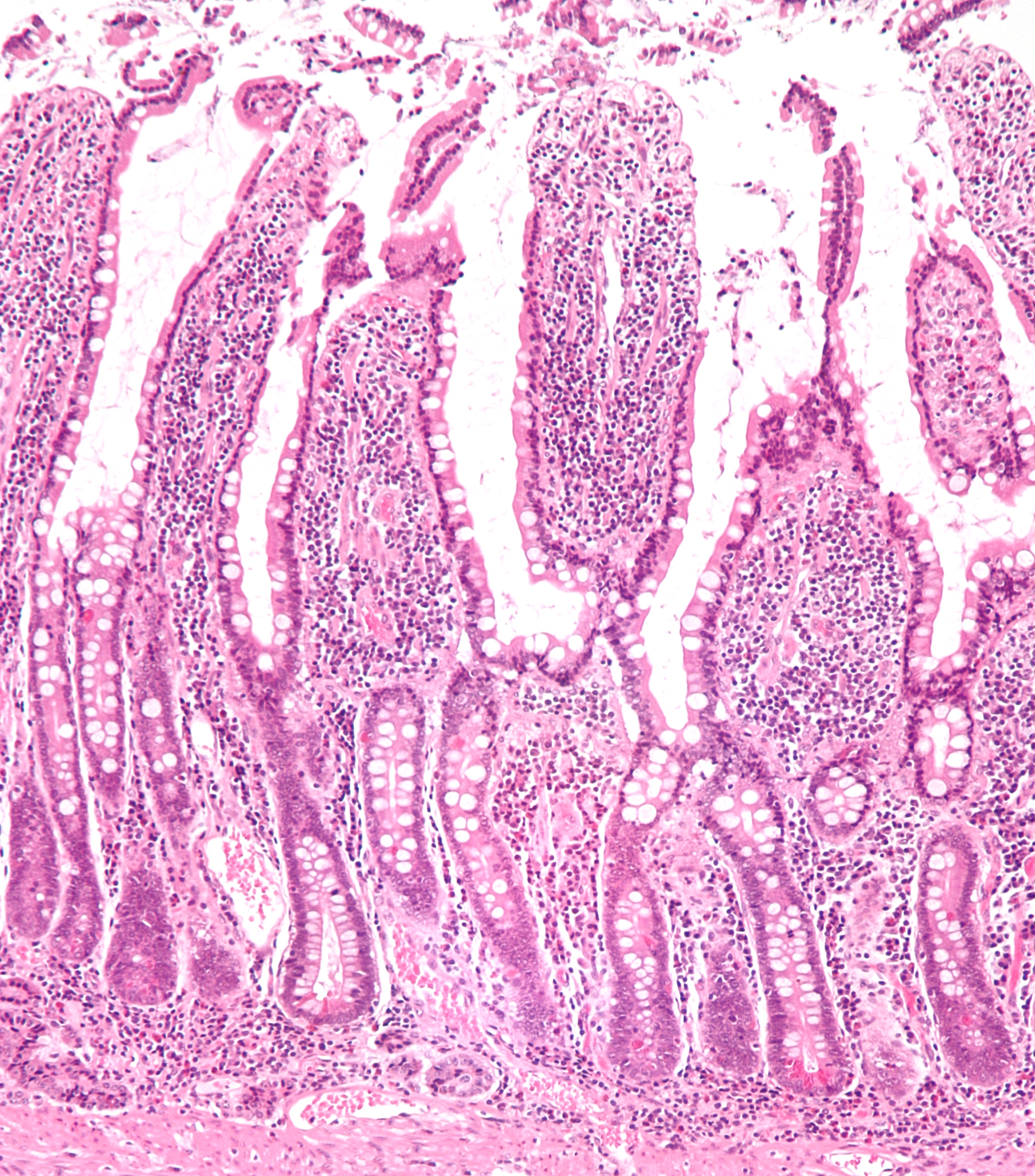
The small intestine or small bowel is an organ in the gastrointestinal tract where most of the absorption of nutrients from food takes place. It lies between the stomach and large intestine, and receives bile and pancreatic juice through the pancreatic duct to aid in digestion. The small intestine is about long and folds many times to fit in the abdomen. Although it is longer than the large intestine, it is called the small intestine because it is narrower in diameter. The small intestine has three distinct regions – the duodenum, jejunum, and ileum. The duodenum, the shortest, is where preparation for absorption through small finger-like protrusions called villi begins. The jejunum is specialized for the absorption through its lining by enterocytes: small nutrient particles which have been previously digested by enzymes in the duodenum. The main function of the ileum is to absorb vitamin B12, bile salts, and whatever products of digestion that were not absorbed by the ... [...More Info...] [...Related Items...] OR: [Wikipedia] [Google] [Baidu] |
Jaundice
Jaundice, also known as icterus, is a yellowish or greenish pigmentation of the skin and sclera due to high bilirubin levels. Jaundice in adults is typically a sign indicating the presence of underlying diseases involving abnormal heme metabolism, liver dysfunction, or biliary-tract obstruction. The prevalence of jaundice in adults is rare, while jaundice in babies is common, with an estimated 80% affected during their first week of life. The most commonly associated symptoms of jaundice are itchiness, pale feces, and dark urine. Normal levels of bilirubin in blood are below 1.0 mg/ dl (17 μmol/ L), while levels over 2–3 mg/dl (34–51 μmol/L) typically result in jaundice. High blood bilirubin is divided into two types – unconjugated and conjugated bilirubin. Causes of jaundice vary from relatively benign to potentially fatal. High unconjugated bilirubin may be due to excess red blood cell breakdown, large bruises, genetic conditions s ... [...More Info...] [...Related Items...] OR: [Wikipedia] [Google] [Baidu] |
Endoscopic Retrograde Cholangiopancreatography
Endoscopic retrograde cholangiopancreatography (ERCP) is a technique that combines the use of endoscopy and fluoroscopy to diagnose and treat certain problems of the biliary or pancreatic ductal systems. It is primarily performed by highly skilled and specialty trained gastroenterologists. Through the endoscope, the physician can see the inside of the stomach and duodenum, and inject a contrast medium into the ducts in the biliary tree and pancreas so they can be seen on radiographs. ERCP is used primarily to diagnose and treat conditions of the bile ducts and main pancreatic duct, including gallstones, inflammatory strictures (scars), leaks (from trauma and surgery), and cancer. ERCP can be performed for diagnostic and therapeutic reasons, although the development of safer and relatively non-invasive investigations such as magnetic resonance cholangiopancreatography (MRCP) and endoscopic ultrasound has meant that ERCP is now rarely performed without therapeutic intent. Medical u ... [...More Info...] [...Related Items...] OR: [Wikipedia] [Google] [Baidu] |
Endoscopic Ultrasound
Endoscopic ultrasound (EUS) or echo-endoscopy is a medical procedure in which endoscopy (insertion of a probe into a hollow organ) is combined with ultrasound to obtain images of the internal organs in the chest, abdomen and colon. It can be used to visualize the walls of these organs, or to look at adjacent structures. Combined with Doppler imaging, nearby blood vessels can also be evaluated. Endoscopic ultrasonography is most commonly used in the upper digestive tract and in the respiratory system. The procedure is performed by gastroenterologists or pulmonologists who have had extensive training. For the patient, the procedure feels almost identical to the endoscopic procedure without the ultrasound part, unless ultrasound-guided biopsy of deeper structures is performed. Digestive tract Upper digestive tract For endoscopic ultrasound of the upper digestive tract, a probe is inserted into the esophagus, stomach, and duodenum during a procedure called esophagogastroduodenoscopy. ... [...More Info...] [...Related Items...] OR: [Wikipedia] [Google] [Baidu] |
Projectional Radiography
Projectional radiography, also known as conventional radiography, is a form of radiography and medical imaging that produces two-dimensional images by x-ray radiation. The image acquisition is generally performed by radiographers, and the images are often examined by radiologists. Both the procedure and any resultant images are often simply called "X-ray". Plain radiography or roentgenography generally refers to projectional radiography (without the use of more advanced techniques such as computed tomography that can generate 3D-images). ''Plain radiography'' can also refer to radiography without a radiocontrast agent or radiography that generates single static images, as contrasted to fluoroscopy, which are technically also projectional. Equipment X-ray generator Projectional radiographs generally use X-rays created by X-ray generators, which generate X-rays from X-ray tubes. Grid An anti-scatter grid may be placed between the patient and the detector to reduce the quanti ... [...More Info...] [...Related Items...] OR: [Wikipedia] [Google] [Baidu] |
Hepatology
Hepatology is the branch of medicine that incorporates the study of liver, gallbladder, biliary tree, and pancreas as well as management of their disorders. Although traditionally considered a sub-specialty of gastroenterology, rapid expansion has led in some countries to doctors specializing solely on this area, who are called hepatologists. Diseases and complications related to viral hepatitis and alcohol are the main reason for seeking specialist advice. More than two billion people have been infected with hepatitis B virus at some point in their life, and approximately 350 million have become persistent carriers. Up to 80% of liver cancers can be attributed to either hepatitis B or hepatitis C virus. In terms of mortality, the former is second only to smoking among known agents causing cancer. With more widespread implementation of vaccination and strict screening before blood transfusion, lower infection rates are expected in the future. In many countries, however, overall ... [...More Info...] [...Related Items...] OR: [Wikipedia] [Google] [Baidu] |


