|
Intracranial Epidermoid Cyst
Intracranial epidermoid cysts develop in the early embryonic phases. The cysts develop when epithelial cells are confined with cells that form the brain. Signs and symptoms Review of past cases, patients often do not exhibit many symptoms or obtain a diagnosis until they are around 20 to 40 years old. If the patient does show symptoms, it is most likely due to pressure from growth of the tumor. Depending on which part the epidermoid is pressing against can result in varying symptoms. * Headaches – often worse in the morning or by changing positions; can be constant and become more severe or more frequent; not your typical headache * Vision problems like blurred vision, double vision, or loss of peripheral vision * Loss of sensation or movement in the arms, legs, or face * Dizziness or difficulty with balance and walking, unsteadiness, vertigo * Speech difficulties * Confusion in everyday matters or disorientation * Seizures, especially in someone who hasn’t had seizures before ... [...More Info...] [...Related Items...] OR: [Wikipedia] [Google] [Baidu] |
Intracranial Epidermoid Cyst - MRI Scans 10
The cranial cavity, also known as intracranial space, is the space within the skull that accommodates the brain. The skull minus the mandible is called the ''cranium''. The cavity is formed by eight cranial bones known as the neurocranium that in humans includes the skull cap and forms the protective case around the brain. The remainder of the skull is called the facial skeleton. Meninges are protective membranes that surround the brain to minimize damage of the brain when there is head trauma. Meningitis is the inflammation of meninges caused by bacterial or viral infections. Structure The capacity of an adult human cranial cavity is 1,200–1,700 cm3. The spaces between meninges and the brain are filled with a clear cerebrospinal fluid, increasing the protection of the brain. Facial bones of the skull are not included in the cranial cavity. There are only eight cranial bones: The occipital, sphenoid, frontal, ethmoid, two parietal, and two temporal bones are fused ... [...More Info...] [...Related Items...] OR: [Wikipedia] [Google] [Baidu] |
Headaches
Headache is the symptom of pain in the face, head, or neck. It can occur as a migraine, tension-type headache, or cluster headache. There is an increased risk of depression in those with severe headaches. Headaches can occur as a result of many conditions. There are a number of different classification systems for headaches. The most well-recognized is that of the International Headache Society, which classifies it into more than 150 types of primary and secondary headaches. Causes of headaches may include dehydration; fatigue; sleep deprivation; stress; the effects of medications (overuse) and recreational drugs, including withdrawal; viral infections; loud noises; head injury; rapid ingestion of a very cold food or beverage; and dental or sinus issues (such as sinusitis). Treatment of a headache depends on the underlying cause, but commonly involves pain medication (especially in case of migraine or cluster headache). A headache is one of the most commonly experienced of ... [...More Info...] [...Related Items...] OR: [Wikipedia] [Google] [Baidu] |
Visual Impairment
Visual impairment, also known as vision impairment, is a medical definition primarily measured based on an individual's better eye visual acuity; in the absence of treatment such as correctable eyewear, assistive devices, and medical treatment– visual impairment may cause the individual difficulties with normal daily tasks including reading and walking. Low vision is a functional definition of visual impairment that is chronic, uncorrectable with treatment or correctable lenses, and impacts daily living. As such low vision can be used as a disability metric and varies based on an individual's experience, environmental demands, accommodations, and access to services. The American Academy of Ophthalmology defines visual impairment as the best-corrected visual acuity of less than 20/40 in the better eye, and the World Health Organization defines it as a presenting acuity of less than 6/12 in the better eye. The term blindness is used for complete or nearly complete vision loss. In ... [...More Info...] [...Related Items...] OR: [Wikipedia] [Google] [Baidu] |
Dizziness
Dizziness is an imprecise term that can refer to a sense of disorientation in space, vertigo, or lightheadedness. It can also refer to disequilibrium or a non-specific feeling, such as giddiness or foolishness. Dizziness is a common medical complaint, affecting 20-30% of persons. Dizziness is broken down into 4 main subtypes: vertigo (~25-50%), disequilibrium (less than ~15%), presyncope (less than ~15%), and nonspecific dizziness (~10%). * Vertigo is the sensation of spinning or having one's surroundings spin about them. Many people find vertigo very disturbing and often report associated nausea and vomiting. * Presyncope describes lightheadedness or feeling faint; the name relates to syncope, which is actually fainting. * Disequilibrium is the sensation of being off balance and is most often characterized by frequent falls in a specific direction. This condition is not often associated with nausea or vomiting. * Non-specific dizziness may be psychiatric in origin. It is ... [...More Info...] [...Related Items...] OR: [Wikipedia] [Google] [Baidu] |
Epidermis
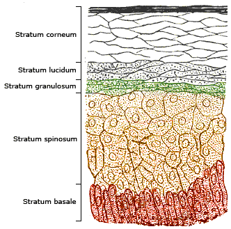
The epidermis is the outermost of the three layers that comprise the skin, the inner layers being the dermis and hypodermis. The epidermis layer provides a barrier to infection from environmental pathogens and regulates the amount of water released from the body into the atmosphere through transepidermal water loss. The epidermis is composed of multiple layers of flattened cells that overlie a base layer (stratum basale) composed of columnar cells arranged perpendicularly. The layers of cells develop from stem cells in the basal layer. The human epidermis is a familiar example of epithelium, particularly a stratified squamous epithelium. The word epidermis is derived through Latin , itself and . Something related to or part of the epidermis is termed epidermal. Structure Cellular components The epidermis primarily consists of keratinocytes ( proliferating basal and differentiated suprabasal), which comprise 90% of its cells, but also contains melanocytes, Langerhans ... [...More Info...] [...Related Items...] OR: [Wikipedia] [Google] [Baidu] |
Neural Tube
In the developing chordate (including vertebrates), the neural tube is the embryonic precursor to the central nervous system, which is made up of the brain and spinal cord. The neural groove gradually deepens as the neural fold become elevated, and ultimately the folds meet and coalesce in the middle line and convert the groove into the closed neural tube. In humans, neural tube closure usually occurs by the fourth week of pregnancy (the 28th day after conception). The ectodermal wall of the tube forms the rudiment of the nervous system. The centre of the tube is the ''neural canal''.It is an important structure for the development of fetus's brain and spine Development The neural tube develops in two ways: primary neurulation and secondary neurulation. Primary neurulation divides the ectoderm into three cell types: * The internally located neural tube * The externally located epidermis * The neural crest cells, which develop in the region between the neural tube and epider ... [...More Info...] [...Related Items...] OR: [Wikipedia] [Google] [Baidu] |
Prenatal Development
Prenatal development () includes the development of the embryo and of the fetus during a viviparous animal's gestation. Prenatal development starts with fertilization, in the germinal stage of embryonic development, and continues in fetal development until birth. In human pregnancy, prenatal development is also called antenatal development. The development of the human embryo follows fertilization, and continues as fetal development. By the end of the tenth week of gestational age the embryo has acquired its basic form and is referred to as a fetus. The next period is that of fetal development where many organs become fully developed. This fetal period is described both topically (by organ) and chronologically (by time) with major occurrences being listed by gestational age. The very early stages of embryonic development are the same in all mammals, but later stages of development, and the length of gestation varies. Terminology In the human: Different terms are used to ... [...More Info...] [...Related Items...] OR: [Wikipedia] [Google] [Baidu] |
Brain Stem
The brainstem (or brain stem) is the posterior stalk-like part of the brain that connects the cerebrum with the spinal cord. In the human brain the brainstem is composed of the midbrain, the pons, and the medulla oblongata. The midbrain is continuous with the thalamus of the diencephalon through the tentorial notch, and sometimes the diencephalon is included in the brainstem. The brainstem is very small, making up around only 2.6 percent of the brain's total weight. It has the critical roles of regulating cardiac, and respiratory function, helping to control heart rate and breathing rate. It also provides the main motor and sensory nerve supply to the face and neck via the cranial nerves. Ten pairs of cranial nerves come from the brainstem. Other roles include the regulation of the central nervous system and the body's sleep cycle. It is also of prime importance in the conveyance of motor and sensory pathways from the rest of the brain to the body, and from the body back to the ... [...More Info...] [...Related Items...] OR: [Wikipedia] [Google] [Baidu] |
Cranial Nerves
Cranial nerves are the nerves that emerge directly from the brain (including the brainstem), of which there are conventionally considered twelve pairs. Cranial nerves relay information between the brain and parts of the body, primarily to and from regions of the head and neck, including the special senses of vision, taste, smell, and hearing. The cranial nerves emerge from the central nervous system above the level of the first vertebra of the vertebral column. Each cranial nerve is paired and is present on both sides. There are conventionally twelve pairs of cranial nerves, which are described with Roman numerals I–XII. Some considered there to be thirteen pairs of cranial nerves, including cranial nerve zero. The numbering of the cranial nerves is based on the order in which they emerge from the brain and brainstem, from front to back. The terminal nerves (0), olfactory nerves (I) and optic nerves (II) emerge from the cerebrum, and the remaining ten pairs arise from the ... [...More Info...] [...Related Items...] OR: [Wikipedia] [Google] [Baidu] |
Cerebellopontine Angle
The cerebellopontine angle (CPA) ( la, angulus cerebellopontinus) is located between the cerebellum and the pons. The cerebellopontine angle is the site of the cerebellopontine angle cistern one of the subarachnoid cisterns that contains cerebrospinal fluid, arachnoid tissue, cranial nerves, and associated vessels. The cerebellopontine angle is also the site of a set of neurological disorders known as the cerebellopontine angle syndrome. Structure The cerebellopontine angle is formed by the cerebellopontine fissure. This fissure is made when the cerebellum folds over to the pons, creating a sharply defined angle between them. The angle formed in turn creates a subarachnoid cistern, the cerebellopontine angle cistern. The pia mater follows the outline of the fissure and the arachnoid mater continues across the divide so that the subarachnoid space is dilated at this area, forming the cerebellopontine angle cistern. The anterior inferior cerebellar artery (AICA) is the principa ... [...More Info...] [...Related Items...] OR: [Wikipedia] [Google] [Baidu] |
Magnetic Resonance Imaging
Magnetic resonance imaging (MRI) is a medical imaging technique used in radiology to form pictures of the anatomy and the physiological processes of the body. MRI scanners use strong magnetic fields, magnetic field gradients, and radio waves to generate images of the organs in the body. MRI does not involve X-rays or the use of ionizing radiation, which distinguishes it from CT and PET scans. MRI is a medical application of nuclear magnetic resonance (NMR) which can also be used for imaging in other NMR applications, such as NMR spectroscopy. MRI is widely used in hospitals and clinics for medical diagnosis, staging and follow-up of disease. Compared to CT, MRI provides better contrast in images of soft-tissues, e.g. in the brain or abdomen. However, it may be perceived as less comfortable by patients, due to the usually longer and louder measurements with the subject in a long, confining tube, though "Open" MRI designs mostly relieve this. Additionally, implants and oth ... [...More Info...] [...Related Items...] OR: [Wikipedia] [Google] [Baidu] |
Computed Tomography
A computed tomography scan (CT scan; formerly called computed axial tomography scan or CAT scan) is a medical imaging technique used to obtain detailed internal images of the body. The personnel that perform CT scans are called radiographers or radiology technologists. CT scanners use a rotating X-ray tube and a row of detectors placed in a gantry to measure X-ray attenuations by different tissues inside the body. The multiple X-ray measurements taken from different angles are then processed on a computer using tomographic reconstruction algorithms to produce tomographic (cross-sectional) images (virtual "slices") of a body. CT scans can be used in patients with metallic implants or pacemakers, for whom magnetic resonance imaging (MRI) is contraindicated. Since its development in the 1970s, CT scanning has proven to be a versatile imaging technique. While CT is most prominently used in medical diagnosis, it can also be used to form images of non-living objects. The 1979 Nob ... [...More Info...] [...Related Items...] OR: [Wikipedia] [Google] [Baidu] |


