|
Infantile Myofibromatosis
Infantile myofibromatosis (IMF) is a rare tumor found in 1 in 150,000 to 1 in 400,000 live births. It is nonetheless the most common tumor derived from fibrous connective tissue that occurs primarily in infants and young children. IMF tumors are benign in the sense that they do not metastasize to distant tissues although when occurring in the viscera, i.e. internal organs, carry guarded to poor prognoses and can be life-threatening, particularly in newborns and young infants. IMF tumors occur in three clinical patterns: 1) solitary IMF tumors (also called myofibromas) which often regress spontaneously and rarely cause serious issues; 2) multiple tumors (no viscera involvement) which consists of numerous (i.e. dozens to >100) IMF lesions most of which are located in the skin and subcutaneous tissues but not visceral organs, may regress spontaneously, and rarely cause serious issues; and 3) multiple tumors (with viscera involvement) (also called generalized myofibromatosis) which rar ... [...More Info...] [...Related Items...] OR: [Wikipedia] [Google] [Baidu] |
Neonatology
Neonatology is a subspecialty of pediatrics that consists of the medical care of newborn infants, especially the ill or premature newborn. It is a hospital-based specialty, and is usually practised in neonatal intensive care units (NICUs). The principal patients of neonatologists are newborn infants who are ill or require special medical care due to prematurity, low birth weight, intrauterine growth restriction, congenital malformations (birth defects), sepsis, pulmonary hypoplasia or birth asphyxia. Historical developments Though high infant mortality rates were recognized by the medical community at least as early as the 1860s, advances in modern neonatal intensive care have led to a significant decline in infant mortality in the modern era. This has been achieved through a combination of technological advances, enhanced understanding of newborn physiology, improved sanitation practices, and development of specialized units for neonatal intensive care. Around the mid-19th ce ... [...More Info...] [...Related Items...] OR: [Wikipedia] [Google] [Baidu] |
Nodule (medicine)
In medicine, nodules are small firm lumps, usually greater than 1 cm in diameter. If filled with fluid they are referred to as cysts. Smaller (less than 0.5 cm) raised soft tissue bumps may be termed papules. The evaluation of a skin nodule includes a description of its appearance, its location, how it feels to touch and any associated symptoms which may give clues to an underlying medical condition. Nodules in skin include dermatofibroma and pyogenic granuloma. Nodules may form on tendons and muscles in response to injury, and are frequently found on vocal cords. They may occur in organs such as the lung, or thyroid, or be a sign in other medical conditions such as rheumatoid arthritis. Characteristics Nodules are small firm lumps usually greater than 1 cm in diameter, found in skin and other organs. If filled with fluid they are usually softer and referred to as cysts. Smaller (less than 0.5 cm) raised soft tissue bumps may be termed papules. Evaluation The evaluation of a ... [...More Info...] [...Related Items...] OR: [Wikipedia] [Google] [Baidu] |
Maxillary Sinus
The pyramid-shaped maxillary sinus (or antrum of Highmore) is the largest of the paranasal sinuses, and drains into the middle meatus of the nose through the osteomeatal complex.Human Anatomy, Jacobs, Elsevier, 2008, page 209-210 Structure It is the largest air sinus in the body. Found in the body of the maxilla, this sinus has three recesses: an alveolar recess pointed inferiorly, bounded by the alveolar process of the maxilla; a zygomatic recess pointed laterally, bounded by the zygomatic bone; and an infraorbital recess pointed superiorly, bounded by the inferior orbital surface of the maxilla. The medial wall is composed primarily of cartilage. The ostia for drainage are located high on the medial wall and open into the semilunar hiatus of the lateral nasal cavity; because of the position of the ostia, gravity cannot drain the maxillary sinus contents when the head is erect (see pathology). The ostium of the maxillary sinus is high up on the medial wall and on average is 2. ... [...More Info...] [...Related Items...] OR: [Wikipedia] [Google] [Baidu] |
Ethmoid Sinus
The ethmoid sinuses or ethmoid air cells of the ethmoid bone are one of the four paired paranasal sinuses. The cells are variable in both size and number in the lateral mass of each of the ethmoid bones and cannot be palpated during an extraoral examination. They are divided into anterior and posterior groups. The ethmoid air cells are numerous thin-walled cavities situated in the ethmoidal labyrinth and completed by the frontal, maxilla, lacrimal, sphenoidal, and palatine bones. They lie between the upper parts of the nasal cavities and the orbits, and are separated from these cavities by thin bony lamellae. Groups of sinuses The groups of the ethmoidal air cells drain into the nasal meatuses.Otorhinolaryngology, Head and Neck Surgery, Anniko, Springer, 2010, page 188 * The posterior group the ''posterior ethmoidal sinus'' drains into the superior meatus above the middle nasal concha; sometimes one or more opens into the sphenoidal sinus. * The anterior group the ''anterior ethmo ... [...More Info...] [...Related Items...] OR: [Wikipedia] [Google] [Baidu] |
Somatic Mutation
A somatic mutation is a change in the DNA sequence of a somatic cell of a multicellular organism with dedicated reproductive cells; that is, any mutation that occurs in a cell other than a gamete, germ cell, or gametocyte. Unlike germline mutations, which can be passed on to the descendants of an organism, somatic mutations are not usually transmitted to descendants. This distinction is blurred in plants, which lack a dedicated germline, and in those animals that can reproduce asexually through mechanisms such as budding, as in members of the cnidarian genus ''Hydra''. While somatic mutations are not passed down to an organism's offspring, somatic mutations will be present in all descendants of a cell within the same organism. Many cancers are the result of accumulated somatic mutations. Fraction of cells affected The term somatic generally refers to the cells of the body, in contrast to the reproductive (germline) cells, which give rise to the egg or sperm. For example, in ... [...More Info...] [...Related Items...] OR: [Wikipedia] [Google] [Baidu] |
Penetrance
Penetrance in genetics is the proportion of individuals carrying a particular variant (or allele) of a gene (the genotype) that also express an associated trait (the phenotype). In medical genetics, the penetrance of a disease-causing mutation is the proportion of individuals with the mutation who exhibit clinical symptoms among all individuals with such mutation. For example, if a mutation in the gene responsible for a particular autosomal dominant disorder has 95% penetrance, then 95% of those with the mutation will develop the disease, while 5% will not. A condition, most commonly inherited in an autosomal dominant manner, is said to show complete penetrance if clinical symptoms are present in all individuals who have the disease-causing mutation. A condition which shows complete penetrance is neurofibromatosis type 1 – every person who has a mutation in the gene will show symptoms of the condition. The penetrance is 100%. Common examples used to show degrees of penetrance are ... [...More Info...] [...Related Items...] OR: [Wikipedia] [Google] [Baidu] |
Dominance (genetics)
In genetics, dominance is the phenomenon of one variant (allele) of a gene on a chromosome masking or overriding the effect of a different variant of the same gene on the other copy of the chromosome. The first variant is termed dominant and the second recessive. This state of having two different variants of the same gene on each chromosome is originally caused by a mutation in one of the genes, either new (''de novo'') or inherited. The terms autosomal dominant or autosomal recessive are used to describe gene variants on non-sex chromosomes ( autosomes) and their associated traits, while those on sex chromosomes (allosomes) are termed X-linked dominant, X-linked recessive or Y-linked; these have an inheritance and presentation pattern that depends on the sex of both the parent and the child (see Sex linkage). Since there is only one copy of the Y chromosome, Y-linked traits cannot be dominant or recessive. Additionally, there are other forms of dominance such as incomplete d ... [...More Info...] [...Related Items...] OR: [Wikipedia] [Google] [Baidu] |
Mutations
In biology, a mutation is an alteration in the nucleic acid sequence of the genome of an organism, virus, or extrachromosomal DNA. Viral genomes contain either DNA or RNA. Mutations result from errors during DNA or viral replication, mitosis, or meiosis or other types of damage to DNA (such as pyrimidine dimers caused by exposure to ultraviolet radiation), which then may undergo error-prone repair (especially microhomology-mediated end joining), cause an error during other forms of repair, or cause an error during replication (translesion synthesis). Mutations may also result from insertion or deletion of segments of DNA due to mobile genetic elements. Mutations may or may not produce detectable changes in the observable characteristics (phenotype) of an organism. Mutations play a part in both normal and abnormal biological processes including: evolution, cancer, and the development of the immune system, including junctional diversity. Mutation is the ultimate source o ... [...More Info...] [...Related Items...] OR: [Wikipedia] [Google] [Baidu] |
Germline Mutation
A germline mutation, or germinal mutation, is any detectable variation within germ cells (cells that, when fully developed, become sperm and ova). Mutations in these cells are the only mutations that can be passed on to offspring, when either a mutated sperm or oocyte come together to form a zygote. After this fertilization event occurs, germ cells divide rapidly to produce all of the cells in the body, causing this mutation to be present in every somatic and germline cell in the offspring; this is also known as a constitutional mutation. Germline mutation is distinct from somatic mutation. Germline mutations can be caused by a variety of endogenous (internal) and exogenous (external) factors, and can occur throughout zygote development. A mutation that arises only in germ cells can result in offspring with a genetic condition that is not present in either parent; this is because the mutation is not present in the rest of the parents' body, only the germline. When mutagenesis oc ... [...More Info...] [...Related Items...] OR: [Wikipedia] [Google] [Baidu] |
Magnetic Resonance Imaging
Magnetic resonance imaging (MRI) is a medical imaging technique used in radiology to form pictures of the anatomy and the physiological processes of the body. MRI scanners use strong magnetic fields, magnetic field gradients, and radio waves to generate images of the organs in the body. MRI does not involve X-rays or the use of ionizing radiation, which distinguishes it from CT and PET scans. MRI is a medical application of nuclear magnetic resonance (NMR) which can also be used for imaging in other NMR applications, such as NMR spectroscopy. MRI is widely used in hospitals and clinics for medical diagnosis, staging and follow-up of disease. Compared to CT, MRI provides better contrast in images of soft-tissues, e.g. in the brain or abdomen. However, it may be perceived as less comfortable by patients, due to the usually longer and louder measurements with the subject in a long, confining tube, though "Open" MRI designs mostly relieve this. Additionally, implants and oth ... [...More Info...] [...Related Items...] OR: [Wikipedia] [Google] [Baidu] |
Spectrogram
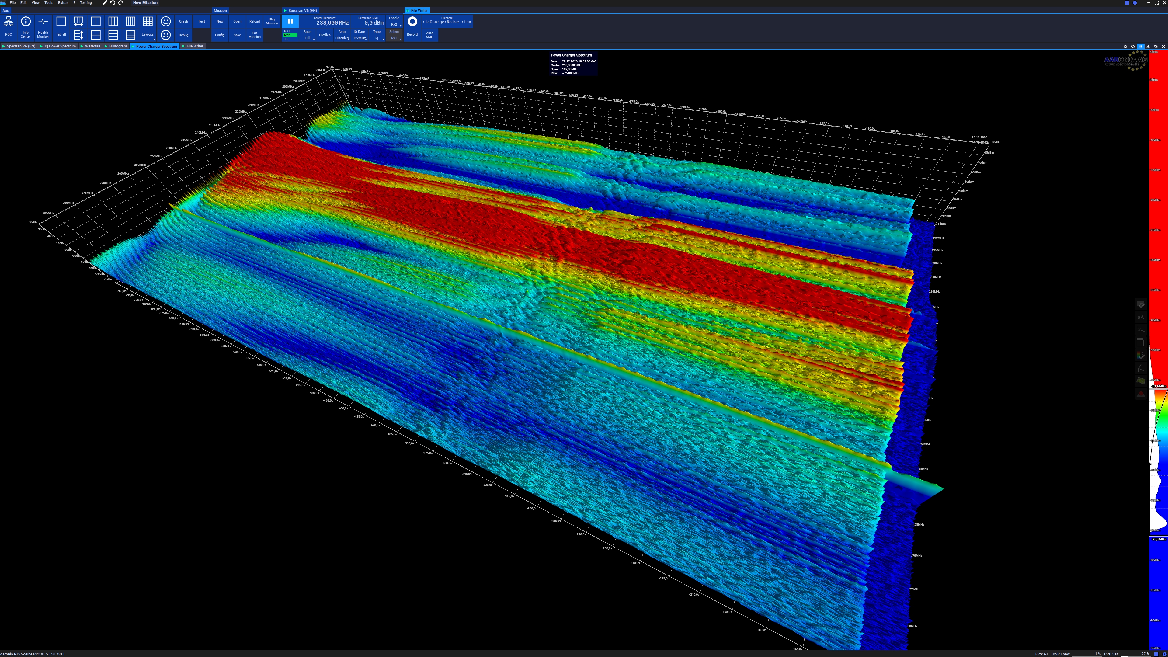
A spectrogram is a visual representation of the spectrum of frequencies of a signal as it varies with time. When applied to an audio signal, spectrograms are sometimes called sonographs, voiceprints, or voicegrams. When the data are represented in a 3D plot they may be called ''waterfall displays''. Spectrograms are used extensively in the fields of music, linguistics, sonar, radar, speech processing, seismology, and others. Spectrograms of audio can be used to identify spoken words phonetically, and to analyse the various calls of animals. A spectrogram can be generated by an optical spectrometer, a bank of band-pass filters, by Fourier transform or by a wavelet transform (in which case it is also known as a scaleogram or scalogram). A spectrogram is usually depicted as a heat map, i.e., as an image with the intensity shown by varying the colour or brightness. Format A common format is a graph with two geometric dimensions: one axis represents time, and the other axis r ... [...More Info...] [...Related Items...] OR: [Wikipedia] [Google] [Baidu] |


